Podcast
Questions and Answers
The apex of the heart is palpated at the left fifth ______ space.
The apex of the heart is palpated at the left fifth ______ space.
intercostal
The left lung's oblique fissure runs along the seventh ______.
The left lung's oblique fissure runs along the seventh ______.
rib
The aortic valve is located at the second ______ space near the sternum.
The aortic valve is located at the second ______ space near the sternum.
intercostal
The mitral valve is located in the fifth intercostal space, lateral to the midpoint of the ______.
The mitral valve is located in the fifth intercostal space, lateral to the midpoint of the ______.
Use 'A Place To ______' to remember the order of aortic, pulmonary, tricuspid, and mitral valves.
Use 'A Place To ______' to remember the order of aortic, pulmonary, tricuspid, and mitral valves.
The heart is located anterior to the ______, flanked by the lungs.
The heart is located anterior to the ______, flanked by the lungs.
The ribcage is formed by the first ten ______.
The ribcage is formed by the first ten ______.
The anterior ribcage includes costal cartilages that articulate with the ______.
The anterior ribcage includes costal cartilages that articulate with the ______.
The ______ runs posteriorly, while the trachea lies anterior.
The ______ runs posteriorly, while the trachea lies anterior.
The sternal angle is the joint between the ______ and sternum body.
The sternal angle is the joint between the ______ and sternum body.
The second costal cartilage indicates the location of the second ______.
The second costal cartilage indicates the location of the second ______.
The ______ major is slightly obscured in women by breast tissue.
The ______ major is slightly obscured in women by breast tissue.
The sternocleidomastoid runs from the neck to the ______.
The sternocleidomastoid runs from the neck to the ______.
Flashcards are hidden until you start studying
Study Notes
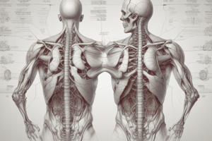
Surface Anatomy of the Thorax
- Surface anatomy studies the body's superficial features and their relation to deeper structures.
- Key thoracic contents include the heart, lungs, trachea, and esophagus, all enclosed by the ribcage formed by the first ten ribs.
Thoracic Structure Overview
- 12 thoracic vertebrae each correspond with a rib.
- Esophagus runs posteriorly, while the trachea, which bifurcates for air delivery, lies anterior.
- The heart is located anterior to the trachea, flanked by the lungs, surrounded by pleural membranes.
Ribcage and Sternum
- Anterior ribcage includes costal cartilages that articulate with the sternum, composed of three parts: manubrium, body, and xiphoid process.
- Clavicle links the manubrium to the scapula, while the coracoid process projects anteriorly from the scapula.
Surface Palpation Techniques
- Locate the jugular notch at the top of the rib cage and feel for aortic pulsations.
- Move laterally to palpate the clavicle and the coracoid process beneath it.
- Sternal angle is the joint between the manubrium and sternum body; a key landmark for anatomical reference.
Intercostal Space Identification
- The second costal cartilage indicates the location of the second rib, with intercostal spaces palpable below.
- Costal margin is demarcated by the tenth rib, costal cartilages, and the xiphoid process.
Musculature of the Thorax
- Palpate the deltoid and pectoralis major; the latter slightly obscured in women by breast tissue.
- The linear alba is a key indentation running vertically along the abdominal muscles.
- Rectus abdominis appears as vertical muscular segments known as the "six-pack."
Important Neck Muscles
- The sternocleidomastoid runs from the neck to the manubrium; palpate it on either side for identification.
Relation to Deeper Structures
- Trachea palpable between sternocleidomastoid muscles; tracheal rings also felt until engulfed by the sternum at the jugular notch.
- Lung apex is 2-3 cm above the clavicle; bases are around the sixth or seventh intercostal space.
- Left lung's oblique fissure runs along the seventh rib; right lung's fissure follows the sixth intercostal space.
Heart Location and Auscultation Points
- The heart is centrally positioned in the thorax, extended into the left thoracic area primarily by the left and right ventricles.
- The apex of the heart is palpated at the left fifth intercostal space.
- Valve auscultation points:
- Aortic valve: second intercostal space near the sternum.
- Pulmonary valve: same horizontal plane as aortic, but left side; locate around the third intercostal space.
- Tricuspid valve: palpate three intercostal spaces down from the aortic valve position.
- Mitral valve: located in the fifth intercostal space, lateral to the midpoint of the clavicle.
Mnemonic for Valve Order
- Use "A Place To Meet" to remember the order of aortic, pulmonary, tricuspid, and mitral valves.
Surface Anatomy of the Thorax
- Surface anatomy examines superficial body features and their connection to deeper structures.
- Thoracic cavity contains crucial organs such as the heart, lungs, trachea, and esophagus.
- The ribcage is formed by the first ten ribs, providing a protective enclosure.
Thoracic Structure Overview
- Comprises 12 thoracic vertebrae, each matching a rib.
- The esophagus is situated posteriorly, while the trachea, bifurcating for air transport, is found anteriorly.
- The heart is located anterior to the trachea, positioned between the lungs and enveloped by pleural membranes.
Ribcage and Sternum
- The anterior ribcage features costal cartilages that connect to the sternum, which consists of three sections: the manubrium, body, and xiphoid process.
- The clavicle connects the manubrium to the scapula, with the coracoid process extending anteriorly from the scapula.
Surface Palpation Techniques
- Identify the jugular notch atop the rib cage to feel aortic pulsations.
- Palpate laterally to locate the clavicle and surrounding coracoid process.
- The sternal angle marks the junction of the manubrium and sternum body, serving as an important anatomical landmark.
Intercostal Space Identification
- The second costal cartilage signals the position of the second rib, with palpable intercostal spaces beneath it.
- The costal margin is defined by the tenth rib, costal cartilages, and xiphoid process.
Musculature of the Thorax
- Palpate visibly the deltoid and pectoralis major muscles, the latter partially obscured by breast tissue in women.
- The linear alba is a prominent vertical tendon along the abdominal muscles.
- Rectus abdominis is identified as vertical muscle segments, commonly referred to as the "six-pack."
Important Neck Muscles
- The sternocleidomastoid muscle runs from the neck to the manubrium, easily palpated on both sides.
Relation to Deeper Structures
- The trachea can be palpated between the sternocleidomastoid muscles, with tracheal rings discernible until obscured by the sternum at the jugular notch.
- The lung apex reaches 2-3 cm above the clavicle, while the bases are around the sixth or seventh intercostal space.
- The left lung's oblique fissure aligns with the seventh rib; the right lung's fissure corresponds to the sixth intercostal space.
Heart Location and Auscultation Points
- Centrally located in the thorax, the heart extends into the left thoracic area due to the left and right ventricles.
- The apex of the heart is palpated at the left fifth intercostal space.
- Valve auscultation points include:
- Aortic valve: located at the second intercostal space near the sternum.
- Pulmonary valve: in the same plane as the aortic but on the left, around the third intercostal space.
- Tricuspid valve: three intercostal spaces below the aortic valve position.
- Mitral valve: situated in the fifth intercostal space, lateral to the clavicle midpoint.
Mnemonic for Valve Order
- Use "A Place To Meet" to remember the sequence of valve auscultation: aortic, pulmonary, tricuspid, and mitral.
Studying That Suits You
Use AI to generate personalized quizzes and flashcards to suit your learning preferences.