Podcast
Questions and Answers
What is the primary imaging purpose of Tc99m-succimer in renal imaging?
What is the primary imaging purpose of Tc99m-succimer in renal imaging?
- To examine ureteral obstruction and reflux
- To assess renal blood flow and perfusion
- To visualize renal cortical outline, size, shape, and contour (correct)
- To identify renal tumors and cysts
Which imaging technique is utilized for normal renal findings with Tc99m-succimer?
Which imaging technique is utilized for normal renal findings with Tc99m-succimer?
- Transverse imaging using ultrasound
- Posterior projection with left and right posterior oblique images (correct)
- Lateral projection only
- Anterior projection with oblique images
What abnormal renal finding is characterized by the kidneys being located anteriorly in the pelvis?
What abnormal renal finding is characterized by the kidneys being located anteriorly in the pelvis?
- Horseshoe kidney (correct)
- Pelvic kidney
- Renal agenesis
- Ectopic kidney
What represents a normal finding when utilizing Tc99m-succimer for renal imaging?
What represents a normal finding when utilizing Tc99m-succimer for renal imaging?
Which of the following is NOT an abnormal finding in renal imaging?
Which of the following is NOT an abnormal finding in renal imaging?
Flashcards
Tc99m-succimer
Tc99m-succimer
A radioactive substance used to visualize the kidneys in static renal imaging.
Static Renal Imaging
Static Renal Imaging
A type of renal imaging that captures a single image of the kidneys.
Horseshoe Kidney
Horseshoe Kidney
A specific congenital abnormality where both kidneys are fused together, forming a 'U' shape.
Ectopic Kidney
Ectopic Kidney
Signup and view all the flashcards
Kidney Absence
Kidney Absence
Signup and view all the flashcards
Study Notes
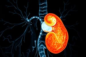
Static Renal Imaging
- Imaging Agent: Tc99m-succimer (1-6 mCi [37-222 MBq])
- Imaging Purpose: Assess renal cortex outline, size, shape, and contour to evaluate healthy renal tissue.
- Imaging Procedure: Posterior projection images, including left and right posterior obliques, taken 2-3 hours after a flow study.
- Normal Findings: Smooth renal outlines, consistent tracer uptake, and consistent tracer distribution throughout both kidneys.
- Abnormal Renal Findings: Congenital abnormalities like horseshoe kidney, ectopic kidney, and absence of one or both kidneys.
Horseshoe Kidney
- Description: Located in the pelvic region, anterior to the normal position, due to incomplete separation of the two kidneys during fetal development.
Studying That Suits You
Use AI to generate personalized quizzes and flashcards to suit your learning preferences.