Podcast
Questions and Answers
Which vertebral group typically articulates with ribs?
Which vertebral group typically articulates with ribs?
- Thoracic (correct)
- Lumbar
- Cervical
- Sacral
Which of the following is a characteristic of 'atypical' vertebrae such as C7?
Which of the following is a characteristic of 'atypical' vertebrae such as C7?
- Full articulation with ribs
- Presence of foramen transversarium
- Absence of foramen transversarium (correct)
- Fusion with the sacrum
The vertebral arch is formed by the:
The vertebral arch is formed by the:
- Lamina and spinous process
- Pedicles and lamina (correct)
- Body and pedicles
- Body and lamina
Which ligament is located anterior to the vertebral bodies and limits extension?
Which ligament is located anterior to the vertebral bodies and limits extension?
Intervertebral discs are described as avascular. How do they receive nutrients?
Intervertebral discs are described as avascular. How do they receive nutrients?
Herniation of an intervertebral disc typically occurs in which direction and why?
Herniation of an intervertebral disc typically occurs in which direction and why?
Destruction of the transverse ligament of the atlas (C1) is most clinically significant in:
Destruction of the transverse ligament of the atlas (C1) is most clinically significant in:
If a disc protrusion occurs at the C3/C4 level, which nerve root is typically compressed?
If a disc protrusion occurs at the C3/C4 level, which nerve root is typically compressed?
The lateral corticospinal tract is primarily responsible for:
The lateral corticospinal tract is primarily responsible for:
In spinal terminology, 'rostral' is synonymous with:
In spinal terminology, 'rostral' is synonymous with:
Which imaging modality is best suited for initial assessment of bony anatomy in trauma situations?
Which imaging modality is best suited for initial assessment of bony anatomy in trauma situations?
In trauma series X-rays, which view is specifically used to visualize the odontoid process and C1-C2 articulation?
In trauma series X-rays, which view is specifically used to visualize the odontoid process and C1-C2 articulation?
For assessing soft tissue structures of the spine, including disc protrusions, which imaging modality is preferred?
For assessing soft tissue structures of the spine, including disc protrusions, which imaging modality is preferred?
What is a key characteristic of a T2-weighted MRI sequence?
What is a key characteristic of a T2-weighted MRI sequence?
In disc prolapse, pain radiating down to the knee, following the L3 dermatome, suggests compression of which nerve root?
In disc prolapse, pain radiating down to the knee, following the L3 dermatome, suggests compression of which nerve root?
Sciatica is a common term for radiculopathy affecting which region of the spine?
Sciatica is a common term for radiculopathy affecting which region of the spine?
Cauda Equina Syndrome is characterized by compression of:
Cauda Equina Syndrome is characterized by compression of:
Which of the following is a 'red flag' symptom suggestive of Cauda Equina Syndrome?
Which of the following is a 'red flag' symptom suggestive of Cauda Equina Syndrome?
Spondylolysis is best described as:
Spondylolysis is best described as:
Spondylolisthesis most commonly occurs at which spinal level?
Spondylolisthesis most commonly occurs at which spinal level?
Spinal stenosis typically involves:
Spinal stenosis typically involves:
Ligamentum flavum thickening is a common cause of:
Ligamentum flavum thickening is a common cause of:
Syringomyelia is defined as:
Syringomyelia is defined as:
In the management of spinal trauma, the ATLS protocol emphasizes immediate immobilization of suspected C-spine injuries. Which of the following is used for this purpose?
In the management of spinal trauma, the ATLS protocol emphasizes immediate immobilization of suspected C-spine injuries. Which of the following is used for this purpose?
According to the ASIA Impairment Scale, a complete spinal cord injury (AIS-A) implies:
According to the ASIA Impairment Scale, a complete spinal cord injury (AIS-A) implies:
In a lateral cervical spine X-ray, what is assessed by tracing the contour lines?
In a lateral cervical spine X-ray, what is assessed by tracing the contour lines?
A Jefferson fracture is a type of fracture affecting which vertebra?
A Jefferson fracture is a type of fracture affecting which vertebra?
Odontoid fractures are fractures of which part of the C2 vertebra?
Odontoid fractures are fractures of which part of the C2 vertebra?
According to the 3-column theory of thoracolumbar spine stability, instability is indicated if how many columns are disrupted?
According to the 3-column theory of thoracolumbar spine stability, instability is indicated if how many columns are disrupted?
Anterior cord syndrome is characterized by:
Anterior cord syndrome is characterized by:
Flashcards
The vertebral column
The vertebral column
Extends from the skull to the pelvis; consists of 33 vertebrae divided into 5 groups.
Atypical Vertebrae
Atypical Vertebrae
C1, C2, C7, T9-T12, Sacrum and Coccyx.
Vertebral Body
Vertebral Body
Located anteriorly, this is the main weight-bearing structure of a vertebra.
Lamina and Pedicles
Lamina and Pedicles
Signup and view all the flashcards
Anterior Longitudinal Ligament
Anterior Longitudinal Ligament
Signup and view all the flashcards
Posterior Longitudinal Ligament
Posterior Longitudinal Ligament
Signup and view all the flashcards
Intervertebral Discs
Intervertebral Discs
Signup and view all the flashcards
Annulus Fibrosus
Annulus Fibrosus
Signup and view all the flashcards
Nucleus Pulposus
Nucleus Pulposus
Signup and view all the flashcards
Rheumatoid Arthritis Spine
Rheumatoid Arthritis Spine
Signup and view all the flashcards
Function of Dorsal Columns
Function of Dorsal Columns
Signup and view all the flashcards
Cephalad
Cephalad
Signup and view all the flashcards
Caudad
Caudad
Signup and view all the flashcards
CT Scan Use
CT Scan Use
Signup and view all the flashcards
MRI Scan Use
MRI Scan Use
Signup and view all the flashcards
ATLS Plain Film X-rays
ATLS Plain Film X-rays
Signup and view all the flashcards
PEG view
PEG view
Signup and view all the flashcards
Disc Prolapse
Disc Prolapse
Signup and view all the flashcards
Lumbar Radiculopathy or Sciatica
Lumbar Radiculopathy or Sciatica
Signup and view all the flashcards
Cauda Equina Syndrome
Cauda Equina Syndrome
Signup and view all the flashcards
Cauda Equina Syndrome - Presentation
Cauda Equina Syndrome - Presentation
Signup and view all the flashcards
Spondylopathy
Spondylopathy
Signup and view all the flashcards
Spondylosis
Spondylosis
Signup and view all the flashcards
Spondylolysis
Spondylolysis
Signup and view all the flashcards
Spondylolisthesis
Spondylolisthesis
Signup and view all the flashcards
Spondyloptosis
Spondyloptosis
Signup and view all the flashcards
Spinal Stenosis
Spinal Stenosis
Signup and view all the flashcards
Syringomyelia
Syringomyelia
Signup and view all the flashcards
Spinal shock
Spinal shock
Signup and view all the flashcards
Central Cord Syndrome
Central Cord Syndrome
Signup and view all the flashcards
Study Notes
Learning Objectives
- It is essential to revise THEP 1 Anatomy & Clinical MSK lectures alongside this one.
- A solid grasp of common spine pathologies is a must.
- Understanding the investigation and management of these pathologies is necessary.
- Understanding spine trauma is required.
Bony Anatomy
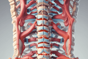
- The spine runs from the skull to the pelvis consisting of 33 vertebrae.
- The spine is divided into 5 groups: 7 Cervical, 12 Thoracic, 5 Lumbar, 5 fused in the Sacrum, and 4 fused in the Coccyx.
- 'Atypical' vertebrae include: C1, C2, C7, T9-T12, Sacrum, and Coccyx.
- C7 lacks a foramen transversarium.
- T9-T12 only partly articulate with ribs.
- A typical vertebra has a vertebral body anteriorly.
- Posteriorly and laterally, lamina and pedicles form an arch.
Intervertebral Discs
- They make up one-quarter of the spinal column's length.
- No intervertebral discs between the Atlas (C1), Axis (C2), Sacrum, and Coccyx.
- Discs are avascular, depending on endplates to diffuse necessary nutrients.
- Discs are fibrocartilaginous cushions, acting as the spine's shock absorbers.
- They allow vertebral motion, including extension and flexion.
- The disc has secondary cartilaginous joints.
- Extension and flexion are also allowed.
- Intervertebral discs consist of the annulus fibrosus and nucleus pulposus.
- Discs usually herniate posteriorly because of a thinner annulus fibrosus.
Transverse Ligament and Rheumatoid Spine
- In Rheumatoid Arthritis, the transverse ligament gets destroyed because of the inflammatory process.
- This destruction leads to Atlantoaxial instability.
- Before intubation, a C-spine XR is important.
Nerve Roots
- Nerve root compression is a common pathology.
- Disc protrusion compresses the nerve root below the level of the disc.
- A C3/4 disc protrusion would compress the C4 nerve root.
- An L4/5 disc protrusion typically compresses the L5 nerve root.
Spinal Cord Pathways
- Descending spinal cord tracts manage motor function.
- Lateral Corticospinal Tract manages motor function.
- Ventral Corticospinal Tract manages motor function.
- Ascending spinal cord tracts manage sensory function.
- Doral Columns deal with deep touch, proprioception, and vibration.
- Lateral Spinothalamic Tract handles pain and temperature.
- Ventral Spinothalamic Tract handles light touch.
Spinal Terminology
- Inter: between.
- Cephalad/Rostral: pertaining to or located towards the head or front.
- Caudad: pertaining to or located towards the tail or posterior.
- Ventral: located on the front side.
- Dorsal: located on the back side.
- Axial/Transverse, Sagittal, and Coronal are body planes.
Imaging Modalities
- X-rays or Radiographs are used for bony anatomy.
- Used in trauma situations.
- Trauma series ATLS Plain film X-rays include C-spine, Erect Chest, and AP pelvis views.
- In emergency departments for major trauma, a CT scan may be done immediately instead of X-rays.
- X-rays are fast and easy but are often inadequate.
- CT scans reveal bony anatomy and subluxation with greater detail utilizing the same language as radiographs.
- MRI is used for soft tissue anatomy, disc protrusion, and collections. – Increased/decreased signal intensity
- All vertebrae must be visible from the skull to the top of T2 in a cervical spine radiograph.
- AP, lateral and PEG views are needed.
- CT scans are increasingly used to assess C-spine injuries.
- Peg views are helpful to viewing lateral mass fractures.
- Increased distance or asymmetry between the peg and lateral masses may indicate fracture or ligament disruption.
- Indications for CT spine include spinal trauma, evaluation of lesions (tumors), and congenital spine abnormalities (scoliosis).
- CT images are described by body part (spine) and axis.
- MRI is indicated for disc disease, tumors, and soft tissues.
- MRI sequences include T1-weighted, T2-weighted, STIR, and T1-weighted fat sat with gadolinium contrast.
- MRI images are described by body part, axis, and sequence.
- MRI sequences are identified by information on the scan.
- T1 and T2-weighted scans are common and easy to distinguish.
- In T1 weighted images CSF (cerebrospinal fluid) appears dark.
- In T2 weighted images CSF (cerebrospinal fluid) appears bright.
- T2-weighted sagittal lumbosacral is a specific MRI type for a particular examination.
Disc Prolapse
- The intervertebral disc herniates into the spinal canal, causing irritation or compression of nerves.
- Signs and symptoms vary depending on structures affected and can include being asymptomatic, nerve root irritation, pain radiating down the leg (e.g., L3 compression to the knee), numbness, and even late paralysis.
- Causes include trauma and degenerative discs.
Disc Prolapse Management
- Non-operative measures include waiting about 4 weeks, bed rest, analgesia, muscle relaxants (e.g., diazepam), and epidural injections
- Operative measures include a discectomy which can be standard or microdiscectomy.
- Complications include nerve injury, infection, spinal abscess, and CSF leak.
Radiculopathy vs Myelopathy
- Radiculopathy: Nerve root compression
- Myelopathy: Spinal cord compression
- Lumbar radiculopathy = sciatica
Cauda Equina Syndrome
- The Cauda Equina Syndrome compression involves some or all nerve roots of the cauda equina.
- Loss of bowel or bladder function is a symptom.
- Emergent MRI is required.
- Early surgery is required to avoid bladder/bowel incontinence and lower limb weakness.
- Admission is important because patients needs counsel on back pain.
Spinal Pathology
- Spondylopathy is a general term for a disorder of the vertebrae.
- Spondylosis refers to osteoarthritis of the vertebrae.
- Spondylolysis refers to a pars inter-articularis defect that can progress to spondylolisthesis.
- Spondylolisthesis is the slipping of one vertebra over another.
- Spondyloptosis is a high-grade (completely slipped) spondylolisthesis.
- Spondylolisthesis is common in adolescents and children.
- It involves the displacement of a vertebral segment over another beneath it
- The most common site is at L4/5 and L5/S1 spinal segments.
- The disease process begins with stress reaction in the pars articularis.
- Spondylolisthesis treatment includes both conservative and surgical management.
- Conservative treatment includes bed rest and a supporting brace.
- Surgical intervention is indicated if symptoms are disabling, particularly in young adults, or if neurological compression is evident.
- Spinal fusion may be necessary.
Spinal Stenosis
- Spinal stenosis involves the narrowing of the spinal canal.
- This can result from thickened ligaments (eg, ligamentum flavum), a congenitally narrow canal, arthritic changes, or compression fractures.
- Symptoms include those of nerve compression, such as pain, paraesthesia, motor deficits, and neurogenic claudication
- Investigations involve XR and MRI imaging
- Non-operative management includes analgesics, NSAIDs, weight loss, and aerobic exercise.
- Surgical options include decompressive laminectomy, and decompression with fusion is multiple levels are involved.
Syrinx & Syringomyelia
- Syringomyelia is a syrinx (fluid-filled cavity) within the spinal cord that progressively expands, which leads to neurologic deficits.
- It is secondary to lesions that partially obstruct CSF flow. – Chiari malformations, Spinal cord trauma, infections, tumours, and Scoliosis
- Treatment is only necessary if symptomatic.
Management of Spinal Trauma
- Activate the trauma team.
- Follow the Advanced Trauma Life Support (ATLS) protocol.
- Suspected C-spine injuries should be immediately immobilized with a hard collar and sandbags.
- Assess airway with suction and airway adjuncts if required
- Assess breathing with bag, mask/intubation, and 100% O2 via non-rebreather mask
- Assess circulation because neurogenic shock is rare, and haemorrhagic shock is likely
- Assess for disability through GCS, motor, and sensory levels
Cervical Spine Examination
- Verify that awake, alert, and not intoxicated.
- No distracting painful injury.
- Rule out neck pain or midline tenderness.
- Remove collar and palpate spine if no pain is present.
- When pain is present, exclude an injury with AP, Lateral, and PEG (open mouth) views.
- CT imaging is indicated.
- Ensure visualization from C1 to T1.
Assessing Lateral Films
- There are several components to assessment:
- The top of T1 should be visible.
- All 3 contour lines should be traced. – Check Vertebral bodies and Intervertebral disc spaces Check soft tissues
- Use a long AP view to check the spinous process alignment and any abnormal widening of the interspinous distance.
- C1 (Atlas) fractures can be burst fractures (Jefferson), which are unstable due to the vertical compression force being transmitted through the occipital condyles to lateral masses
- Instability is dictated by transverse ligament involvement
- Posterior arch fractures of C1 are more stable, but potentially very dangerous.
- Hangman fractures of the pedicle or the odontoid PEG fractures are fractures of C2.
- Other C2 fractures include anterior wedge and spinous process.
- In Type 1 fractures, the location is typically specified as the upper dens.
- The orientation or pattern of the fracture is noted as being oblique.
- In Type II fractures, the zone is defined as the base of dens, showcasing the transverse direction or orientation of the fracture pattern.
- Type III fractures manifests in Body of axis, involving the facets.
- Thoraco-lumbar spine instability results if 2 out of 3 columns are disrupted.
Thoraco Lumbar Spine
- Compression fractures exhibit a distinct pattern identified as a wedge or anterior
- Accounts for 50-70% of all TL fractures, generally stable, usually affecting one column.
- Approximately 15% of all TL injuries, exhibiting an unstable nature, and these fractures effect over one vertebral column.
- There are Flexion-distraction (lap belt) injuries
- Comprising about 10% of all TL spinal column injuries.
Spinal Shock
- A misnomer that does not relate to hemodynamic compromise.
- Involves a loss of cord function and reflex activity below the level of injury.
- Above T6 injuries may cause neurogenic shock, hypotension, bradycardia and peripheral vasodilation.
- The four stages of spinal shock:
- Areflexia (first 24hrs)
- Initial reflex return (1-3 days)
- Hyperreflexia (1-4 weeks)
- Hyperreflexia/spasticity (4 weeks +)
Cord Syndromes
- Cord syndromes include central cord syndrome, anterior cord syndrome, and Brown-Sequard syndrome.
- Central cord syndrome is common in incomplete injuries with neck extension
- Hand motor fibers are more central, and hands are more affected than legs.
- Brown Sequard syndrome is rare.
- Is the result of ipsilateral loss of motor function, vibration, and proprioception.
- Includes contralateral loss of pain and temperature sensation.
- Anterior Cord Syndrome is the result of direct or indirect injury to the anterior spinal cord.
- Spinothalamic tract injury can be characterized loss of motor, pain, light touch, and temperature sensation.
- Don't forget spinal hematomas which can be intradural, epidural, the result of trauma and related to being elderly.
- Differences between BP, Pulse, Motor, Reflexes in Spinal Shock, Hypovolemic Shock and Neurogenic shock are important to recall.
Studying That Suits You
Use AI to generate personalized quizzes and flashcards to suit your learning preferences.


