Podcast
Questions and Answers
Dorsal root ganglia contain the cell bodies of motor neurons.
Dorsal root ganglia contain the cell bodies of motor neurons.
False (B)
Each dorsal root is typically divided into 10-12 rootlets.
Each dorsal root is typically divided into 10-12 rootlets.
False (B)
The lateral section of the dorsal root conveys fine touch and vibration sensations.
The lateral section of the dorsal root conveys fine touch and vibration sensations.
False (B)
Aδ and C fibers enter the dorsal horn at the level of the Lissauer dorsolateral tract.
Aδ and C fibers enter the dorsal horn at the level of the Lissauer dorsolateral tract.
Second-order neurons in lamina I receive input from Aβ fibers.
Second-order neurons in lamina I receive input from Aβ fibers.
The green neuron in the substantia gelatinosa is primarily involved in the direct transmission of pain signals.
The green neuron in the substantia gelatinosa is primarily involved in the direct transmission of pain signals.
The medial section of the dorsal root feeds into the spinothalamic tract.
The medial section of the dorsal root feeds into the spinothalamic tract.
The ascending branch of Aβ fibers within the dorsal funiculus terminates in the cuneate and gracilis nuclei.
The ascending branch of Aβ fibers within the dorsal funiculus terminates in the cuneate and gracilis nuclei.
The spinocerebellar tract transmits conscious proprioceptive information to the cerebellum.
The spinocerebellar tract transmits conscious proprioceptive information to the cerebellum.
Mossy fibers of the cerebellum are directly connected to the axons of proprioceptive neurons.
Mossy fibers of the cerebellum are directly connected to the axons of proprioceptive neurons.
The intermedio-lateral column contains preganglionic parasympathetic neurons primarily within the T1-L2 segments.
The intermedio-lateral column contains preganglionic parasympathetic neurons primarily within the T1-L2 segments.
The intermedio-medial column primarily receives afferent input from the ventral roots related to motor reflexes.
The intermedio-medial column primarily receives afferent input from the ventral roots related to motor reflexes.
Lamina VIII interneurons project exclusively ipsilaterally to laminae 7 and 9.
Lamina VIII interneurons project exclusively ipsilaterally to laminae 7 and 9.
The phrenic nucleus, located in the C2-C5 ventromedial region, controls the muscles of the limbs.
The phrenic nucleus, located in the C2-C5 ventromedial region, controls the muscles of the limbs.
The Onuf nucleus, situated in the S1-S2 ventrolateral region, innervates muscles for sphincters.
The Onuf nucleus, situated in the S1-S2 ventrolateral region, innervates muscles for sphincters.
The accessory spinal nucleus, located in the C1-C5 lateral area, receives bilateral inputs from the corticospinal tract to control lateral neck muscles.
The accessory spinal nucleus, located in the C1-C5 lateral area, receives bilateral inputs from the corticospinal tract to control lateral neck muscles.
Rexed lamina X is exclusive to the posterior horn of the spinal cord.
Rexed lamina X is exclusive to the posterior horn of the spinal cord.
The gray commissure is divided into an anterior and a posterior part by the central nucleus.
The gray commissure is divided into an anterior and a posterior part by the central nucleus.
The intermediate zone of the spinal cord gray matter predominantly consists of Rexed laminae VI, VII, and IX.
The intermediate zone of the spinal cord gray matter predominantly consists of Rexed laminae VI, VII, and IX.
The fasciculus proprius is predominantly found within the lateral columns of the white matter.
The fasciculus proprius is predominantly found within the lateral columns of the white matter.
The vestibulospinal, reticulospinal, and tectospinal tracts are all ascending sensory pathways.
The vestibulospinal, reticulospinal, and tectospinal tracts are all ascending sensory pathways.
The reticulospinal tract originates from the superior colliculus.
The reticulospinal tract originates from the superior colliculus.
Medial descending tracts connect with the ventromedial gray matter to control distal limb muscles.
Medial descending tracts connect with the ventromedial gray matter to control distal limb muscles.
The tectospinal tract has ipsilateral projections and is involved in posture.
The tectospinal tract has ipsilateral projections and is involved in posture.
Lateral corticospinal and rubrospinal tracts control distal muscles by targeting the dorsolateral grey matter of the ventral laminae.
Lateral corticospinal and rubrospinal tracts control distal muscles by targeting the dorsolateral grey matter of the ventral laminae.
Corticospinal projections are 60% contralateral and 40% ipsilateral, primarily targeting motor neurons in the spinal cord.
Corticospinal projections are 60% contralateral and 40% ipsilateral, primarily targeting motor neurons in the spinal cord.
The rubrospinal tract projects ipsilaterally onto interneurons.
The rubrospinal tract projects ipsilaterally onto interneurons.
Aminergic tracts from the brainstem use only serotonin and noradrenaline as neurotransmitters in the spinal cord for modulation.
Aminergic tracts from the brainstem use only serotonin and noradrenaline as neurotransmitters in the spinal cord for modulation.
The raphe-spinal system uses noradrenaline as a neurotransmitter and the coeruleus-spinal tract uses serotonin.
The raphe-spinal system uses noradrenaline as a neurotransmitter and the coeruleus-spinal tract uses serotonin.
Vegetative projections from the upper centers target the spinal cord preganglionic neurons.
Vegetative projections from the upper centers target the spinal cord preganglionic neurons.
The periaqueductal grey, once activated, will primarily activate the neurons which are insensitive to endorphins.
The periaqueductal grey, once activated, will primarily activate the neurons which are insensitive to endorphins.
Neurons in the periaqueductal grey project to the locus coeruleus and the raphe nucleus, which project down to first order neurons.
Neurons in the periaqueductal grey project to the locus coeruleus and the raphe nucleus, which project down to first order neurons.
Morphine and enkephalins act by randomly diffusing inside our body, targeting second order neurons.
Morphine and enkephalins act by randomly diffusing inside our body, targeting second order neurons.
Lamina I receives afferent fibers carrying only pain and temperature sensations.
Lamina I receives afferent fibers carrying only pain and temperature sensations.
The dorsomarginal nucleus, also known as Rexed lamina I, only contributes fibers for the lateral spinothalamic tract.
The dorsomarginal nucleus, also known as Rexed lamina I, only contributes fibers for the lateral spinothalamic tract.
The substantia gelatinosa of Rolando is primarily composed of Golgi type I neurons.
The substantia gelatinosa of Rolando is primarily composed of Golgi type I neurons.
The gelatinosa cells receive sensory input exclusively from the central fibers of unipolar neurons of the dorsal root ganglia.
The gelatinosa cells receive sensory input exclusively from the central fibers of unipolar neurons of the dorsal root ganglia.
In the Substantia Gelatinosa, inhibitory interneurons synapse on Lamina VI.
In the Substantia Gelatinosa, inhibitory interneurons synapse on Lamina VI.
Efferent neurons from Lamina II project only to more cranial segments of the spinal cord.
Efferent neurons from Lamina II project only to more cranial segments of the spinal cord.
Descending fibers from higher centers form only excitatory synapses with the gelatinosa cells.
Descending fibers from higher centers form only excitatory synapses with the gelatinosa cells.
Lamina I directly sends second order neurons to the lateral geniculate nucleus of the thalamus.
Lamina I directly sends second order neurons to the lateral geniculate nucleus of the thalamus.
The reticular formation, specifically the locus coeruleus and raphe magnus, project down to affect enkephalinergic pain modulating neurons in Lamina I.
The reticular formation, specifically the locus coeruleus and raphe magnus, project down to affect enkephalinergic pain modulating neurons in Lamina I.
The unmyelinated axons of gelatinosa cells are sparsely branched.
The unmyelinated axons of gelatinosa cells are sparsely branched.
Flashcards
Dorsal roots
Dorsal roots
Nerve fibers that transmit sensory information to the spinal cord.
Lateral section of dorsal roots
Lateral section of dorsal roots
Part of dorsal roots carrying nociceptive and thermal signals via Aδ and C fibers.
Aδ and C fibers
Aδ and C fibers
Nerve fibers responsible for transmitting sharp pain (Aδ) and dull pain (C).
Lissauer's tract
Lissauer's tract
Signup and view all the flashcards
Substantia gelatinosa
Substantia gelatinosa
Signup and view all the flashcards
DCML system
DCML system
Signup and view all the flashcards
Aβ fibers
Aβ fibers
Signup and view all the flashcards
Dorsal funiculus
Dorsal funiculus
Signup and view all the flashcards
Vestibulospinal tract
Vestibulospinal tract
Signup and view all the flashcards
Reticulospinal tract
Reticulospinal tract
Signup and view all the flashcards
Tectospinal tract
Tectospinal tract
Signup and view all the flashcards
Corticospinal tract
Corticospinal tract
Signup and view all the flashcards
Rubrospinal tract
Rubrospinal tract
Signup and view all the flashcards
Aminergic pathways
Aminergic pathways
Signup and view all the flashcards
Periaqueductal grey
Periaqueductal grey
Signup and view all the flashcards
Endorphins
Endorphins
Signup and view all the flashcards
Morphine
Morphine
Signup and view all the flashcards
Interneurons
Interneurons
Signup and view all the flashcards
Spinocerebellar Tract
Spinocerebellar Tract
Signup and view all the flashcards
Intermedio-lateral Column
Intermedio-lateral Column
Signup and view all the flashcards
Intermediolateral Cell Column
Intermediolateral Cell Column
Signup and view all the flashcards
Intermedio-medial Column
Intermedio-medial Column
Signup and view all the flashcards
Vegetative Sacral Nucleus
Vegetative Sacral Nucleus
Signup and view all the flashcards
Phrenic Nucleus
Phrenic Nucleus
Signup and view all the flashcards
Onuf Nucleus
Onuf Nucleus
Signup and view all the flashcards
Accessory Spinal Nucleus
Accessory Spinal Nucleus
Signup and view all the flashcards
Lamina X
Lamina X
Signup and view all the flashcards
Gray Commissure
Gray Commissure
Signup and view all the flashcards
Intermediate Zone
Intermediate Zone
Signup and view all the flashcards
Intraspinal System
Intraspinal System
Signup and view all the flashcards
Fasciculus Proprius
Fasciculus Proprius
Signup and view all the flashcards
Descending System
Descending System
Signup and view all the flashcards
Ascending Sensory System
Ascending Sensory System
Signup and view all the flashcards
Motorneurons
Motorneurons
Signup and view all the flashcards
Commissural grey
Commissural grey
Signup and view all the flashcards
Nucleus proprius
Nucleus proprius
Signup and view all the flashcards
Dorsolateral tract
Dorsolateral tract
Signup and view all the flashcards
Gelatinosa cells
Gelatinosa cells
Signup and view all the flashcards
Spinothalamic tract
Spinothalamic tract
Signup and view all the flashcards
Study Notes
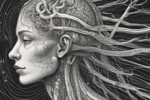
Spinal Cord to Cortex
- The posterior aspect of the spinal cord receives sensory fibers originating in the dorsal root ganglia. Each dorsal root is divided into 6-8 rootlets, with medial and lateral sections.
- Sensory fibers are categorized by their function, myelin sheath presence, conduction velocity (m/s), and fiber diameter (μm).
- Nociception and thermal inputs are carried by Ad and C fibers which enter the spinal cord through the Lissauer dorsolateral tract.
- These fibers branch into ascending and descending branches, terminating in the dorsal horn at the same level of entry or 3/4 segments higher.
- Medial sections of the spinal cord convey fine touch and vibration sensations through Aβ fibers.
- These Aβ fibers enter and divide into ascending and descending branches, forming the dorsal funiculus (dorsal column).
- Descending branches further divide into the septomarginalis and interfascicular fasciculi.
- Neurons in these fasciculi relay proprioceptive and tactile information, mediating reflexes.
- Ascending branches terminate at the gracilis and cuneate nuclei.
Spinal Cord Internal Structure
- The white matter is divided into three funiculi (columns): ventral, dorsal, and lateral columns. The spinal cord's grey matter section comprises the ventral horn, dorsal horn, lateral horn, and the grey commissure.
- The cervical, thoracic, lumbar, and sacral regions of the spinal cord exhibit specific functional subdivisions.
- Cervical and lumbar enlargements contain larger numbers of motor neurons for hands and face.
Functional Subdivision of Spinal Cord
- Longitudinally, cells are structured into laminae/columns, which includes non-myelinated fibers. Axons are organized into white matter columns.
- Transversely, neurons are arranged in an anterior-posterior manner (afferents posterior, efferents anterior) and in a lateral-lateral manner.
- Somatotopically, each area corresponds to a body part..
Functional Organization of Gray Matter
- The grey matter is composed of laminae (10) from I to X.
- Lamina I (marginalis, Waldeyer) contains neurons receiving pain, temperature, and light touch sensations
- Lamina II (substantia gelatinosa, Rolando) contains Golgi type II neurons, receiving pain, temperature, and light touch.
- Lamina III-VI (nucleus proprius): relays pain, light touch, and temperature.
- Lamina VII (intermediolateral cell column, Clarke's column): conveys proprioceptive information from muscle spindles and Golgi tendon organs.
- Lamina VIII (interneurons): links other laminae, including sensory and motor neurons.
- Lamina IX (motorneurons): controls voluntary muscle movements.
- Lamina X (commissural grey): connects the left and right hemispheres of the spinal cord.
Other Details
- Descending fibres from higher brain centers (eg cerebral cortex) modulate pain and temperature sensations in the spinal cord.
- Pain signals can be modulated by interneurons in the substantia gelatinosa.
- Nociceptive information can be modulated via descending projections (i.e.raphe magnus).
- The spinal cord displays no adaptation to chronic pain, but shows sensitization.
- The trigeminal nerve, a cranial nerve, innervates sensory areas of the face and head; sensory neurons are located within the Gasserian ganglion, not the brainstem.
- The nucleus proprius (laminae IV and V) contains large nerve cells that receive inputs from the dorsal root ganglia.
Studying That Suits You
Use AI to generate personalized quizzes and flashcards to suit your learning preferences.