Podcast
Questions and Answers
What is the approximate duration of the process of spermatogenesis?
What is the approximate duration of the process of spermatogenesis?
- 72 days
- 64 days (correct)
- 48 days
- 96 days
What is the function of the acrosome in a spermatozoon?
What is the function of the acrosome in a spermatozoon?
- To provide structural support to the spermatozoon
- To generate energy for swimming
- To regulate spermatozoon motility
- To contain hydrolytic enzymes (correct)
What is the number of primary oocytes present in the ovaries by birth?
What is the number of primary oocytes present in the ovaries by birth?
- 100,000 - 200,000
- 1 million - 2 million
- 400,000 - 500,000
- 700,000 - 2 million (correct)
What is the structure that contains large, helical mitochondria and generates energy for swimming in a spermatozoon?
What is the structure that contains large, helical mitochondria and generates energy for swimming in a spermatozoon?
During which stage of development do the primary oocytes undergo meiosis?
During which stage of development do the primary oocytes undergo meiosis?
What is the result of the fusion of the sperm cell nucleus with the egg cell nucleus?
What is the result of the fusion of the sperm cell nucleus with the egg cell nucleus?
What is the purpose of the acrosome reaction during fertilization?
What is the purpose of the acrosome reaction during fertilization?
What is the consequence of having less than 50% normal spermatozoa?
What is the consequence of having less than 50% normal spermatozoa?
What is the primary function of capacitation during fertilization?
What is the primary function of capacitation during fertilization?
What is the result of the cortical granular reaction during fertilization?
What is the result of the cortical granular reaction during fertilization?
What is the consequence of having a high number of abnormal spermatozoa?
What is the consequence of having a high number of abnormal spermatozoa?
What is a characteristic of cleavage during embryonic development?
What is a characteristic of cleavage during embryonic development?
What is the result of the fluid absorption by the morula?
What is the result of the fluid absorption by the morula?
What is the last step in the process of implantation?
What is the last step in the process of implantation?
What is a distinguishing feature of cleavage from normal mitoses?
What is a distinguishing feature of cleavage from normal mitoses?
At what stage does the morula transform into a blastocyst?
At what stage does the morula transform into a blastocyst?
What is the function of Syncytiotrophoblast?
What is the function of Syncytiotrophoblast?
What is the term for the process of the blastocyst hatching from the zona pellucida?
What is the term for the process of the blastocyst hatching from the zona pellucida?
What is the outcome of the differentiation of Embryoblast?
What is the outcome of the differentiation of Embryoblast?
What is the function of HCG?
What is the function of HCG?
What are the two cavities formed during the 2nd week of development?
What are the two cavities formed during the 2nd week of development?
What is the primary function of the lacunae formed in the syncytiotrophoblast on Day 9?
What is the primary function of the lacunae formed in the syncytiotrophoblast on Day 9?
What is the result of the fusion of adjacent lacunae on Days 11 and 13?
What is the result of the fusion of adjacent lacunae on Days 11 and 13?
Which layer of the bilaminar embryonic disc gives rise to the amniotic sac?
Which layer of the bilaminar embryonic disc gives rise to the amniotic sac?
What is the origin of the yolk sac?
What is the origin of the yolk sac?
What is the purpose of the lacunar network in the syncytiotrophoblast?
What is the purpose of the lacunar network in the syncytiotrophoblast?
What is the primary result of gastrulation?
What is the primary result of gastrulation?
What is the significance of the end of week 8 in embryonic development?
What is the significance of the end of week 8 in embryonic development?
What is the role of the primitive node during gastrulation?
What is the role of the primitive node during gastrulation?
What is the result of the craniocaudal and lateral folding during the 3rd week of development?
What is the result of the craniocaudal and lateral folding during the 3rd week of development?
During which stage of development do the three primary germ layers form?
During which stage of development do the three primary germ layers form?
Which region of the node do cells migrate to form the prechordal plate and notochord?
Which region of the node do cells migrate to form the prechordal plate and notochord?
What is the purpose of the prechordal plate in the cephalic direction?
What is the purpose of the prechordal plate in the cephalic direction?
What is the result of the differentiation of the ectoderm into neuroectoderm?
What is the result of the differentiation of the ectoderm into neuroectoderm?
What is the role of the notochord in the formation of vertebrae?
What is the role of the notochord in the formation of vertebrae?
What is the significance of the cloacal membrane in the caudal end?
What is the significance of the cloacal membrane in the caudal end?
What is the role of the allantois in humans?
What is the role of the allantois in humans?
Which body axis is established by the anterior visceral endoderm?
Which body axis is established by the anterior visceral endoderm?
What structure is responsible for establishing the left—right body axis?
What structure is responsible for establishing the left—right body axis?
Which molecule is involved in the molecular mechanism of body axis establishment?
Which molecule is involved in the molecular mechanism of body axis establishment?
What is the location of the anterior visceral endoderm in the hypoblast layer?
What is the location of the anterior visceral endoderm in the hypoblast layer?
What is the fate of the anterior visceral endoderm?
What is the fate of the anterior visceral endoderm?
What is the result of the differentiation of the ectoderm into neuroectoderm?
What is the result of the differentiation of the ectoderm into neuroectoderm?
What is the significance of the neural crest cells?
What is the significance of the neural crest cells?
What is the approximate day of closure of the anterior neuropore?
What is the approximate day of closure of the anterior neuropore?
What is the result of the fusion of the neural plate in the midline?
What is the result of the fusion of the neural plate in the midline?
What is the significance of the neural crest cells in relation to birth defects?
What is the significance of the neural crest cells in relation to birth defects?
What is the fate of the caudal portion of the neural tube?
What is the fate of the caudal portion of the neural tube?
What is the origin of mesoderm cells?
What is the origin of mesoderm cells?
What is the primary function of ectoderm?
What is the primary function of ectoderm?
What is the result of the failure of the neural tube to close in the cranial region?
What is the result of the failure of the neural tube to close in the cranial region?
What is the recommended daily intake of folic acid to prevent neural tube defects?
What is the recommended daily intake of folic acid to prevent neural tube defects?
What is the most common site for spina bifida to occur?
What is the most common site for spina bifida to occur?
Which of the following is NOT a structure that arises from ectoderm?
Which of the following is NOT a structure that arises from ectoderm?
What is the name of the cavity that forms as a result of the mesodermal germ layer development?
What is the name of the cavity that forms as a result of the mesodermal germ layer development?
What is the future derivative of the paraxial mesoderm?
What is the future derivative of the paraxial mesoderm?
Which of the following is a derivative of the lateral plate mesoderm?
Which of the following is a derivative of the lateral plate mesoderm?
What is the process by which blood vessels arise from blood islands?
What is the process by which blood vessels arise from blood islands?
What is the segmental muscle component of a somite?
What is the segmental muscle component of a somite?
What is the connection between the parietal and lateral plate mesoderm?
What is the connection between the parietal and lateral plate mesoderm?
What is the derivative of the endodermal germ layer that gives rise to the epithelial lining of the respiratory tract?
What is the derivative of the endodermal germ layer that gives rise to the epithelial lining of the respiratory tract?
What is the origin of the epithelial lining of the urinary bladder and urethra?
What is the origin of the epithelial lining of the urinary bladder and urethra?
What is the derivative of the endodermal germ layer that forms the tympanic cavity and auditory tube?
What is the derivative of the endodermal germ layer that forms the tympanic cavity and auditory tube?
Which structure is NOT formed from the endodermal germ layer?
Which structure is NOT formed from the endodermal germ layer?
What is the fate of the primitive gut?
What is the fate of the primitive gut?
Flashcards are hidden until you start studying
Study Notes
Spermatogenesis
- Spermatogenesis takes approximately 64 days to complete
- During fetal period, Primordial Germ Cells (PGCs) undergo mitosis to form spermatogonia
- At puberty, spermatogonia undergo meiosis to form primary spermatocytes
- Primary spermatocytes divide into secondary spermatocytes (haploid, 2n) which then develop into spermatids
- Spermatids eventually mature into spermatozoa (22+X or 22+Y)
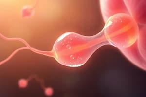
Spermatozoon Structure
- Spermatozoon length: 60-65μm
- Head of spermatozoon consists of condensed nucleus capped by an apical vesicle (acrosome) containing hydrolytic enzymes
- Neck of spermatozoon is short
- Tail of spermatozoon consists of midpiece and long tail
- Midpiece contains large, helical mitochondria which generate energy for swimming
- Long tail contains microtubules that enable swimming
Oogenesis
- During fetal life, PGCs differentiate into oogonia
- By 5th month of pregnancy, oogonia develop into primary oocytes in ovaries
- By birth, approximately 700,000 to 2 million primary oocytes are present
- By puberty, approximately 400,000 primary oocytes remain
Human Oogenesis and Follicular Development
- Follicle consists of oocyte and follicular cells/granulosa cells
- Mature follicle develops from primary oocyte (P.O) to secondary oocyte (S.O)
Ovulation and Menstrual Cycle
- Ovulation is a crucial part of the menstrual cycle that leads to fertilization
Fertilization
- Fertilization is the fusion of the sperm cell nucleus with the egg cell nucleus to produce a zygote (fertilized egg)
Before Fertilization
- Spermatozoa must undergo capacitation to fertilize an egg
- Capacitation involves the destabilization of the acrosomal sperm head membrane
- Capacitation is facilitated by the removal of steroids (e.g. cholesterol) and glycoproteins
- Capacitation increases permeability to Ca2+
Acrosome Reaction
- Release of enzymes (acrosin- & trypsin-like substances) needed to penetrate the zona pellucida
Fertilization Steps
- Acrosome reaction
- Cortical granular reaction/ penetration of the corona radiata
- Penetration of the zona pellucida
- Fusion of the oocyte and sperm cell membranes
- The oocyte resumes meiosis, resulting in a definitive oocyte
Fertilization Results
- Restoration of the diploid number of chromosomes
- Determination of the sex of the new individual
- Initiation of cleavage
Spermatozoa Abnormalities
- Spermatozoa with abnormalities can affect fertility
- Abnormalities include:
- Small, narrow, or piriform (pear-shaped) heads
- Double or triple heads
- Acrosomal defects
- Double tails
- If < 50% of spermatozoa are normal, fertility decreases
- If abnormal spermatozoa are present in high numbers, infertility increases significantly
Embryonic Development
- Cleavage is the first phase of embryonic development, characterized by a rapid series of mitotic divisions immediately after fertilization.
- The purpose of cleavage is to generate a large number of cells, resulting in an increase in the nucleus-to-cytoplasmic ratio.
Cleavage vs. Normal Mitoses
- Cleavage differs from normal mitoses in two key ways:
- Blastomeres do not grow in size between successive cell divisions.
- Cleavage occurs very rapidly.
Blastocyst Formation
- By day 4, the morula, consisting of approximately 30 cells, begins to absorb fluid.
- As the hydrostatic pressure of the fluid increases, a large cavity called the blastocyst cavity (blastocoel) forms.
- The morula develops a fluid-filled cavity and is transformed into a blastocyst.
Implantation
- The blastocyst attaches to the surface of the endometrium.
- Trophoblast cells invade the endometrium.
- Uteroplacental circulation is established.
Implantation
- The blastocyst hatches from the zona pellucida before implanting in the uterine wall
- Human chorionic gonadotropin (HCG) is produced during implantation
2nd Week: Development of the Bilaminar Embryonic Disc
- The embryoblast (inner cell mass) differentiates into the bilaminar embryonic disc, consisting of:
- Epiblast
- Hypoblast
- The trophoblast differentiates into:
- Cytotrophoblast
- Syncytiotrophoblast, which erodes blood vessels of the endometrium and establishes a haematotrophic circulation
- Cavities formation occurs, including:
- Amniotic cavity
- Yolk sac
UteroPlacenta Circulatory System
- On Day 9, isolated spaces called lacunae are formed in the syncytiotrophoblast, which later fill with nutrition secretions from eroded endometrial glands and maternal blood from ruptured maternal capillaries.
Lacunar Network Formation
- By Day 11 and 13, adjacent lacunae fuse to form a lacunar network, allowing maternal vessels to open and maternal blood to flow through.
Embryonic Development
2nd Week: Bilaminar Embryonic Disc
- The embryonic disc consists of two layers: inner cell mass (Epiblast) and Hypoblast.
Formation of Fluid-Filled Sacs
- The Epiblast forms the Amniotic sac, which is fluid-filled.
- The Hypoblast forms the Yolk sac, which is also fluid-filled.
Gastrulation and Embryonic Development
- During the 3rd week, gastrulation occurs, marked by the formation of the primitive streak on the surface of the epiblast.
- The primitive streak is a narrow groove with a slightly bulging side, containing the primitive node at its cephalic end.
- The primitive node is an elevated area surrounding the primitive pit, where cells of the epiblast migrate.
Consequences of Gastrulation
- Cells that invaginate displace the hypoblast, forming the endoderm.
- Cells that come to lie between the epiblast and the newly created endoderm form the mesoderm.
- Remaining cells in the epiblast form the ectoderm.
- The mesoderm spreads laterally and cranially, establishing the fate map.
Neurulation and Embryonic Development
- Neurulation occurs after gastrulation, characterized by craniocaudal folding and lateral folding, transforming the 2D disk into a 3D cylinder.
- By the end of week 8, the embryonic period ends, and all major organ systems have developed, although their function may be minimal.
- The embryo has a distinct human appearance by the end of week 8.
Fate Map Established During Gastrulation
- Cells from the primitive streak contribute to various regions:
- Cranial region of the node → prechordal plate and notochord
- Lateral edges of the node → paraxial mesoderm
- Midstreak region → intermediate mesoderm
- More caudal part → lateral plate mesoderm
- Caudal most part → extraembryonic mesoderm
Notochord Formation
- Notochord forms from mesoderm and endoderm via notochordal plate detaching from endoderm
- Definitive notochord differentiates from ectoderm into neuroectoderm, forming neural plate
- Notochord plays a role in vertebrae formation
Other Formations in Trilaminar Embryonic Disc
Cephalic Direction
- Prechordal plate induces neural development
- Oropharyngeal membrane forms the future opening of the oral cavity
- Primitive pit: neurenteric canal temporarily connects amniotic and yolk sac cavities
Caudal End
- Cloacal membrane forms the future opening of the anal and urogenital systems
- Allantois, though rudimentary in humans, may be involved in abnormalities of bladder development
Body Axes
- Three axes are established in the body: anterior-posterior (A-P, also known as craniocaudal), dorso-ventral (D-V), and left-right (L-R)
- The anterior visceral endoderm (AVE) at the cranial end of the hypoblast layer will develop into the head region
- The primitive node and notochord (midline) are involved in determining the left-right (L-R) axis
- SHH and 5-HT are molecules involved in the molecular mechanism of axis establishment
Ectodermal Germ Layer
- Induction of neuroectoderm from overlying ectoderm by notochord and prechordal mesoderm
- Neuroectoderm formation marks the initial event of neurulation
Neurulation
- Formation of neural plate, folds, and groove
- Elevation of lateral edges of neural plate to form neural folds
- Fusion of neural folds to form neural tube
- Communication with amniotic cavity through:
- Anterior (cranial) neuropore
- Posterior (caudal) neuropore
Closure of Neural Tube
- Closure of anterior neuropore: around day 25
- Closure of posterior neuropore: around day 28
Future Central Nervous System
- Narrow caudal portion: develops into spinal cord
- Cephalic portion: develops into brain vesicles
Neural Crest Cells
- Located at lateral border of neuroectoderm
- Undergoes epithelial-to-mesenchymal transition
- Fundamentally important and contributes to many organs and tissues
- Sometimes referred to as the fourth germ layer
- Related to one-third of all birth defects
- Related to many cancers, such as melanomas, neuroblastomas, and others
Embryonic Tissues
- Mesoderm: derived from epiblast and extraembryonic tissues
- Mesenchyme: loosely organized embryonic connective tissue, regardless of origin
Ectoderm
- Gives rise to organs and structures that maintain contact with the outside world:
- Central nervous system
- Peripheral nervous system
- Sensory epithelium of the ear, nose, and eye
- Epidermis, including hair and nails
- Additionally gives rise to:
- Subcutaneous glands
- Mammary glands
- Pituitary gland
- Enamel of the teeth
Neural Tube Defects (NTDs)
- Caused by failure of neural tube to close
- Effects vary depending on location of lesion:
- Cranial region: anencephaly (most of the brain fails to form)
- Cervical region caudally: spina bifida
- Most common site: lumbosacral region
- Lose neurological function based on level and severity of lesion
- Prevention:
- 400 μg of folic acid daily
- 50% to 70% of NTDs can be prevented
- Take folic acid 3 months prior to conception and throughout pregnancy
Mesodermal Germ Layer
- The mesodermal germ layer divides into four regions: paraxial, lateral plate, parietal, and visceral mesoderm
- Paraxial mesoderm forms close to the midline and thickens
- Lateral plate mesoderm forms laterally
- Parietal mesoderm is continuous with the mesoderm covering the amnion
- Visceral mesoderm is continuous with the mesoderm covering the yolk sac
- These layers line the intraembryonic cavity
- Intermediate mesoderm connects parietal and lateral plate mesoderm
Development of Mesodermal Germ Layer
- Paraxial mesoderm gives rise to future somites
- Intermediate mesoderm develops into future excretory units
- Lateral plate mesoderm differentiates into parietal and visceral mesoderm layers lining the intraembryonic cavity
Somite Differentiation
- Each somite gives rise to three distinct components: sclerotome, myotome, and dermatome
- Sclerotome forms tendon, cartilage, and bone
- Myotome forms segmental muscle
- Dermatome forms the dermis of the back
- Each myotome and dermatome has its own segmental nerve component
Blood and Blood Vessels
- Blood cells and blood vessels arise from mesoderm
- Vasculogenesis: blood vessels form from blood islands
- Angiogenesis: new vessels sprout from existing vessels
Endodermal Germ Layer Derivatives
- The endodermal germ layer gives rise to the primitive gut, which further develops into the gastrointestinal tract and respiratory tract.
- The primitive gut is divided into two parts: the foregut, which forms the oropharyngeal membrane, and the hindgut, which forms the cloacal membrane.
Epithelial Lining and Organs
- The epithelial lining of the primitive gut, allantois, and vitelline duct (intraembryonic) originates from the endoderm.
- The endoderm forms the epithelial lining of several organs, including:
- Respiratory tract
- Urinary bladder and urethra
- Tympanic cavity and auditory tube
- The endoderm also gives rise to the parenchyma of:
- Thyroid gland
- Parathyroid glands
- Liver
- Pancreas
- Additionally, the endoderm forms the reticular stroma of:
- Tonsils
- Thymus
Studying That Suits You
Use AI to generate personalized quizzes and flashcards to suit your learning preferences.