Podcast
Questions and Answers
What is the primary function of the epimysium in skeletal muscle?
What is the primary function of the epimysium in skeletal muscle?
- To connect muscle to bone
- To provide form and a smooth surface for muscles to glide against each other (correct)
- To surround individual muscle fibres
- To contain myofilaments for muscle contraction
What does the perimysium primarily surround in skeletal muscle?
What does the perimysium primarily surround in skeletal muscle?
- The entire muscle belly
- Single muscle fibres
- Myofibrils within muscle fibres
- Bundles of muscle fibres (fascicles) (correct)
The basic functional contractile unit of myofibrils is known as what?
The basic functional contractile unit of myofibrils is known as what?
- Myofibril
- Muscle fibre
- Sarcomere (correct)
- Fascicle
Which statement correctly describes myofilaments?
Which statement correctly describes myofilaments?
What happens to muscle fibres with an increase in myofibrils?
What happens to muscle fibres with an increase in myofibrils?
Flashcards
Epimysium
Epimysium
The outermost layer of connective tissue surrounding the entire muscle.
Perimysium
Perimysium
Connective tissue that surrounds bundles of muscle fibers, forming compartments within the muscle.
Endomysium
Endomysium
A delicate connective tissue layer that wraps around individual muscle fibers.
Sarcomere
Sarcomere
Signup and view all the flashcards
Muscle Fiber
Muscle Fiber
Signup and view all the flashcards
Study Notes
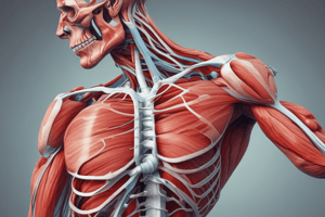
Skeletal Muscle Structure
- Epimysium: Outer connective tissue layer surrounding the entire muscle, providing shape and a smooth surface for muscles to move against each other.
- Perimysium: Connective tissue that surrounds bundles of muscle fibers (fascicles).
- Endomysium: Connective tissue surrounding individual muscle fibers.
- Muscle Fiber (Muscle Cell): Long, cylindrical fibers within a bundle (fascicle), lying parallel and capable of stretching up to 50cm. Thousands of these fibers are present in each bundle.
- Myofibril: Cylindrical structures within muscle fibers exhibiting light and dark bands (striations). These bands are due to the presence of myofilaments (actin and myosin) responsible for muscle contraction. Myofibril increase or decrease which leads to muscle hypertrophy or atrophy.
- Sarcomere: The basic functional unit of a myofibril. Thousands of sarcomeres are arranged in series. They are bordered by Z-lines, dense protein bands where actin filaments attach. Contains both actin and myosin myofilaments.
- Actin Filament: Thin filaments that interact with myosin for muscle contraction and relaxation.
- Myosin Filament: Thick filaments that work with actin to bring about muscle contraction and relaxation.
Studying That Suits You
Use AI to generate personalized quizzes and flashcards to suit your learning preferences.