Podcast
Questions and Answers
What is the primary function of skeletal muscle?
What is the primary function of skeletal muscle?
- Facilitating digestion
- Pumping blood throughout the body
- Locomotion (correct)
- Regulating blood pressure
Which structure is the cell membrane of an individual muscle fiber?
Which structure is the cell membrane of an individual muscle fiber?
- Sarcolemma (correct)
- Sarcoplasm
- Sarcomere
- Myofibril
Which type of muscle fiber is characterized by being multinucleated?
Which type of muscle fiber is characterized by being multinucleated?
- Cardiac muscle fiber
- Skeletal muscle fiber (correct)
- Striated muscle fiber
- Smooth muscle fiber
What is a sarcomere?
What is a sarcomere?
Which proteins are classified as contractile proteins in muscle fibers?
Which proteins are classified as contractile proteins in muscle fibers?
What is the diameter range of skeletal muscle fibers?
What is the diameter range of skeletal muscle fibers?
What type of muscle utilizes gap junctions for communication?
What type of muscle utilizes gap junctions for communication?
Which filament is primarily responsible for muscle contraction?
Which filament is primarily responsible for muscle contraction?
Which nervous system primarily controls skeletal muscle?
Which nervous system primarily controls skeletal muscle?
What component of skeletal muscle is crucial for action potential transmission?
What component of skeletal muscle is crucial for action potential transmission?
What are the components of myosin II?
What are the components of myosin II?
What role does troponin C play in muscle contraction?
What role does troponin C play in muscle contraction?
What is the primary function of the sliding filament theory?
What is the primary function of the sliding filament theory?
Which of the following statements about actin is true?
Which of the following statements about actin is true?
What initiates muscle relaxation?
What initiates muscle relaxation?
Which type of troponin inhibits the interaction of myosin with actin?
Which type of troponin inhibits the interaction of myosin with actin?
What is the role of ATP in muscle contraction?
What is the role of ATP in muscle contraction?
What is the composition of the tail region of myosin?
What is the composition of the tail region of myosin?
Which of the following best describes the relationship between muscle contraction and membrane action potentials?
Which of the following best describes the relationship between muscle contraction and membrane action potentials?
Which of the following accurately describes the head of myosin?
Which of the following accurately describes the head of myosin?
Flashcards
Skeletal Muscle Fiber
Skeletal Muscle Fiber
Skeletal muscle fibers are large, multinucleated cells controlled by the somatic nervous system.
Cardiac Muscle Fiber
Cardiac Muscle Fiber
Cardiac muscle fibers are smaller, have one or two nuclei, and are controlled by the autonomic nervous system. They are interconnected through gap junctions.
Smooth Muscle Cell
Smooth Muscle Cell
Smooth muscle cells are the smallest, found in bundles or sheets, and are controlled by the autonomic nervous system. They also have gap junctions.
Fasciculus
Fasciculus
Signup and view all the flashcards
Sarcolemma
Sarcolemma
Signup and view all the flashcards
Myofibril
Myofibril
Signup and view all the flashcards
Sarcomere
Sarcomere
Signup and view all the flashcards
Thin Filament
Thin Filament
Signup and view all the flashcards
Thick Filament
Thick Filament
Signup and view all the flashcards
I Band
I Band
Signup and view all the flashcards
What is the structure of Myosin II?
What is the structure of Myosin II?
Signup and view all the flashcards
Why is the connection between the head and tail of Myosin II flexible?
Why is the connection between the head and tail of Myosin II flexible?
Signup and view all the flashcards
Describe the structure of an actin filament.
Describe the structure of an actin filament.
Signup and view all the flashcards
What is the role of tropomyosin?
What is the role of tropomyosin?
Signup and view all the flashcards
What are the components and functions of troponin?
What are the components and functions of troponin?
Signup and view all the flashcards
What is the sliding filament theory?
What is the sliding filament theory?
Signup and view all the flashcards
What are the key components of muscle contraction?
What are the key components of muscle contraction?
Signup and view all the flashcards
What is the role of cross-bridge formation in muscle contraction?
What is the role of cross-bridge formation in muscle contraction?
Signup and view all the flashcards
What is the role of calcium ions in muscle contraction?
What is the role of calcium ions in muscle contraction?
Signup and view all the flashcards
How does muscle relaxation occur?
How does muscle relaxation occur?
Signup and view all the flashcards
Study Notes
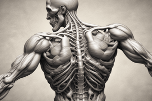
Skeletal Muscle Structure and Function
- Skeletal muscle is responsible for locomotion, body defense, shaping the body, and skilled activities like writing and speaking.
- Muscles also aid in respiration (intercostal muscles and diaphragm), blood pumping, blood pressure and blood flow regulation, digestion, urine storage and expulsion, and childbirth.
- Muscle cells can be excited chemically, electrically, and mechanically.
- Action potentials are transmitted along muscle cell membranes.
- Muscles are specialized for contraction and generating force.
Muscle Types
- Skeletal muscle fibers (10-100 µm diameter), have multiple nuclei and are part of the somatic nervous system.
- Cardiac muscle fibers (10-15 µm diameter) have one or two nuclei, gap junctions, and are part of the autonomic (parasympathetic and sympathetic) nervous system.
- Smooth muscle cells (2-15 µm in diameter), are arranged in bundles or sheets, have gap junctions, and are part of the autonomic nervous system.
Muscle Classification
- Voluntary muscle (Skeletal)
- Involuntary muscle (Cardiac and Smooth)
Comparison of Muscle Types
| Feature | Skeletal Muscle | Smooth Muscle | Cardiac Muscle |
|---|---|---|---|
| Histological | Cylindrical, striated, unbranched | Spindle shaped, unstriated | Striated, branched |
| Functional | Voluntary | Involuntary | Involuntary |
| Nerve supply | Somatic nerves | Autonomic nerves | Autonomic nerves |
| Hormones on excitability & contraction | No | Yes | Yes |
Muscle Properties
| Feature | Skeletal Muscle | Smooth Muscle | Cardiac Muscle |
|---|---|---|---|
| Excitability | Yes | Yes | Yes |
| Conductivity | Yes | Yes | Yes |
| Contractility | Yes | Yes | Yes |
| Rhythmicity | Absent | Irregular | Regular |
| Tonicity | Present | Present | Absent |
Muscle Tissue Properties
- Excitability
- Contractility
- Elasticity
- Extensibility
Neuromuscular Junction
- Motor axon terminals contain vesicles containing acetylcholine (ACh).
- Acetylcholinesterase is present on the muscle fiber.
- Nerve impulse arrival at the axon terminal initiates the release of acetylcholine.
- Acetylcholine binds to receptors on the muscle fiber.
- This leads to an end-plate potential (EPP) and then to an action potential along the muscle fiber.
Muscle Contraction Types
- Isometric contraction: Muscle length remains constant; tension increases.
- Isotonic contraction: Muscle shortens; tension remains constant.
Skeletal Muscle Structure
-
Muscle belly consists of fascicles.
-
Fascicles consist of muscle fibers.
-
Sarcolemma: The cell membrane of the muscle fiber.
-
Myofibrils are found within each muscle fiber.
-
Myofibrils are divided into compartments by Z lines.
-
Sarcomere: The portion of the myofibril between two adjacent Z lines.
-
Sarcomere contains two types of filaments: Thin filaments (actin, tropomyosin, troponin) and Thick filaments (myosin).
-
Thin and thick filaments are contractile proteins.
-
Regulatory proteins (tropomyosin and troponin) also play a vital role in muscle contraction.
Thick Filaments
- Myosin II consists of globular heads and a long tail.
- The head has binding sites for actin and ATP.
- Thick filaments are composed of heavy chains and light chains.
Thin Filaments
- Actin filaments form a double helix.
- Tropomyosin molecules lie in the groove between actin chains.
- Troponin molecules are located along tropomyosin at intervals.
Troponin
- This protein has three components (T, I, and C).
- T binds troponin to tropomyosin.
- I inhibits the interaction of myosin with actin.
- C binds calcium and initiates muscle contraction
Sliding Filament Theory
- Actin and myosin filaments slide past each other during muscle contraction.
- This shortening of the sarcomere results in overall muscle contraction.
Sarcotubular System
-
Membrane surrounds muscle fibrils
-
T-tubules and sarcoplasmic reticulum (L-tubules).
-
T-tubules help transmit action potentials to the interior of the muscle fiber.
-
L-tubules are involved in calcium ion storage and release.
-
Triad: A junction of the T-tubule and two dilated regions of the sarcoplasmic reticulum.
-
A triad contains receptors (dihydropyridine receptors and ryanodine receptors) for regulating calcium ion release into the cytoplasm.
Muscle Contraction
- The sliding filament theory and the role of calcium ions regulate muscle contraction, which causes shortening in sarcomeres and overall muscle movement.
- ATP is used during the process.
Key Concepts
- Muscles consist of overlapping actin and myosin protein filaments, which slide to produce force during contraction.
- This process uses ATP and involves cross-bridge formation.
- Calcium ions are essential for mediating the coupling between membrane action potentials and muscle contraction.
- Relaxation occurs when calcium ions are removed.
Studying That Suits You
Use AI to generate personalized quizzes and flashcards to suit your learning preferences.