Podcast
Questions and Answers
What is the average diameter of muscle fibers?
What is the average diameter of muscle fibers?
- 250 μm
- 100 μm (correct)
- 50 μm
- 200 μm
Which type of protein is classified as a contractile protein in muscle fibers?
Which type of protein is classified as a contractile protein in muscle fibers?
- Myosin (correct)
- Dystrophin
- Tropomyosin
- Actin
What are the light bands in muscle fibers called?
What are the light bands in muscle fibers called?
- Z bands
- H bands
- A bands
- I bands (correct)
How many actin filaments surround one myosin filament in a sarcomere?
How many actin filaments surround one myosin filament in a sarcomere?
Which component reflects the myosin filament length within a sarcomere and does not change size during muscle contraction?
Which component reflects the myosin filament length within a sarcomere and does not change size during muscle contraction?
What happens to I bands during muscle contraction?
What happens to I bands during muscle contraction?
Which of the following proteins is the largest known protein in muscle fibers?
Which of the following proteins is the largest known protein in muscle fibers?
What defines muscle contraction?
What defines muscle contraction?
What is the primary component of skeletal muscle in adults by body weight percentage?
What is the primary component of skeletal muscle in adults by body weight percentage?
Which of the following is NOT a function of skeletal muscle?
Which of the following is NOT a function of skeletal muscle?
What is the role of ATP in the cross-bridge cycling of skeletal muscle?
What is the role of ATP in the cross-bridge cycling of skeletal muscle?
What is the primary role of calcium in skeletal muscle contraction?
What is the primary role of calcium in skeletal muscle contraction?
What is one common type of muscular dystrophy mentioned?
What is one common type of muscular dystrophy mentioned?
Which type of muscle is responsible for eye movement?
Which type of muscle is responsible for eye movement?
What is thermogenesis in the context of skeletal muscle?
What is thermogenesis in the context of skeletal muscle?
What is the average diameter of a muscle fiber in skeletal muscle?
What is the average diameter of a muscle fiber in skeletal muscle?
What does muscle contraction refer to in the context of skeletal muscles?
What does muscle contraction refer to in the context of skeletal muscles?
What proteins compose the thick myofilaments in skeletal muscle?
What proteins compose the thick myofilaments in skeletal muscle?
What role does calcium play in muscle contraction?
What role does calcium play in muscle contraction?
Which statement describes the role of tropomyosin in muscle contraction?
Which statement describes the role of tropomyosin in muscle contraction?
What happens during rigor mortis in relation to muscle contraction?
What happens during rigor mortis in relation to muscle contraction?
What is the purpose of the cross-bridge cycle in muscle fibers?
What is the purpose of the cross-bridge cycle in muscle fibers?
Which of the following describes the interaction between myosin heads and actin during muscle contraction?
Which of the following describes the interaction between myosin heads and actin during muscle contraction?
What prevents myosin from binding to actin in a relaxed muscle fiber?
What prevents myosin from binding to actin in a relaxed muscle fiber?
Flashcards
Muscle Fiber
Muscle Fiber
A single muscle cell, long and cylindrical, responsible for muscle contraction. It's covered by a plasma membrane called sarcolemma and contains multiple nuclei.
Myofibrils
Myofibrils
Densely packed subunits within a muscle fiber, running its entire length. They are responsible for muscle contraction and are composed of thick and thin myofilaments.
Thick Myofilaments
Thick Myofilaments
Made of the protein myosin, responsible for pulling actin filaments during muscle contraction. They are located in the center of the sarcomere.
Thin Myofilaments
Thin Myofilaments
Signup and view all the flashcards
Sarcomere
Sarcomere
Signup and view all the flashcards
Striations
Striations
Signup and view all the flashcards
Sliding Filament Theory
Sliding Filament Theory
Signup and view all the flashcards
A Bands
A Bands
Signup and view all the flashcards
Muscle Contraction Definition
Muscle Contraction Definition
Signup and view all the flashcards
What are myofilaments?
What are myofilaments?
Signup and view all the flashcards
What's special about myosin?
What's special about myosin?
Signup and view all the flashcards
What do tropomyosin and troponin do?
What do tropomyosin and troponin do?
Signup and view all the flashcards
How are cross-bridges formed?
How are cross-bridges formed?
Signup and view all the flashcards
What is the cross-bridge cycle?
What is the cross-bridge cycle?
Signup and view all the flashcards
What happens in rigor mortis?
What happens in rigor mortis?
Signup and view all the flashcards
How much myosin interacts with actin?
How much myosin interacts with actin?
Signup and view all the flashcards
What are the 3 main types of muscle?
What are the 3 main types of muscle?
Signup and view all the flashcards
What makes muscles 'excitable'?
What makes muscles 'excitable'?
Signup and view all the flashcards
What is the function of the diaphragm?
What is the function of the diaphragm?
Signup and view all the flashcards
What are the 2 types of thermogenesis?
What are the 2 types of thermogenesis?
Signup and view all the flashcards
What is Duchenne Muscular Dystrophy?
What is Duchenne Muscular Dystrophy?
Signup and view all the flashcards
What is Myasthenia Gravis?
What is Myasthenia Gravis?
Signup and view all the flashcards
What is Sarcopenia?
What is Sarcopenia?
Signup and view all the flashcards
How does skeletal muscle contribute to blood glucose regulation?
How does skeletal muscle contribute to blood glucose regulation?
Signup and view all the flashcards
Study Notes
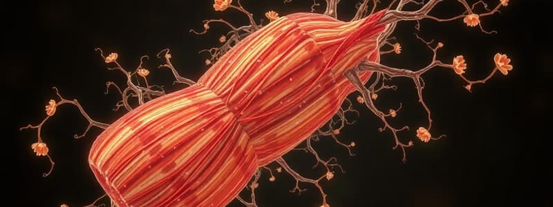
Skeletal Muscle Structure and Function
- Skeletal muscle is made of muscle fibers, averaging 100 µm in diameter and up to several centimeters long.
- Muscle fibers have plasma membranes called sarcolemma.
- Muscle fibers are multinucleated, containing multiple nuclei.
- Muscle fibers are striated (striped), with alternating light (I bands) and dark (A bands) regions.
- Myofibrils are densely packed subunits within muscle fibers.
- Myofibrils are composed of thick (myosin) and thin (actin) myofilaments.
- Myofilaments are organized into sarcomeres, the functional units of muscle contraction.
- A sarcomere extends from Z-line to Z-line.
- Sarcomeres contain thick and thin filaments that overlap to create the striations.
- The I band contains only thin filaments, while the A band contains both thick and thin filaments.
- The H zone is the central region of the A band where only thick filaments are present.
- The M line is the center of the sarcomere, connecting the thick filaments.
- Muscle fibers contain myofibrils arranged in a parallel manner, allowing for coordinated contraction.
- Periosteum covers the bone, and tendons connect muscle to bone.
Skeletal Muscle Proteins
- Contractile proteins: Myosin and Actin
- Regulatory proteins: Troponin and Tropomyosin
- Structural proteins: Titin, Nebulin, Alpha-actin, Myomesin, Dystrophin
- Titin (3700 kDa) is the largest protein in the sarcomere and is responsible for providing elasticity.
- Dystrophin (427 kDa) is vital for muscle cell stability.
Sliding Filament Mechanism
- Muscle contraction is not necessarily shortening; it's the activation of force-generating sites within muscle fibers (cross-bridges).
- During muscle contraction, sarcomeres shorten.
- The I band shortens.
- The H zone shortens.
- The A band remains the same length.
- During the sliding filament mechanism, thin filaments slide towards the H zone.
Cross-Bridge Cycle
- Myosin heads act as ATPase enzymes, splitting ATP into ADP and phosphate.
- This allows the myosin head to bind to actin when the muscle is stimulated.
- The myosin head then undergoes a power stroke, pulling the thin filament.
- The myosin head releases ADP and phosphate, returning to its original conformation.
- A new ATP molecule binds to the myosin head, causing detachment from actin.
Regulation of Contraction
- Tropomyosin physically blocks cross-bridges.
- Troponin complex:
- Troponin I inhibits myosin binding.
- Troponin T binds to tropomyosin.
- Troponin C binds to calcium.
- Calcium is released inside muscle fiber when stimulated.
- Calcium binding to troponin C causes a conformational change in troponin and tropomyosin.
- This allows myosin access to form cross-bridges.
Role of ATP and Calcium
- ATP is essential for the detachment of myosin from actin.
- Calcium is crucial for initiating the cross-bridge cycle.
- When calcium levels are low, tropomyosin blocks actin's binding sites, preventing myosin from binding and stopping contraction.
Studying That Suits You
Use AI to generate personalized quizzes and flashcards to suit your learning preferences.


