Podcast
Questions and Answers
What proportion of the human body mass is typically comprised of skeletal muscle?
What proportion of the human body mass is typically comprised of skeletal muscle?
- Approximately 25%
- Approximately 60%
- Approximately 10% (correct)
- Approximately 40%
In skeletal muscle physiology, what is the function of titin?
In skeletal muscle physiology, what is the function of titin?
- Catalyzing ATP hydrolysis for muscle contraction.
- Maintaining the alignment of actin and myosin filaments and providing elasticity. (correct)
- Transmitting action potentials from the sarcolemma to the sarcoplasmic reticulum.
- Initiating the release of calcium ions from the sarcoplasmic reticulum.
What is the role of the M line in the structure of a sarcomere?
What is the role of the M line in the structure of a sarcomere?
- Serves as the boundary between sarcomeres.
- Connects and aligns the myosin filaments in the center of the sarcomere.
- Anchors the actin filaments. (correct)
- Binds calcium ions to initiate muscle contraction.
Within the context of skeletal muscle contraction, what event is directly triggered by the depolarization of the muscle fiber membrane?
Within the context of skeletal muscle contraction, what event is directly triggered by the depolarization of the muscle fiber membrane?
During muscle contraction, what prevents the binding of myosin to actin in a resting muscle?
During muscle contraction, what prevents the binding of myosin to actin in a resting muscle?
What is the primary function of the sarcoplasmic reticulum in muscle cells?
What is the primary function of the sarcoplasmic reticulum in muscle cells?
In the context of the 'walk-along' theory of muscle contraction, what directly causes the power stroke?
In the context of the 'walk-along' theory of muscle contraction, what directly causes the power stroke?
Which structural component of myofibrils contains the M line?
Which structural component of myofibrils contains the M line?
How does the action potential propagate along the muscle fiber membrane?
How does the action potential propagate along the muscle fiber membrane?
Which of the following subunits of troponin has a strong affinity for calcium ions?
Which of the following subunits of troponin has a strong affinity for calcium ions?
What event immediately follows the tilting of the myosin head during the walk-along theory of muscle contraction?
What event immediately follows the tilting of the myosin head during the walk-along theory of muscle contraction?
What is the approximate number of actin filaments present in each myofibril?
What is the approximate number of actin filaments present in each myofibril?
What is the primary characteristic of the I bands in a sarcomere?
What is the primary characteristic of the I bands in a sarcomere?
What is the role of acetylcholine-gated cation channels in muscle contraction?
What is the role of acetylcholine-gated cation channels in muscle contraction?
In a relaxed muscle, what molecule physically blocks the active sites on actin, preventing myosin binding?
In a relaxed muscle, what molecule physically blocks the active sites on actin, preventing myosin binding?
Flashcards
Sarcomere
Sarcomere
The functional unit of skeletal muscle, lying between two Z disks.
Sarcolemma
Sarcolemma
The true cell membrane of the muscle fiber, consisting of plasmalemma
Sarcoplasm
Sarcoplasm
The intracellular fluid between myofibrils, containing potassium, magnesium, phosphate, and mitochondria.
Sarcoplasmic Reticulum
Sarcoplasmic Reticulum
Signup and view all the flashcards
Myofibrils
Myofibrils
Signup and view all the flashcards
Thick Filaments
Thick Filaments
Signup and view all the flashcards
Thin Filaments
Thin Filaments
Signup and view all the flashcards
Titin
Titin
Signup and view all the flashcards
Acetylcholine (ACh)
Acetylcholine (ACh)
Signup and view all the flashcards
Muscle Contraction
Muscle Contraction
Signup and view all the flashcards
Myosin Filaments
Myosin Filaments
Signup and view all the flashcards
Actin Filaments
Actin Filaments
Signup and view all the flashcards
Tropomyosin
Tropomyosin
Signup and view all the flashcards
Troponin
Troponin
Signup and view all the flashcards
Walk-Along Theory
Walk-Along Theory
Signup and view all the flashcards
Study Notes
Skeletal Muscle Contraction
- Skeletal muscle makes up about 40% of the body's mass
- Cardiac and smooth muscle account for 10% of body mass
- Basic contraction principles apply to all three muscle types
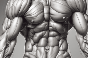
Physiologic Anatomy of Skeletal Muscle
- Skeletal muscles contain numerous fibers ranging from 10-80 micrometers in diameter
- Each fiber comprises successively smaller subunits
- Most skeletal muscle fibers extend the entire length of the muscle
Sarcomere
- The M line is located at the center
- The Z disk connects two actin filaments.
- H bands contain thick filaments not overlapped by thin filaments
Sarcolemma
- The sarcolemma consists of a true cell membrane known as the plasmalemma
- The sarcolemma also features an outer coat of polysaccharide material containing collagen fibrils
- At a muscle fiber's end, the sarcolemma's surface layer fuses with a tendon fiber
- Tendon fibers bundle together to form muscle tendons which then insert into bones
Myofibrils
- Myofibrils are composed of actin and myosin filaments
- Each muscle fiber contains several hundred to several thousand myofibrils
- A myofibril consists of approximately 1,500 myosin and 3,000 actin filaments
- Thick filaments consist of myosin
- Thin filaments consist of actin
- Myosin and actin interdigitation creates alternating light and dark bands
- Actin composes light bands (I bands) and is isotropic to polarized light
- Myosin composes thick bands (A bands) and is anisotropic to polarized light
- They give skeletal and cardiac muscle a striated appearance
- A sarcomere is the portion of a myofibril between two successive Z disks
Titin Filamentous Molecule
- Titin maintains the side-by-side relationship of actin and myosin
- Each titin molecule has a molecular weight of 3 million, making it the largest protein molecule in the body
- Titin is filamentous and springy which acts as a framework and holds actin and myosin filaments in place
- One titin end is attached to the Z disk
- One titin end is tethered to the M filament
Sarcoplasm
- Sarcoplasm is the intracellular fluid between myofibrils
- Sarcoplasm includes large amounts of potassium, magnesium, and phosphate
- Sarcoplasm contains mitochondria
Sarcoplasmic Reticulum
- The sarcoplasmic reticulum is a specialized endoplasmic reticulum (ER) of skeletal muscle
Muscle Contraction
- An action potential travels along a motor nerve to its endings on muscle fibers
- The nerve endings secrete a small amount of acetylcholine (a neurotransmitter)
- Acetylcholine acts on the muscle fiber, opening acetylcholine-gated cation channels
- Channel opening allows large quantities of sodium ions to diffuse into the muscle fiber membrane, causing depolarization
- This opens voltage-gated sodium channels, initiating an action potential in the membrane
- The action potential travels along the muscle fiber membrane in the same manner as in nerve fiber membranes
- The action potential depolarizes the muscle membrane and causes the sarcoplasmic reticulum to release stored calcium ions
- Calcium ions initiate attractive forces between actin and myosin filaments, leading to the filaments sliding alongside each other, which is a contractile process
- Calcium ions are pumped back to the sarcoplasmic reticulum by a calcium membrane pump after a fraction of a second, remaining stored until a new action potential arrives
- The removal of calcium ions from the myofibrils ceases muscle contraction
Contractile Filaments
- Myosin filaments are composed of multiple myosin molecules
- A myosin molecule has 6 polypeptide chains
- Two heavy chains wrap spirally to form a helix- tail
- One end of each heavy chain is folded bilaterally into a globular polypeptide, forming a myosin head
- There are also 4 light chains
- The function of each head is to control muscle contraction
Actin Filaments
- The backbone of each actin filament is a double-stranded F-actin molecule
- Two strands are wound in a helix, similarly to myosin
- Each strand consists of G-actin molecules
- One molecule of ADP is attached to each G-actin molecule
- ADP are active site on actin, when myosin filaments interact to cause muscle contraction
Tropomyosin
- Tropomyosin is wrapped around the sides of the f-actin helix
- Tropomyosin lies on top of the active sites in a resting state to prevent attraction between actin and myosin
- No contraction occurs
Troponin
- Troponin are small proteins attached along the sides of tropomyosin
- Troponin is a complex of 3 loosely bound protein subunits with roles in muscle contraction
- Troponin I has a strong affinity for actin
- Troponin T binds to tropomyosin
- Troponin C binds calcium ions
Muscle Contraction (Actin and Myosin)
- A pure actin filament without the troponin-tropomyosin complex binds instantly with myosin filament heads
- When the troponin-tropomyosin (T-T) complex is added to actin, myosin and actin cannot bind
- Active sites on relaxed muscle actin are inhibited or physically covered by the T-T complex
- In the presence of large amounts of calcium ions, the T-T complex effect is inhibited
- Calcium ions bind with Troponin-C, which triggers a change in the troponin complex
- The tropomyosin molecule is then tugged on
- This uncovers the active sites of the actin, which causes attractions to activate in myosin heads, resulting in contraction
Walk-Along (Ratchet) Theory of Contraction
- Two cross-bridge heads attach to and engage from actin's active sites
- When a head attaches to an active site, the head tilts toward the arm, dragging the actin filament
- The tilt of the head is known as the power stroke
- The head breaks immediately after tilting
- The head binds to the next active site along the actin filament
- The head then tilts again to produce another power stroke
Assignment topics
- Characteristics of whole muscle contraction
- Isometric vs isotonic
- Fast vs slow muscle fibers
- Summation
- Tetanization
Studying That Suits You
Use AI to generate personalized quizzes and flashcards to suit your learning preferences.
Related Documents
Description
Overview of skeletal muscle contraction, including physiologic anatomy and the roles of the sarcolemma, sarcomere and myofibrils. Skeletal muscle comprises approximately 40% of the body's mass. Basic contraction principles apply to all three muscle types.