Podcast
Questions and Answers
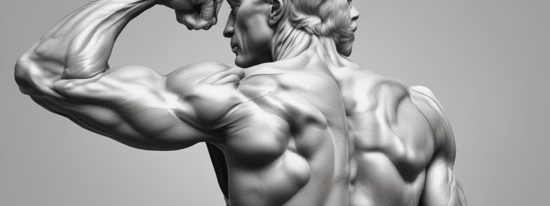
(A) ______ cuff muscles of shoulder, posterior view.
(A) ______ cuff muscles of shoulder, posterior view.
Rotator
Frontalis is a muscle in the ______.
Frontalis is a muscle in the ______.
face
The joint capsule surrounds the ______.
The joint capsule surrounds the ______.
joint
Olecranon bursitis affects the ______.
Olecranon bursitis affects the ______.
Epicondylitis is inflammation of the ______.
Epicondylitis is inflammation of the ______.
The hip is a ______ joint, permitting movement of the femur on many axes
The hip is a ______ joint, permitting movement of the femur on many axes
The knee consists of four bones: femur, tibia, fibula, and ______
The knee consists of four bones: femur, tibia, fibula, and ______
Fibrocartilaginous disks, medial and lateral ______, cushion the tibia and femur
Fibrocartilaginous disks, medial and lateral ______, cushion the tibia and femur
Collateral ligaments give medial and lateral stability to the ______
Collateral ligaments give medial and lateral stability to the ______
There are four separate bursae in the anterior knee that help to reduce the friction of knee ______
There are four separate bursae in the anterior knee that help to reduce the friction of knee ______
The rotator cuff muscles include supraspinatus, infraspinatus, teres minor, and ______
The rotator cuff muscles include supraspinatus, infraspinatus, teres minor, and ______
The shoulder joint is a ball-and-socket joint that permits movement of the humerus in several ______
The shoulder joint is a ball-and-socket joint that permits movement of the humerus in several ______
The ______ is a bony prominence at the tip of the elbow
The ______ is a bony prominence at the tip of the elbow
Inflammation of the olecranon bursa can result in a condition known as ______
Inflammation of the olecranon bursa can result in a condition known as ______
Epicondylitis is a condition characterized by inflammation of the ______ of the elbow
Epicondylitis is a condition characterized by inflammation of the ______ of the elbow
To palpate the biceps groove, rotate the arm and forearm ______.
To palpate the biceps groove, rotate the arm and forearm ______.
Palpate the muscle insertions of the supraspinatus, infraspinatus, and teres minor near the greater tuberosity of the humerus by lifting the elbow ______ to extend the shoulder.
Palpate the muscle insertions of the supraspinatus, infraspinatus, and teres minor near the greater tuberosity of the humerus by lifting the elbow ______ to extend the shoulder.
Examine the range of motion of the shoulders by asking the patient to perform the following movements: Shrug the ______.
Examine the range of motion of the shoulders by asking the patient to perform the following movements: Shrug the ______.
Raise both arms forward and straight up over the ______.
Raise both arms forward and straight up over the ______.
Place both arms behind the ______, elbows out.
Place both arms behind the ______, elbows out.
The ______ is a bursa located at the tip of the elbow.
The ______ is a bursa located at the tip of the elbow.
_____________ is inflammation or degeneration of the tendons where they attach to the epicondyles of the humerus.
_____________ is inflammation or degeneration of the tendons where they attach to the epicondyles of the humerus.
The _______________ is a ligament that encircles the head of the radius, holding it against the ulna at the proximal radioulnar joint.
The _______________ is a ligament that encircles the head of the radius, holding it against the ulna at the proximal radioulnar joint.
The ____________ joint is formed by the articulation of the humerus, radius, and ulna.
The ____________ joint is formed by the articulation of the humerus, radius, and ulna.
The _______________ joint is the primary articulation of the shoulder, connecting the humerus with the scapula.
The _______________ joint is the primary articulation of the shoulder, connecting the humerus with the scapula.
______ bursa is a fluid-filled sac that acts as a cushion between the skin and the olecranon process.
______ bursa is a fluid-filled sac that acts as a cushion between the skin and the olecranon process.
Suspect ______ or tendonitis when a point tenderness exists at the lateral epicondyle or along the grooves of the olecranon process.
Suspect ______ or tendonitis when a point tenderness exists at the lateral epicondyle or along the grooves of the olecranon process.
Examine the ______’s range of motion by asking the patient to perform the following movements: With the ______ fully extended at 0 degrees, bend and straighten the ______. Expect flexion of 160 degrees and extension returning to 0 degrees or 180 degrees of full extension.
Examine the ______’s range of motion by asking the patient to perform the following movements: With the ______ fully extended at 0 degrees, bend and straighten the ______. Expect flexion of 160 degrees and extension returning to 0 degrees or 180 degrees of full extension.
With the elbow flexed at a right angle, rotate the ______ from palm side down to palm side up. Expect pronation of 90 degrees and supination of 90 degrees.
With the elbow flexed at a right angle, rotate the ______ from palm side down to palm side up. Expect pronation of 90 degrees and supination of 90 degrees.
Have the patient maintain flexion and extension while you apply opposing force to evaluate the strength of the elbow ______.
Have the patient maintain flexion and extension while you apply opposing force to evaluate the strength of the elbow ______.
Inspect the contour of the shoulders, the shoulder girdle, the clavicles, and scapulae, and the surrounding ______.
Inspect the contour of the shoulders, the shoulder girdle, the clavicles, and scapulae, and the surrounding ______.
When the shoulder contour is asymmetric and one shoulder has hollows in the rounding contour, suspect a shoulder ______.
When the shoulder contour is asymmetric and one shoulder has hollows in the rounding contour, suspect a shoulder ______.
Observe for a winged scapula, an outward prominence of the scapula, indicating injury to the nerve of the anterior ______ muscle.
Observe for a winged scapula, an outward prominence of the scapula, indicating injury to the nerve of the anterior ______ muscle.
Olecranon ______ results in swelling and tenderness of the bursa.
Olecranon ______ results in swelling and tenderness of the bursa.
Expect increased pain with pronation and supination of the ______.
Expect increased pain with pronation and supination of the ______.
To perform the passive painful arc maneuver (Neer test), forward flex the patient’s arm up to 150 degrees while depressing the scapula. This presses the greater tuberosity and supraspinatus muscle against the anteroinferior acromion. Increased shoulder pain is associated with rotator cuff inflammation or a tear (Fig. 22.43A). The Hawkins-Kennedy test is performed by abducting the shoulder to 90 degrees, flexing the elbow to 90 degrees, and then internally rotating the arm to its limit. Increased shoulder pain is associated with rotator cuff inflammation or a possible tear (Fig. 22.43B). To test the strength of the rotator cuff muscles, perform the following maneuvers. A normal result is indicated by the absence of pain or weakness with the following maneuvers: To assess the supraspinatus muscle of the rotator cuff, have the patient place the arm in 90 degrees of abduction, 30 degrees of forward flexion, and internally rotated (thumbs pointing down). Apply downward pressure on the arm against patient resistance. To assess the subscapularis muscle, have the patient hold the arm at the side, elbow flexed 90 degrees, and rotate the forearm medially against resistance. To evaluate the infraspinatus and teres minor muscles, have the patient hold the arm at the side, elbow flexed 90 degrees, and rotate the arm laterally against resistance. Pain and weakness with opposing force is an unexpected finding and may be associated with inflammation or a tear.
To perform the passive painful arc maneuver (Neer test), forward flex the patient’s arm up to 150 degrees while depressing the scapula. This presses the greater tuberosity and supraspinatus muscle against the anteroinferior acromion. Increased shoulder pain is associated with rotator cuff inflammation or a tear (Fig. 22.43A). The Hawkins-Kennedy test is performed by abducting the shoulder to 90 degrees, flexing the elbow to 90 degrees, and then internally rotating the arm to its limit. Increased shoulder pain is associated with rotator cuff inflammation or a possible tear (Fig. 22.43B). To test the strength of the rotator cuff muscles, perform the following maneuvers. A normal result is indicated by the absence of pain or weakness with the following maneuvers: To assess the supraspinatus muscle of the rotator cuff, have the patient place the arm in 90 degrees of abduction, 30 degrees of forward flexion, and internally rotated (thumbs pointing down). Apply downward pressure on the arm against patient resistance. To assess the subscapularis muscle, have the patient hold the arm at the side, elbow flexed 90 degrees, and rotate the forearm medially against resistance. To evaluate the infraspinatus and teres minor muscles, have the patient hold the arm at the side, elbow flexed 90 degrees, and rotate the arm laterally against resistance. Pain and weakness with opposing force is an unexpected finding and may be associated with inflammation or a tear.
The Hawkins-Kennedy test is performed by abducting the shoulder to 90 degrees, flexing the elbow to 90 degrees, and then internally rotating the arm to its limit. Increased shoulder pain is associated with rotator cuff inflammation or a possible tear (Fig. 22.43B). To test the strength of the rotator cuff muscles, perform the following maneuvers. A normal result is indicated by the absence of pain or weakness with the following maneuvers: To assess the supraspinatus muscle of the rotator cuff, have the patient place the arm in 90 degrees of abduction, 30 degrees of forward flexion, and internally rotated (thumbs pointing down). Apply downward pressure on the arm against patient resistance. To assess the subscapularis muscle, have the patient hold the arm at the side, elbow flexed 90 degrees, and rotate the forearm medially against resistance. To evaluate the infraspinatus and teres minor muscles, have the patient hold the arm at the side, elbow flexed 90 degrees, and rotate the arm laterally against resistance. Pain and weakness with opposing force is an unexpected finding and may be associated with inflammation or a tear.
The Hawkins-Kennedy test is performed by abducting the shoulder to 90 degrees, flexing the elbow to 90 degrees, and then internally rotating the arm to its limit. Increased shoulder pain is associated with rotator cuff inflammation or a possible tear (Fig. 22.43B). To test the strength of the rotator cuff muscles, perform the following maneuvers. A normal result is indicated by the absence of pain or weakness with the following maneuvers: To assess the supraspinatus muscle of the rotator cuff, have the patient place the arm in 90 degrees of abduction, 30 degrees of forward flexion, and internally rotated (thumbs pointing down). Apply downward pressure on the arm against patient resistance. To assess the subscapularis muscle, have the patient hold the arm at the side, elbow flexed 90 degrees, and rotate the forearm medially against resistance. To evaluate the infraspinatus and teres minor muscles, have the patient hold the arm at the side, elbow flexed 90 degrees, and rotate the arm laterally against resistance. Pain and weakness with opposing force is an unexpected finding and may be associated with inflammation or a tear.
To assess the supraspinatus muscle of the ______ cuff, have the patient place the arm in 90 degrees of abduction, 30 degrees of forward flexion, and internally rotated (thumbs pointing down). Apply downward pressure on the arm against patient resistance. To assess the subscapularis muscle, have the patient hold the arm at the side, elbow flexed 90 degrees, and rotate the forearm medially against resistance. To evaluate the infraspinatus and teres minor muscles, have the patient hold the arm at the side, elbow flexed 90 degrees, and rotate the arm laterally against resistance. Pain and weakness with opposing force is an unexpected finding and may be associated with inflammation or a tear.
To assess the supraspinatus muscle of the ______ cuff, have the patient place the arm in 90 degrees of abduction, 30 degrees of forward flexion, and internally rotated (thumbs pointing down). Apply downward pressure on the arm against patient resistance. To assess the subscapularis muscle, have the patient hold the arm at the side, elbow flexed 90 degrees, and rotate the forearm medially against resistance. To evaluate the infraspinatus and teres minor muscles, have the patient hold the arm at the side, elbow flexed 90 degrees, and rotate the arm laterally against resistance. Pain and weakness with opposing force is an unexpected finding and may be associated with inflammation or a tear.
To evaluate the infraspinatus and ______ minor muscles, have the patient hold the arm at the side, elbow flexed 90 degrees, and rotate the arm laterally against resistance. Pain and weakness with opposing force is an unexpected finding and may be associated with inflammation or a tear.
To evaluate the infraspinatus and ______ minor muscles, have the patient hold the arm at the side, elbow flexed 90 degrees, and rotate the arm laterally against resistance. Pain and weakness with opposing force is an unexpected finding and may be associated with inflammation or a tear.
Pain and weakness with opposing force is an unexpected finding and may be associated with inflammation or a tear.
Pain and weakness with opposing force is an unexpected finding and may be associated with inflammation or a tear.
Flashcards
Temporomandibular Joint (TMJ)
Temporomandibular Joint (TMJ)
A ball-and-socket joint allowing movement of the mandible and articular disk.
Intervertebral Disks
Intervertebral Disks
Separate vertebrae and act as shock absorbers in the spine.
Hip Joint
Hip Joint
A ball-and-socket joint facilitating multi-axis femur movement.
Acetabulum
Acetabulum
Signup and view all the flashcards
Knee Joint
Knee Joint
Signup and view all the flashcards
Menisci
Menisci
Signup and view all the flashcards
Collateral Ligaments (Knee)
Collateral Ligaments (Knee)
Signup and view all the flashcards
Cruciate Ligaments (Knee)
Cruciate Ligaments (Knee)
Signup and view all the flashcards
Shoulder Joint
Shoulder Joint
Signup and view all the flashcards
Rotator Cuff Muscles
Rotator Cuff Muscles
Signup and view all the flashcards
Elbow Joint
Elbow Joint
Signup and view all the flashcards
Olecranon Bursa
Olecranon Bursa
Signup and view all the flashcards
Muscles of the Upper Arm
Muscles of the Upper Arm
Signup and view all the flashcards
Extensor Carpi Radialis Longus/Brevis
Extensor Carpi Radialis Longus/Brevis
Signup and view all the flashcards
Muscles of the Buttock
Muscles of the Buttock
Signup and view all the flashcards
Muscles of the Thigh (Adductors)
Muscles of the Thigh (Adductors)
Signup and view all the flashcards
Muscles of the Lower Leg
Muscles of the Lower Leg
Signup and view all the flashcards
Study Notes
Muscles of the Face and Head
- Auricularis anterior, frontalis, orbicularis oculi, procerus, nasalis, levator labii superioris, and zygomaticus minor are muscles of the face and head
- Posterior auricular, sternocleidomastoid, trapezius, and platysma are also muscles of the face and head
Temporomandibular Joint
- The temporomandibular joint (TMJ) is a ball-and-socket joint that allows for movement of the mandible (lower jawbone) and the articular disk
- The TMJ is supported by the styloid process and the stylomandibular ligament
- The joint capsule surrounds the joint and is lined with synovial membrane
Vertebral Joints
- Intervertebral disks separate the vertebrae and act as shock absorbers
- The joint capsule surrounds the joint and is lined with synovial membrane
- The sacrum and coccyx are part of the vertebral column
Hip Joint
- The hip joint is a ball-and-socket joint that allows for movement of the femur (thigh bone) in multiple axes
- The acetabulum is the socket of the pelvis that forms the hip joint
- The joint is supported by three strong ligaments and contains multiple bursae to reduce friction
Knee Joint
- The knee joint is a synovial hinge joint that connects the femur and tibia (shin bone)
- The joint has four bones (femur, tibia, fibula, and patella) and three separate articulating compartments
- Fibrocartilaginous disks (menisci) cushion the tibia and femur
- Collateral ligaments provide medial and lateral stability to the knee
- Cruciate ligaments (anterior and posterior) add anterior and posterior stability
- The knee joint has four separate bursae to reduce friction
Shoulder Joint
- The shoulder joint is a ball-and-socket joint that allows for movement of the humerus (upper arm bone) in multiple axes
- The rotator cuff muscles (supraspinatus, infraspinatus, teres minor, and subscapularis) and their tendons reinforce the glenohumeral joint
- The joint is supported by multiple ligaments and contains a subacromial bursa to reduce friction
Elbow Joint
- The elbow joint is a hinge joint that connects the humerus and radius (forearm bone)
- The joint has three bones (humerus, radius, and ulna) and two articulating compartments
- The olecranon bursa is a fluid-filled sac that acts as a cushion between the skin and the olecranon process
- Suspect epicondylitis or tendonitis when there is point tenderness at the lateral epicondyle or along the grooves of the olecranon process and epicondyles
Muscles of the Upper Extremities
- Deltoid, biceps brachii, brachialis, and triceps are muscles of the upper arm
- Extensor carpi radialis longus, extensor carpi radialis brevis, and extensor digitorum communis are muscles of the forearm
- Abductor pollicis brevis, abductor pollicis longus, and extensor pollicis brevis are muscles of the hand
Muscles of the Lower Extremities
- Gluteus maximus, gluteus medius, and gluteus minimus are muscles of the buttock
- Adductor magnus, adductor longus, and adductor brevis are muscles of the thigh
- Tibialis anterior, extensor digitorum longus, and peroneus tertius are muscles of the lower leg
Studying That Suits You
Use AI to generate personalized quizzes and flashcards to suit your learning preferences.


