Podcast
Questions and Answers
What is the term for the internal organs of the body, specifically those within the chest or abdomen?
What is the term for the internal organs of the body, specifically those within the chest or abdomen?
- Visceral (correct)
- Parietal
- Organic
- Vicus
What is the term for the wall of the body cavity?
What is the term for the wall of the body cavity?
- Parietal (correct)
- Visceral
- Cavity
- Vicus
What is the singular of 'viscera' in Latin?
What is the singular of 'viscera' in Latin?
- Organ
- Viscus (correct)
- Viscera
- Vicus
What is the figurative meaning of 'visceral'?
What is the figurative meaning of 'visceral'?
What does 'visceral' refer to in the context of feelings or emotions?
What does 'visceral' refer to in the context of feelings or emotions?
The term 'visceral' refers to the wall of the body cavity.
The term 'visceral' refers to the wall of the body cavity.
The term 'parietal' is used to describe the heart or lungs.
The term 'parietal' is used to describe the heart or lungs.
The Latin word 'viscus' means 'an organ of the body'.
The Latin word 'viscus' means 'an organ of the body'.
The term 'visceral' is only used to describe physical organs and not emotions.
The term 'visceral' is only used to describe physical organs and not emotions.
The terms 'visceral' and 'parietal' are interchangeable.
The terms 'visceral' and 'parietal' are interchangeable.
What is the primary function of the intercostal muscles?
What is the primary function of the intercostal muscles?
What is the purpose of the diaphragm?
What is the purpose of the diaphragm?
What type of bones make up the ribs?
What type of bones make up the ribs?
What is the role of the thoracic vertebrae in the thoracic cavity?
What is the role of the thoracic vertebrae in the thoracic cavity?
What is the connection between the sternum and the ribs?
What is the connection between the sternum and the ribs?
What is the primary function of the diaphragm in the thoracic cavity?
What is the primary function of the diaphragm in the thoracic cavity?
What is the primary function of the intercostal muscles?
What is the primary function of the intercostal muscles?
What provides structure to the thorax and prevents collapse during inspiration?
What provides structure to the thorax and prevents collapse during inspiration?
What is bounded by the thoracic vertebrae?
What is bounded by the thoracic vertebrae?
What type of connections do the ribs have dorsally and ventrally?
What type of connections do the ribs have dorsally and ventrally?
What is the function of the diaphragm during contraction?
What is the function of the diaphragm during contraction?
What is the primary purpose of the ribs and intercostal muscles in the thoracic cavity?
What is the primary purpose of the ribs and intercostal muscles in the thoracic cavity?
What is the function of the thoracic vertebrae in the thoracic cavity?
What is the function of the thoracic vertebrae in the thoracic cavity?
What is the characteristic of the ribs that allows for movement of the thorax?
What is the characteristic of the ribs that allows for movement of the thorax?
What is the connection between the sternum and the ribs?
What is the connection between the sternum and the ribs?
What is the main function of the diaphragm in the thoracic cavity?
What is the main function of the diaphragm in the thoracic cavity?
What is the main purpose of the ribs and intercostal muscles in the thoracic cavity?
What is the main purpose of the ribs and intercostal muscles in the thoracic cavity?
What is the area between the lungs that contains various structures?
What is the area between the lungs that contains various structures?
What is bounded by the thoracic vertebrae?
What is bounded by the thoracic vertebrae?
What type of connections do the ribs have?
What type of connections do the ribs have?
The diaphragm is a thick, flat muscle that separates the thorax and abdomen.
The diaphragm is a thick, flat muscle that separates the thorax and abdomen.
The thoracic vertebrae bound the thoracic cavity cranially.
The thoracic vertebrae bound the thoracic cavity cranially.
The intercostal muscles are located between the sternum and the ribs.
The intercostal muscles are located between the sternum and the ribs.
The mediastinum is the area between the lungs that contains the heart, oesophagus, and blood vessels.
The mediastinum is the area between the lungs that contains the heart, oesophagus, and blood vessels.
The ribs are flexible, thin bones with cartilage connections dorsally and ventrally.
The ribs are flexible, thin bones with cartilage connections dorsally and ventrally.
What is the space between the parietal and visceral pleura called?
What is the space between the parietal and visceral pleura called?
Which layer of the pleura lines the walls of the thoracic cavity?
Which layer of the pleura lines the walls of the thoracic cavity?
What is the outer layer of the pleura attached to?
What is the outer layer of the pleura attached to?
Which layer of the pleura is attached to the surface of the lungs?
Which layer of the pleura is attached to the surface of the lungs?
What are the two layers of the pleura?
What are the two layers of the pleura?
What is the term for the space between the parietal and visceral pleura?
What is the term for the space between the parietal and visceral pleura?
Which layer of the pleura lines the surface of the lungs?
Which layer of the pleura lines the surface of the lungs?
What is the outer layer of the pleura attached to?
What is the outer layer of the pleura attached to?
How many layers of the pleura are there?
How many layers of the pleura are there?
What is the name of the inner layer of the pleura?
What is the name of the inner layer of the pleura?
What is the function of the space between the parietal and visceral pleura?
What is the function of the space between the parietal and visceral pleura?
Which layer of the pleura is in contact with the ribs?
Which layer of the pleura is in contact with the ribs?
What is the purpose of the visceral pleura?
What is the purpose of the visceral pleura?
What is the relationship between the parietal and visceral pleura?
What is the relationship between the parietal and visceral pleura?
What is the characteristic of the pleural cavity?
What is the characteristic of the pleural cavity?
The visceral pleura is attached to the walls of the thoracic cavity.
The visceral pleura is attached to the walls of the thoracic cavity.
There is only one layer of the pleura.
There is only one layer of the pleura.
The pleural cavity is the space outside the thoracic cavity.
The pleural cavity is the space outside the thoracic cavity.
The parietal pleura is attached to the surface of the lungs.
The parietal pleura is attached to the surface of the lungs.
The pleural cavity is a layer of the pleura.
The pleural cavity is a layer of the pleura.
Flashcards are hidden until you start studying
Study Notes
Respiratory Anatomy
- Parietal refers to the wall of the body cavity.
- Visceral refers to the internal organs of the body, specifically those within the chest (e.g., heart or lungs) or abdomen (e.g., liver, pancreas, or intestines).
Viscera
- The plural form of "viscera" refers to the internal organs of the body.
- The singular form of "viscera" is "viscus", which is Latin for "an organ of the body".
- In a figurative sense, "visceral" refers to a feeling that is deep-seated or intense, often described as a "gut feeling".
Thoracic Cavity
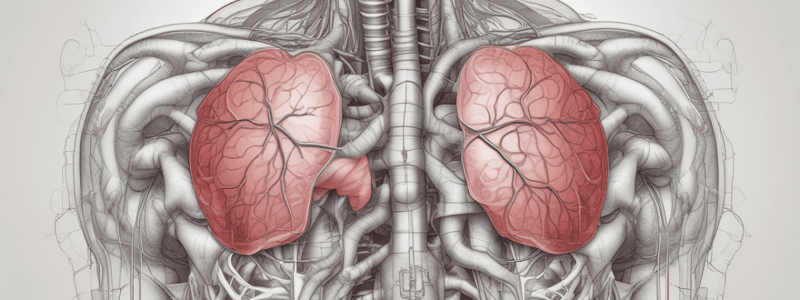
- The thoracic cavity, also known as the "chest", is the region where the lungs are located.
- It is bounded caudally (inferiorly) by the diaphragm, a thin, flat muscle that separates the thorax and abdomen.
Diaphragm
- Contraction of the diaphragm results in inhalation.
Ribs and Intercostal Muscles
- The thoracic cavity is bounded laterally by the ribs and intercostal muscles.
- The ribs are flexible, thin bones with cartilage connections dorsally and ventrally.
- This allows for movement of the thorax while providing structure and preventing collapse during inspiration.
- The ribs also provide protection to the heart and lungs, which are vital organs.
Intercostal Muscles
- Intercostal muscles are the muscles between the ribs.
- They assist in ventilation.
Thoracic Vertebrae
- The thoracic cavity is bounded dorsally by the thoracic vertebrae, which form the spinal column.
Sternum
- The thoracic cavity is bounded ventrally by the sternum, which is connected to the ribs by cartilage.
Thoracic Cavity
- The thoracic cavity, also known as the chest, is where the lungs are located.
- It is bounded caudally by the diaphragm, a thin, flat muscle that separates the thorax and abdomen.
- Contraction of the diaphragm results in inhalation.
Thoracic Cavity Boundaries
- Laterally, the thoracic cavity is bounded by the ribs and intercostal muscles.
- The ribs are flexible, thin bones with cartilage connections dorsally and ventrally, allowing for movement of the thorax while providing structure and protection to the heart and lungs.
- The intercostal muscles, located between the ribs, assist in ventilation.
Thoracic Cavity Boundaries (continued)
- Dorsally, the thoracic cavity is bounded by the thoracic vertebrae, which form the spinal column.
- Ventrally, it is bounded by the sternum, which is connected to the ribs by cartilage.
- Cranially, the thoracic cavity is bounded by the thoracic inlet, where all the structures pass between the neck and thorax, including blood vessels, oesophagus, trachea, and others.
Mediastinum
- The mediastinum is the area between the lungs, containing the trachea, heart, oesophagus, blood vessels, and connective tissue.
- It is divided into cranial (pink) and caudal (purple) mediastinum.
Pleura
- Consists of two serous membranes covering the outer layer of the lung lobes and inside of the thoracic wall.
- Has a space between the two layers, known as the pleural cavity.
Layers of the Pleura
- Parietal Pleura (Outer Layer)
- Also known as the "wall" layer
- Lines the walls of the thoracic cavity
- Attached to the ribs
- Visceral Pleura (Inner Layer)
- Also known as the "organ" layer
- Attached to the surface of the lungs
Studying That Suits You
Use AI to generate personalized quizzes and flashcards to suit your learning preferences.


