Podcast
Questions and Answers
Which of the following accurately describes the relationship between the renal artery and the kidneys?
Which of the following accurately describes the relationship between the renal artery and the kidneys?
- The right renal artery is longer because it needs to pass behind the IVC to reach the right kidney. (correct)
- Both renal arteries are the same length, ensuring equal blood flow to each kidney.
- The left renal artery is longer because it branches directly from the aorta.
- The left renal artery is longer because it needs to pass the IVC to reach the left kidney.
A histological slide of the kidney shows a vessel arching between the cortex and medulla. This vessel is MOST likely the:
A histological slide of the kidney shows a vessel arching between the cortex and medulla. This vessel is MOST likely the:
- Interlobar artery
- Segmental artery
- Interlobular artery
- Arcuate artery (correct)
In a healthy kidney, what BEST describes the sequence of structures through which filtrate passes?
In a healthy kidney, what BEST describes the sequence of structures through which filtrate passes?
- Proximal convoluted tubule → Loop of Henle → Bowman's space → Distal convoluted tubule → Collecting duct
- Bowman's space → Proximal convoluted tubule → Loop of Henle → Distal convoluted tubule → Collecting duct (correct)
- Bowman's space → Distal convoluted tubule → Loop of Henle → Proximal convoluted tubule → Collecting duct
- Proximal convoluted tubule → Distal convoluted tubule → Loop of Henle → Bowman's space → Collecting duct
What characteristic BEST differentiates the thick ascending limb of the loop of Henle from the thick descending limb under a microscope?
What characteristic BEST differentiates the thick ascending limb of the loop of Henle from the thick descending limb under a microscope?
Which statement accurately describes the role and location of the macula densa?
Which statement accurately describes the role and location of the macula densa?
Damage to the glomerular filtration barrier, specifically affecting the podocytes, would MOST likely result in:
Damage to the glomerular filtration barrier, specifically affecting the podocytes, would MOST likely result in:
The administration of an ACE inhibitor would MOST directly affect:
The administration of an ACE inhibitor would MOST directly affect:
In a patient experiencing the Syndrome of Inappropriate ADH secretion, what is the PRIMARY mechanism by which excess ADH impacts kidney function?
In a patient experiencing the Syndrome of Inappropriate ADH secretion, what is the PRIMARY mechanism by which excess ADH impacts kidney function?
A patient is diagnosed with a kidney stone lodged in the renal pelvis. Microscopically, what type of epithelial lining would you expect to observe?
A patient is diagnosed with a kidney stone lodged in the renal pelvis. Microscopically, what type of epithelial lining would you expect to observe?
Where is erythropoietin primarily produced in the kidneys?
Where is erythropoietin primarily produced in the kidneys?
Flashcards
Kidney Function
Kidney Function
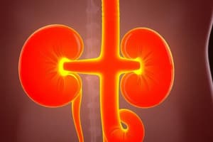
Main function is to purify, filter, and reabsorb blood to produce urine.
Renal Artery
Renal Artery
Where unpurified blood enters the kidney.
Renal Vein
Renal Vein
Where purified blood exits the kidney to re-enter circulation.
Arcuate Artery
Arcuate Artery
Signup and view all the flashcards
Glomerular Capillaries
Glomerular Capillaries
Signup and view all the flashcards
Peritubular Capillaries
Peritubular Capillaries
Signup and view all the flashcards
Nephron
Nephron
Signup and view all the flashcards
Renal Corpuscle
Renal Corpuscle
Signup and view all the flashcards
Macula Densa
Macula Densa
Signup and view all the flashcards
Juxtaglomerular Apparatus (JGA)
Juxtaglomerular Apparatus (JGA)
Signup and view all the flashcards
Study Notes
- Kidneys purify, filter, and reabsorb, each 10-12 cm long.
- Renal circulation originates from the renal artery, a branch of the abdominal aorta.
- Renal artery carries unpurified blood; the renal vein returns purified blood to circulation
Renal Circulation
- Segmental artery pierces renal columns towards the corticomedullary junction.
- Renal columns are extensions of the cortex between renal lobes (cortex and medulla).
- Interlobar arteries run through renal columns between lobes.
- Arcuate arteries arch over interlobar arteries at the corticomedullary junction.
- Interlobular arteries arise from arcuate arteries, running toward the cortex.
- Presence of interlobular arteries confirms location in the cortex.
- Afferent arterioles supply glomeruli for filtration.
- Glomerular capillaries are the main filtration site in Bowman's capsule.
- Filtrate drains through the proximal convoluted tubule, Loop of Henle, distal convoluted tubule, and collecting duct.
- Efferent arterioles receive filtered blood from the glomerulus, branching into peritubular capillaries or vasa recta.
- Peritubular capillaries accompany cortical convoluted tubules, draining into the renal vein.
- Vasa recta accompany the Loop of Henle in the medulla, also draining into the renal vein.
- Interlobular veins "complement" the arterial system, running parallel.
Nephron and Collecting Tubule
- Nephron: kidney's main functional unit
- Cortical nephrons are located in the renal cortex.
- Juxtamedullary nephrons span the renal cortex and medulla
Renal Corpuscle
- Consists of Bowman's capsule and the glomerulus
- Bowman's space is located between the two.
Glomerulus
- Filtration unit within the kidney
- Glomerular filtration barrier includes:
- Endothelial layer: Innermost layer with fenestrated cells, permeable to water, ions, and small molecules but not cells.
- Glomerular Basement Membrane (GBM): Negatively charged layer of collagen IV, laminin, and proteoglycans restricting large proteins like albumin.
- Podocyte Layer: Outermost layer of podocytes forming filtration slits covered by a slit diaphragm to regulate filtration and prevent protein leakage.
Juxtaglomerular Apparatus (JGA)
- It regulates blood pressure and glomerular filtration rate.
- Components include:
- Macula Densa: Located at the junction of the vascular pole and distal tubule, osmoreceptors detecting filtrate osmolarity and recognizing hypo-osmolarity.
- Juxtaglomerular Granular (JG) Cells: Located in the afferent arteriole, baroreceptors detecting low blood pressure or osmolarity, release renin to regulate blood pressure and sodium balance.
- Extraglomerular Mesangial Cells: Located between the macula densa and afferent/efferent arterioles.
Proximal Convoluted Tubules (PCT)
- Reabsorb 60-65% of filtrate, secreting urea, and certain drugs
- Eosinophilic due to many mitochondria.
- Characterized by a prominent brush border for reabsorption and fuzzy lumen from pinocytosis.
Loop of Henle
- Divided into thick and thin descending limbs and a thick ascending limb.
- Thin descending limb is simple squamous epithelium and very permeable to water.
- Thick ascending limb includes active Na+-K+ ATPase maintaining the hyperosmolar medullary interstitium.
Distal Convoluted Tubule (DCT)
- Has a clearer lumen and more basophilic cuboidal cells compared to PCT.
Collecting Duct
- Contains principal cells for sodium reabsorption and potassium secretion, rich in aquaporins for water reabsorption.
- Consists of intercalated cells that secrete or reabsorb hydrogen or bicarbonate ions to maintain acid-base balance.
- Heavily influenced by antidiuretic hormone (ADH), promoting aquaporin opening for water reabsorption
Urine Formation - Glomerular Filtration
- High blood pressure drives filtration through glomerular capillaries, the glomerular basement membrane, and filtration slits.
- Glomerular capillaries: Fenestrated, serving as a size barrier to exclude large molecules.
- Glomerular basement membrane: Negatively charged, serving as a charge barrier.
- Filtration slits: Formed by podocyte secondary processes, permeable to small substances.
Renin-Angiotensin-Aldosterone System
- If the urine is too dilute, it indicates decreased GFR.
- Low salt stimulates macula densa, and decreased blood pressure stimulates JG cells, triggering renin release.
- Renin converts angiotensinogen to angiotensin I, then ACE converts it to angiotensin II
- Angiotensin II, a vasoconstrictor, increases blood pressure and stimulates aldosterone release.
- Aldosterone stimulates DCT to reabsorb sodium.
Urine Excretion
- Urine flows from minor to major calyces, to the renal pelvis, ureter, and urinary bladder.
- Bladder capacity ranges from 500cc to 1L.
- Urothelium protects urinary tract, contains umbrella cells
- Uroplakins on umbrella cells prevent hyperosmotic urine ingress.
Studying That Suits You
Use AI to generate personalized quizzes and flashcards to suit your learning preferences.