Podcast
Questions and Answers
What is a common appearance of a cyst on a radiograph?
What is a common appearance of a cyst on a radiograph?
- Radiopaque shadow with fuzzy edges
- Radiolucency with sharp borders (correct)
- Uniform gray area with no distinct outline
- Dense white area with irregular borders
Which imaging technique is often necessary for a complete assessment of intrabony lesions?
Which imaging technique is often necessary for a complete assessment of intrabony lesions?
- Ultrasound imaging
- Standard dental X-ray
- Fluoroscopy
- Computed tomography (CT) (correct)
In what scenario might a dentist consider using radiopaque dyes or markers?
In what scenario might a dentist consider using radiopaque dyes or markers?
- To enhance image contrast in standard X-rays
- During sialography to clarify gland architecture (correct)
- To examine soft tissue lesions without involving bone
- When extracting teeth to visualize the root structure
What could indicate a malignant lesion on a radiograph?
What could indicate a malignant lesion on a radiograph?
Which lesion could potentially be a manifestation of hyperparathyroidism?
Which lesion could potentially be a manifestation of hyperparathyroidism?
What is the primary purpose of using laboratory tests in the context of oral lesions?
What is the primary purpose of using laboratory tests in the context of oral lesions?
Why is it important to determine whether a deviation in radiographic appearance is pathologic?
Why is it important to determine whether a deviation in radiographic appearance is pathologic?
Which imaging modality provides indirect images of gland structure in sialography?
Which imaging modality provides indirect images of gland structure in sialography?
What misleading appearance might be prevalent in radiographs of the maxilla and mandible?
What misleading appearance might be prevalent in radiographs of the maxilla and mandible?
What is a potential use for radiopaque markers in dental radiography?
What is a potential use for radiopaque markers in dental radiography?
Flashcards are hidden until you start studying
Study Notes
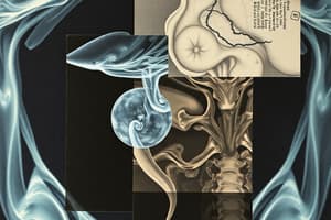
Radiographic Examination
- Radiographs are essential for diagnosing lesions near or within bone after clinical assessments.
- They can indicate osseous reactions, erosion, or intraosseous origins of soft tissue lesions.
- Standard radiographic techniques include periapical, occlusal, and panoramic views; specialized imaging may require computed tomography (CT) or magnetic resonance imaging (MRI).
- Cysts typically present as radiolucencies with clear borders, while aggressive lesions feature ragged, irregular borders.
- Deviation from normal structures in radiographs necessitates distinguishing between pathologic changes and atypical normal anatomy, especially in complex areas like the maxilla and mandible.
- Radiopaque dyes (e.g., sialography) can help evaluate gland structures and pathologic lesions.
- Injection of radiopaque substances into cysts can enhance determination of anatomical boundaries.
- Localization of foreign objects or lesions may involve radiopaque markers like needles or metal spheres.
Laboratory Investigation
- Laboratory tests can be crucial for identifying the systemic disease-related oral lesions.
- Conditions such as hyperparathyroidism, multiple myeloma, leukemia, and lymphomas may manifest as oral lesions.
- For instance, multiple lytic lesions and loss of the lamina dura may indicate hyperparathyroidism, which can be confirmed with serum calcium, phosphorus, and alkaline phosphatase tests.
- References for test requests and guidelines are available in oral and maxillofacial pathology textbooks.
Studying That Suits You
Use AI to generate personalized quizzes and flashcards to suit your learning preferences.