Podcast
Questions and Answers
What is the primary purpose of radiographic anatomy?
What is the primary purpose of radiographic anatomy?
- To diagnose mental health disorders
- To visualize the surface of the body
- To identify normal and abnormal anatomical structures (correct)
- To perform interventional procedures and surgeries
Which radiographic view is obtained when the X-ray beam passes from side to side?
Which radiographic view is obtained when the X-ray beam passes from side to side?
- Anteroposterior (AP) view
- Oblique view
- Lateral view (correct)
- Posteroanterior (PA) view
Which body region includes the stomach and intestines?
Which body region includes the stomach and intestines?
- Cranium
- Abdomen (correct)
- Pelvis
- Thorax
What appears as a dense, white structure on a radiograph?
What appears as a dense, white structure on a radiograph?
What is the term for abnormal breaks or abnormalities in bone structure?
What is the term for abnormal breaks or abnormalities in bone structure?
What type of contrast medium is used for CT scans to enhance visibility of soft tissues?
What type of contrast medium is used for CT scans to enhance visibility of soft tissues?
What appears as a dark area on a radiograph due to air content?
What appears as a dark area on a radiograph due to air content?
Which radiographic view is obtained when the X-ray beam passes from back to front?
Which radiographic view is obtained when the X-ray beam passes from back to front?
What is the term for abnormal masses or densities on a radiograph?
What is the term for abnormal masses or densities on a radiograph?
What type of bone appears white on a radiograph?
What type of bone appears white on a radiograph?
Flashcards are hidden until you start studying
Study Notes
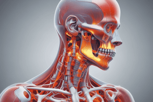
Radiographic Anatomy
Radiographic anatomy is the study of the internal structure of the body using medical imaging techniques, such as X-rays, CT scans, and MRI scans.
Importance of Radiographic Anatomy
- Essential for accurate diagnosis and treatment of diseases
- Helps in identifying normal and abnormal anatomical structures
- Guides interventional procedures and surgeries
Radiographic Views
- Anteroposterior (AP) view: X-ray beam passes from front to back
- Posteroanterior (PA) view: X-ray beam passes from back to front
- Lateral view: X-ray beam passes from side to side
- Oblique view: X-ray beam passes at an angle
Body Regions
- Cranium: skull and brain
- Thorax: chest and lungs
- Abdomen: stomach and intestines
- Pelvis: hips and reproductive organs
- Upper limb: shoulder, arm, and hand
- Lower limb: hip, leg, and foot
Radiographic Appearance of Bones
- Cortical bone: dense, compact bone appears white on radiographs
- Cancellous bone: spongy, porous bone appears grey on radiographs
- Joint spaces: appear as dark areas between bones
Radiographic Appearance of Soft Tissues
- Lungs: appear dark due to air content
- Heart: appears as a dense, white structure
- Liver: appears as a dense, right-sided structure
- Spleen: appears as a dense, left-sided structure
Radiographic Pathologies
- Fractures: appear as breaks or abnormalities in bone structure
- Tumors: appear as abnormal masses or densities
- Infections: appear as abnormal opacities or densities
Contrast Media
- Iodine-based contrast: used for CT scans to enhance visibility of soft tissues
- Barium-based contrast: used for fluoroscopy and CT scans to visualize gastrointestinal tract
- Gadolinium-based contrast: used for MRI scans to enhance visibility of soft tissues
Radiographic Anatomy
- Radiographic anatomy is the study of internal body structures using medical imaging techniques like X-rays, CT scans, and MRI scans.
Importance of Radiographic Anatomy
- Accurate diagnosis and treatment of diseases rely on radiographic anatomy.
- It helps identify normal and abnormal anatomical structures.
- Guides interventional procedures and surgeries.
Radiographic Views
Types of Radiographic Views
- Anteroposterior (AP) view: X-ray beam passes from front to back.
- Posteroanterior (PA) view: X-ray beam passes from back to front.
- Lateral view: X-ray beam passes from side to side.
- Oblique view: X-ray beam passes at an angle.
Body Regions
- Cranium: skull and brain.
- Thorax: chest and lungs.
- Abdomen: stomach and intestines.
- Pelvis: hips and reproductive organs.
- Upper limb: shoulder, arm, and hand.
- Lower limb: hip, leg, and foot.
Radiographic Appearance of Bones
- Cortical bone: dense, compact bone appears white on radiographs.
- Cancellous bone: spongy, porous bone appears grey on radiographs.
- Joint spaces: appear as dark areas between bones.
Radiographic Appearance of Soft Tissues
- Lungs: appear dark due to air content.
- Heart: appears as a dense, white structure.
- Liver: appears as a dense, right-sided structure.
- Spleen: appears as a dense, left-sided structure.
Radiographic Pathologies
- Fractures: appear as breaks or abnormalities in bone structure.
- Tumors: appear as abnormal masses or densities.
- Infections: appear as abnormal opacities or densities.
Contrast Media
- Iodine-based contrast: used for CT scans to enhance visibility of soft tissues.
- Barium-based contrast: used for fluoroscopy and CT scans to visualize gastrointestinal tract.
- Gadolinium-based contrast: used for MRI scans to enhance visibility of soft tissues.
Studying That Suits You
Use AI to generate personalized quizzes and flashcards to suit your learning preferences.