Podcast
Questions and Answers
Which of the following describes tidal volume?
Which of the following describes tidal volume?
- The amount of air remaining in the lungs after a normal breath
- The maximum amount of air the lungs can hold
- Air moving in or out during breathing (correct)
- The amount of air that can be forcefully exhaled after a maximal inhalation
What is vital capacity?
What is vital capacity?
- The volume of air remaining in the lungs after a normal exhalation.
- Air moving in or out during breathing
- The total volume of air the lungs can hold.
- Amount of air exhaled after maximal inhalation (correct)
What happens to lung compliance as pressures rise and lung function declines?
What happens to lung compliance as pressures rise and lung function declines?
- Lung compliance is not affected by pressure
- Lung compliance increases
- Lung compliance remains constant
- Lung compliance decreases (correct)
What is the normal partial pressure of oxygen (PO2) in the mixed venous blood?
What is the normal partial pressure of oxygen (PO2) in the mixed venous blood?
What is the normal partial pressure of carbon dioxide (PCO2) in the end capillary?
What is the normal partial pressure of carbon dioxide (PCO2) in the end capillary?
What is the role of surfactant in the alveoli?
What is the role of surfactant in the alveoli?
Which condition does NOT cause decreased lung compliance?
Which condition does NOT cause decreased lung compliance?
What effect does aging have on the respiratory system?
What effect does aging have on the respiratory system?
How does emphysema impact gas exchange?
How does emphysema impact gas exchange?
How long does it take for O2 and CO2 diffusion to reach equilibrium?
How long does it take for O2 and CO2 diffusion to reach equilibrium?
What is the normal blood flow time through the alveolar capillary system during rest?
What is the normal blood flow time through the alveolar capillary system during rest?
What generally happens to gas exchange at the base of the lungs due to gravity?
What generally happens to gas exchange at the base of the lungs due to gravity?
What is normal ventilation/perfusion ratio (V/Q)?
What is normal ventilation/perfusion ratio (V/Q)?
A shunt occurs when blood flows from the right side of the heart to the left side of the heart:
A shunt occurs when blood flows from the right side of the heart to the left side of the heart:
What is a capillary shunt?
What is a capillary shunt?
Lung tissue affected by a shuntlike effect:
Lung tissue affected by a shuntlike effect:
What does pulmonary vascular resistance (PVR) represent?
What does pulmonary vascular resistance (PVR) represent?
A normal PaO2 level is typically:
A normal PaO2 level is typically:
A normal SaO2 is:
A normal SaO2 is:
What is capnometry?
What is capnometry?
What is normal range of ETCO2 (end-tidal CO2)?
What is normal range of ETCO2 (end-tidal CO2)?
If the colorimetric capnography paper turns purple, what does this indicate?
If the colorimetric capnography paper turns purple, what does this indicate?
What is the formula for calculating minute ventilation (VE)?
What is the formula for calculating minute ventilation (VE)?
What is the normal range for minute ventilation?
What is the normal range for minute ventilation?
What is the term for the removal of an artificial airway?
What is the term for the removal of an artificial airway?
Flashcards
Tidal Volume
Tidal Volume
Air moving in or out during breathing.
Vital Capacity
Vital Capacity
Amount of air exhaled after maximal inhalation.
Lung Compliance
Lung Compliance
Describes how easily the lungs expand. Worsening function requires more ventilator pressure.
Lung Compliance on Ventilation
Lung Compliance on Ventilation
Signup and view all the flashcards
Surfactant Deficiency
Surfactant Deficiency
Signup and view all the flashcards
Factors Affecting Surfactant
Factors Affecting Surfactant
Signup and view all the flashcards
Aging on Ventilation
Aging on Ventilation
Signup and view all the flashcards
Surface Area (Gas Exchange)
Surface Area (Gas Exchange)
Signup and view all the flashcards
Thickness (Gas exchange)
Thickness (Gas exchange)
Signup and view all the flashcards
Conditions that Thicken Membranes
Conditions that Thicken Membranes
Signup and view all the flashcards
Exposure Length
Exposure Length
Signup and view all the flashcards
Aging and Gas Exchange
Aging and Gas Exchange
Signup and view all the flashcards
Damaged Alveoli
Damaged Alveoli
Signup and view all the flashcards
Perfusion Definition
Perfusion Definition
Signup and view all the flashcards
Systemic System (Perfusion)
Systemic System (Perfusion)
Signup and view all the flashcards
Pulmonary System (Perfusion)
Pulmonary System (Perfusion)
Signup and view all the flashcards
Cardiac Output (CO)
Cardiac Output (CO)
Signup and view all the flashcards
Gravity (Perfusion)
Gravity (Perfusion)
Signup and view all the flashcards
Ventilation-Perfusion Relationship
Ventilation-Perfusion Relationship
Signup and view all the flashcards
Shunt Definition
Shunt Definition
Signup and view all the flashcards
Anatomic Shunt
Anatomic Shunt
Signup and view all the flashcards
Capillary Shunt
Capillary Shunt
Signup and view all the flashcards
Shuntlike effect
Shuntlike effect
Signup and view all the flashcards
Venous Admixture
Venous Admixture
Signup and view all the flashcards
Pulmonary Vascular Resistance (PVR)
Pulmonary Vascular Resistance (PVR)
Signup and view all the flashcards
Study Notes
Pulmonary Mechanics
- Tidal volume refers to the air moving in or out during breathing.
- Vital capacity is the amount of air exhaled after maximal inhalation.
- Lung compliance worsens as pressures rise, leading to increased ventilator pressure per breath.
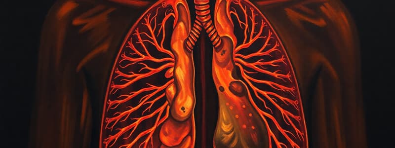
Alveolar-Capillary Interface
- The alveolar-capillary interface is where gas exchange occurs.
- Unoxygenated blood flows from the right heart to the lungs, then to the left atrium.
- PaO2 is the partial pressure of oxygen in the venous side.
- PAO2 is the partial pressure of oxygen in the alveoli where gas exchange is occurring.
Factors Affecting Pulmonary Gas Diffusion
- Lung compliance on ventilation is related to ease of expansion and sensitivity to conditions affecting lung tissues.
- Decreased compliance with surfactant deficiency occurs when surfactant helps alveoli open during inspiration and can cause collapse during expiration.
- Decreased compliance increases the work of breathing
- Decreased compliance causes decreased tidal volume.
- Factors affecting surfactant include premature birth, aging, smoking, acute lung injury, and adult respiratory distress disorder.
- Aging on ventilation includes diaphragm flattening, chest wall rigidity, respiratory muscle weakening, and increased anterior-posterior diameter, which leads to:
- Decreased lung compliance
- Altered pulmonary mechanics
- Air trapping which decreases gas exchange
Affecting Gas Exchange
- A large lung surface area increases gas exchange.
- Emphysema destroys alveolar capillary membranes, surface area is reduced.
- Thinner membranes lead to more rapid diffusion.
- Conditions that thicken the membrane include fluid in alveoli and/or interstitial spaces.
- Pulmonary edema from heart failure and pneumonia
- Inflammatory processes involving alveoli
- Fibrosis of lungs
- Diffusion of O2 and CO2 takes 0.25 seconds to reach equilibrium.
- Blood flows in 0.75 seconds through the alveolar capillary system at rest.
- Increased CO concentrations decrease exposure time, leading to hypoxemia.
- Indicates high CO states like sepsis
- Aging decreases total lung surface area
- Aging increases alveolar-capillary membrane thickness.
- Alveoli destroyed can also decrease diffusion or alter ventilation-perfusion relationships to make gas exchange less efficient
Pulmonary Perfusion
-
Perfusion is the pumping of blood to tissues and organs.
-
The two circulatory systems include systemic and pulmonary systems.
-
Systemic circulation is a vast system from the aorta to the right side of the heart.
-
Pulmonary circulation is a smaller system moving blood from the pulmonary artery in the right ventricle to the lungs and then to the left ventricle.
-
Adequate perfusion depends on systemic circulation.
-
CO, which is between 4-8 L/min, is calculated as HR x SV.
-
SV is the function of ventricular preload, afterload, and contractility. -A MAP between 60-100, and less than 60 indicates MODS
-
Blood distribution relies on gravity, with the greatest gas exchange occurring in the bases.
-
Ventilation is 4 L/min of air flow in and out of alveoli
-
Perfusion is 5 L/min of blood flow in and out of alveoli.
-
The normal ventilation/perfusion ratio (V/Q) is 4:5 (0.8).
-
Heart failure slows perfusion and respiratory failure impairs ventilation.
-
Altered ventilation-perfusion ratios can cause unoxygenated blood to return to the left side of the heart, resulting in hypoxemia.
Shunt
- A shunt is a percentage of CO that flows from the right heart to the left heart without undergoing pulmonary gas exchange.
- This is a major cause of hypoxemia primarily in acutely ill patients.
- There are 2 True shunts which include Anatomic shunt and Capillary shunt
- Anatomic shunts involve blood moving from the right heart to the left heart without coming into contact with the alveoli.
-Normal result when emptying bronchial or venous system.
- Abnormal when there are heart or lung problems, ventricular septal defects, traumatic injuries, or lung tumors
- Capillary shunts are normal blood flow past completely unventilated alveoli. -Conditions include consolidation, atelectasis, and fluid in alveoli Other Shunts include Absolute which are combined Anatomic + capillary shunts
- Lung tissue affected, which is refracted by O2 therapy
- No matter how much O2 is administered, diffusion cannot take place if alveoli are bypassed or non-functioning.
Shuntlike Effect
- Shuntlike effect is not a true shunt because the shunting is not complete.
- Alveolar ventilation is reduced, but, although some alveoli are still functioning, hypoxemia secondary to this effect is responsive to O2 therapy.
- Common causes include bronchospasm, hypoventilation, and pooling of secretions.
- Venous admixture is the effect a pulmonary shunt has on the contents of the blood as it enters the left heart and system as arterial blood.
- Mixture of oxygenated and partially oxygenated blood
- Normal > Venous admixture > shunt
Pulmonary Vascular Resistance (PVR)
- PVR is the resistance to blood flow in the pulmonary vascular system
- Represents the right ventricular afterload.
- Factors affecting PVR include the length, radius and viscosity of the vessel.
- PVR is much lower SVR is shorter
Arterial Blood Gases
- Arterial blood gases interpreted with compensatory status
- PaO2 is the partial pressure of oxygen dissolved in arterial blood, not what is available.
- Normal range: 80-100 mm Hg.
SaO2 and SpO2
- SO2 is the percentage of O2 bound to hemoglobin in blood.
- SaO2 is arterial blood oxygen saturation.
- SpO2 is oxygen saturation with pulse oximetry.
- Normal range: 95%+.
- Hemoglobin (Hgb/Hb) is a major component of RBCs.
- Normal for women: 12-15 g/dL
- Normal for men: 13.5-16 g/dL
Arterial Blood Gas Normals
- ABGs provide information on acid-base and oxygenation status.
- Normal conditions include sea level (760 mm Hg partial pressures), room air (21% oxygen), and blood temperature of 37°C/98.6°F.
- Age can affect ABG readings.
- 30-80 year olds may have a 25-30% decrease in PaO2.
Interpretation
- Interpret ABGs as trends, not single points.
- Interpretation includes determination of acid-base state, level of compensation, and oxygenation status.
- Alveolar ventilation
- Amount of oxygen available in arterial blood for possible tissue use.
- Oxygen-carrying capacity - Oxygen transport
Hypoxemia
- Mild hypoxemia has a PaO2 of 60-75 mm Hg.
- Moderate hypoxemia has a PaO2 of 45-59 mm Hg.
- Severe hypoxemia has a PaO2 of less than 45 mm Hg.
- Normal pH ranges from 7.35-7.45.
- Normal PaCO2 ranges from 35-45 mm Hg.
- Normal HCO3 ranges from 24-28 mEq/L.
- Normal BE is +/- 2 mEq/L.
- Normal PaO2 ranges from 80-100 mm Hg.
- Normal SaO2 is 95%+.
- Hemoglobin measurements range from 13.5-18 g/dL for men and 12-15 g/dL for women.
Capnography
- Capnometry involves a numeric measurement of CO2.
- Capnography, also known as end-tidal CO2 monitoring, uses a non-invasive graphic display of CO2 concentration exhaled by the patient captured by a capnogram.
- Clinical uses include surgical and procedural anesthesia, post-op recovery, post-op PCA recipients, critical care units, and EDs.
- Capnography confirms endotracheal and enteric feeding tube placement.
- Also helps in the detection of mechanical ventilator problems
- ETCO2 monitoring assesses ventilation status and it is an early warning sign for ventilation changes with acceptable range between 30-44 mm Hg:
- Abnormally low ETCO2 (Less than 30 mm Hg) due to Hyperventilation , Hypothermia, PE, or Decreased CO;
- Increased ETCO2 (Greater than 44 mm Hg) with Increased CO2 production from Fever or increased CO or Hypoventilation from Respiratory center depression or neuromuscular disease
- Capnography might not be accurate in morbid obesity, severe pulmonary edema or ventilation-perfusion abnormalities
- Colorimetric capnography uses pH-sensitive paper that changes color based on exhaled CO2 to represent a range of ETCO2.
- Not precise for ET tube but more for ET tube placement. Process
- The CO2 detector is attached to an ET tube.
- The patient breathes six times.
- The device is ready to full expiration.
- It then responds with three color ranges
- Purple indicates ET tube absence in the trachea -Brown or Yellow indicate the ET tube placement in the Esophagus or low pulmonary blood flow or properly in the Trachea, respectively.
Studying That Suits You
Use AI to generate personalized quizzes and flashcards to suit your learning preferences.


