Podcast
Questions and Answers
Emphysema reduces lung function primarily by affecting which of the following factors crucial for pulmonary gas exchange?
Emphysema reduces lung function primarily by affecting which of the following factors crucial for pulmonary gas exchange?
- Lung surface area (correct)
- Pressure gradient between alveoli and capillaries
- Partial pressures of respiratory gases
- Alveolar-capillary membrane thickness
During periods of high cardiac output, such as heavy exercise, what is the primary concern regarding the length of gas exposure in the alveolar-capillary system?
During periods of high cardiac output, such as heavy exercise, what is the primary concern regarding the length of gas exposure in the alveolar-capillary system?
- Carbon dioxide elimination may be excessively prolonged
- Oxygen exchange is usually impaired, leading to hyperoxemia
- The rate of gas diffusion significantly increases, leading to rapid equilibrium
- Oxygen exchange may be impaired in the presence of diffusion abnormalities (correct)
An ideal ventilation-perfusion (V/Q) ratio is approximately 0.8. What does this value represent?
An ideal ventilation-perfusion (V/Q) ratio is approximately 0.8. What does this value represent?
- For every 1 liter of air entering the alveoli, about 0.8 liters of blood flow past them
- For every 4 liters of air entering the alveoli, about 5 liters of blood flow past them (correct)
- For every 5 liters of air entering the alveoli, about 4 liters of blood flow past them
- For every 8 liters of air entering the alveoli, 10 liters of blood flow past them
Which of the following best describes a pulmonary shunt?
Which of the following best describes a pulmonary shunt?
What does capnography measure?
What does capnography measure?
A colorimetric capnography device attached to an endotracheal tube turns purple after several breaths. What does this indicate?
A colorimetric capnography device attached to an endotracheal tube turns purple after several breaths. What does this indicate?
What is the normal range for minute ventilation?
What is the normal range for minute ventilation?
Which of the following ventilator modes assumes that the ventilator and circuitry are insensitive to a patient's respiratory effort or response?
Which of the following ventilator modes assumes that the ventilator and circuitry are insensitive to a patient's respiratory effort or response?
Which of the following is a complication specifically associated with AC mode ventilation?
Which of the following is a complication specifically associated with AC mode ventilation?
A patient on positive pressure ventilation develops increased agitation, coughing, and subcutaneous emphysema. Which complication should the nurse suspect?
A patient on positive pressure ventilation develops increased agitation, coughing, and subcutaneous emphysema. Which complication should the nurse suspect?
What is the calculation for Mean Arterial Pressure (MAP)?
What is the calculation for Mean Arterial Pressure (MAP)?
Which of the following values of the Rapid Shallow Breathing Index (RSBI) indicates a greater likelihood that a patient will wean successfully?
Which of the following values of the Rapid Shallow Breathing Index (RSBI) indicates a greater likelihood that a patient will wean successfully?
You are called to assess a patient on mechanical ventilation with high peak inspiratory pressures. After confirming tube placement and patient sedation, you notice a marked decrease in air entry on the left side of the chest, along with tracheal deviation to the right. What life-threatening complication do you urgently suspect?
You are called to assess a patient on mechanical ventilation with high peak inspiratory pressures. After confirming tube placement and patient sedation, you notice a marked decrease in air entry on the left side of the chest, along with tracheal deviation to the right. What life-threatening complication do you urgently suspect?
A patient with severe ARDS is proned to improve oxygenation. Which physiological mechanism explains why proning often increases PaO2 in these patients?
A patient with severe ARDS is proned to improve oxygenation. Which physiological mechanism explains why proning often increases PaO2 in these patients?
An ICU patient receiving mechanical ventilation suddenly develops dilated pupils, bradycardia, and an increased ICP. What is the MOST likely cause?
An ICU patient receiving mechanical ventilation suddenly develops dilated pupils, bradycardia, and an increased ICP. What is the MOST likely cause?
Flashcards
Partial Pressures of Gases
Partial Pressures of Gases
The difference in partial pressures of oxygen (O2) and carbon dioxide (CO2) between the alveoli and the pulmonary capillaries.
Pressure Gradient
Pressure Gradient
The difference between partial pressures of gases in the alveoli and pulmonary capillaries. Dictates gas flow rate.
Lung Surface Area
Lung Surface Area
The total functional surface area of the lungs available for gas exchange.
Alveolar-Capillary Membrane Thickness
Alveolar-Capillary Membrane Thickness
Signup and view all the flashcards
Length of Gas Exposure
Length of Gas Exposure
Signup and view all the flashcards
Pulmonary Perfusion
Pulmonary Perfusion
Signup and view all the flashcards
Cardiac Output (CO)
Cardiac Output (CO)
Signup and view all the flashcards
Ventilation-Perfusion (V/Q) Relationship
Ventilation-Perfusion (V/Q) Relationship
Signup and view all the flashcards
Pulmonary Shunt
Pulmonary Shunt
Signup and view all the flashcards
Pulmonary Vascular Resistance (PVR)
Pulmonary Vascular Resistance (PVR)
Signup and view all the flashcards
Capnography
Capnography
Signup and view all the flashcards
Colorimetric Capnography
Colorimetric Capnography
Signup and view all the flashcards
Minute Ventilation (VE)
Minute Ventilation (VE)
Signup and view all the flashcards
Tests to Consider for Weaning
Tests to Consider for Weaning
Signup and view all the flashcards
Control Mode Ventilation (CMV)
Control Mode Ventilation (CMV)
Signup and view all the flashcards
Study Notes
- Factors impacting pulmonary gas diffusion in the alveoli involve oxygen availability in the atmosphere and hypoventilation.
Partial Pressures of Gases
- Differences in the partial pressures of O2 and CO2 between the alveoli and pulmonary capillaries drive gas diffusion
- Oxygen moves from high to low pressure, while CO2 moves in the opposite direction
Pressure Gradient
- The difference in gas partial pressures between alveoli and pulmonary capillary blood determines the rate and direction of gas flow
- A greater pressure difference results in more rapid gas flow and greater pressure
Lung Surface Area
- The total functional surface area of the lungs facilitates gas exchange
- Lung disorders that decrease lung surface area, such as pneumonia, emphysema, lung tumors, pneumothorax, and pneumonectomy, impair gas exchange
- A large lung surface increases available alveolar-capillary membrane surface and oxygen and carbon dioxide diffusion
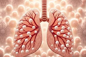
- Emphysema destroys the alveolar-capillary membrane, therefore reducing surface area.
Alveolar-Capillary Membrane Thickness
- The thickness of the membrane enhances rapid gas diffusion
- Conditions that increase membrane thickness (pulmonary edema, pneumonia, and lung fibrosis) decrease the rate of diffusion
Length of Gas Exposure
- During high cardiac output, blood flows faster through the alveolar-capillary system decreasing exposure time for gas exchange
- Oxygen exchange is usually unimpaired in healthy lungs, but hypoxemia may result with diffusion abnormalities
- Diffusion of oxygen and carbon dioxide requires 0.25 seconds to reach equilibrium
- During body rest, blood flows in 0.75 seconds through alveolar- capillary system
- High cardiac output decreases exposure time, resulting in hypoxemia, which includes sepsis.
Aging Effects on Diffusion
- Total lung surface area decreases
- Capillary membrane thickness increases
- Alveoli are destroyed
- Result: Decreased diffusion across the alveolar-capillary membrane + Alters ventilation-to-perfusion relationship = Less efficient gas exchange
Pulmonary Perfusion Components
- Pulmonary perfusion, the flow of blood through the pulmonary circulation, and the following components are essential for gas exchange:
Cardiac Output (CO)
- Cardiac output, or the amount of blood the heart pumps per minute, is calculated by multiplying stroke volume and heart rate
- Normal cardiac output is 4 to 8 liters per minute
- Adequate cardiac output is crucial for maintaining sufficient blood flow through the pulmonary circulation
Gravity
- Blood accumulates in dependent lung areas due to gravity and influences the ventilation-perfusion relationship
Ventilation-Perfusion (V/Q) Relationship
- Ratio of alveolar ventilation (airflow into the alveoli) to pulmonary capillary perfusion (blood flow through the pulmonary capillaries
- An ideal V/Q ratio is approximately 0.8 (for every 4 liters of air entering the alveoli, about 5 liters of blood flow past them)
- Imbalances in this ratio can impair gas exchange
Pulmonary Shunt
- A pulmonary shunt occurs when blood flows from the right side of the heart to the left without participating in gas exchange.
- Anatomic Shunt: Blood bypasses the alveoli entirely, like in congenital heart defects or lung tumors
- Capillary Shunt: Blood flows past unventilated alveoli, as seen in conditions like atelectasis or pulmonary edema
Pulmonary Vascular Resistance (PVR)
- The resistance to blood flow within the pulmonary circulation
- Influenced by the length and radius of the blood vessels and the viscosity of the blood
- Hypoxia, hypercapnia, acidosis, and lung inflation can increase PVR, affecting right ventricular afterload
Mean Arterial Pressure (MAP)
- Measure of perfusion adequacy calculated as (2 x diastolic blood pressure + systolic blood pressure)/3
- A MAP of 70 to 100 mm Hg is perfusion adequacy
- MAP below 60 mm Hg is inadequate.
- The components work together to ensure that blood is adequately perfused through the lungs, allowing for efficient gas exchange and oxygenation of the blood
Capnography
- Measures the concentration of carbon dioxide (CO2) in exhaled air, with several clinical applications:
- Monitoring Ventilation: Used to monitor adequacy in surgical and procedural anesthesia, postoperative recovery, critical care, and emergency departments
- Confirming Endotracheal Tube Placement: A normal capnographic waveform indicates that the tube is in the trachea, not the esophagus
- Assessing CPR Effectiveness: Increased end-tidal CO2 (ETCO2) levels suggest effective compressions and improved cardiac output during CPR
- Detecting Respiratory Depression: Recommended for monitoring patients receiving patient-controlled analgesia (PCA) to detect early signs of respiratory depression
- Identifying Ventilator Problems: Can detect issues such as disconnections, leaks, or malfunctions, by showing changes in the capnographic waveform
- Monitoring Sedation: Used to ensure adequate ventilation and detect early signs of hypoventilation or apnea during procedural sedation.
- Evaluating Metabolic Status: Increased ETCO2 levels indicate conditions such as fever or increased cardiac output, while decreased ETCO2 levels may suggest hyperventilation, hypothermia, or pulmonary embolism
- Confirming Enteric Feeding Tube Placement: Can help to verify accurate placement of feeding tubes, reducing risk of associated misplacement and complications.
Calorimetric Capnography
- Method used to estimate the concentration of carbon dioxide (CO2) in exhaled air using a color-changing device
- Particularly useful in emergency and critical care settings for confirmation of endotracheal tube placement and assessing ventilation status
- Device Composition: Contains pH-sensitive paper that changes color in response to the CO2 levels in exhaled air
- Attachment: Attached to the end of an endotracheal tube or other airway management devices
- Color Change: The CO2 in the breath reacts with the pH-sensitive paper, causing a color change that corresponds to different ranges of CO2 concentration
- Interpretation: Color change used to estimate end-tidal CO2 (ETCO2) levels:
- Purple: Indicates low/no CO2 suggesting the tube is not in the trachea (Nellcor EASYCAP II, using the following color ranges: i.e. 0.03% to less than 0.5% ETCO2, less than 4 mmHg CO2)
- Brown: Indicates moderate CO2 levels, and may suggest the tube is in the esophagus or the patient has low pulmonary blood flow(Nellcor EASYCAP II, using the following color ranges: i.e. 0.5% to less than 2% ETCO2, 4 to less than 15 mmHg CO2)
- Yellow: Indicates normal CO2 levels confirming proper tracheal placement of the tube.
Clinical Applications of Capnography
- Confirming Endotracheal Tube Placement: Used in emergency settings to confirm placement
- Assessing Ventilation: It provides a rapid, visual indication of the patient's ventilatory status in pre-hospital settings, emergency departments, and during transport
- Monitoring During CPR: Can help assess effectiveness of chest compressions during CPR
- Verifying Feeding Tube Placement: Can help ensure they are not mistakenly placed in the respiratory tract
Advantages of Capnography
- Quick and Easy to Use: Provides immediate feedback on CO2 levels
- Noninvasive: Complex equipment or blood sampling is not required
- Portable: Useful in various settings, including pre-hospital care and emergency departments
Limitations of Capnography
- Less Precise: Provides an estimate rather than an exact measurement of ETCO2
- Limited Use: Primarily used for confirming tube placement rather than continuous monitoring
- Not Suitable for All Patients: May not be accurate in patients with severe ventilation-perfusion abnormalities or those with low pulmonary blood flow
Minute Ventilation (VE)
- Total volume of expired air in one minute
- VE = TV x frequency
- Frequency is the same as breaths per minute
- Normal 5 to 10 L/min
Endotracheal Tube
- Guidelines: use orotracheal opposed to nasotracheal tube
- Soft-cuffed ET tubes (in a variety of sizes)
- Stylet - to guide the tube in place by keeping it stiff (orotracheal route only)
- Topical anesthetic - to promote comfort and local anesthesia
- Laryngoscope handle with blade attached and functional light source to open and visualize the airway
- Magill forceps – to assist with guiding the tube past the pharyngeal area
- Suction machine or set up to wall-mounted suction head/ Suction catheter kit with sterile gloves
- Yankauer (oral) suction catheter to clear the airway of secretions
- Syringe for cuff inflation/Water-soluble lubricant – to apply to the ET tube to facilitate insertion
- Endotracheal tube-holding device (or adhesive tape if device not available) - to secure the ET tube following insertion Personal protective equipment/ Sedative medication (under guidance of credentialed provider) and intravenous access for patient comfort and safety and facilitating insertion process/ Stethoscope - to auscultate bilateral lung sounds post insertion/CO2 detector – to aid in identification of tube location
Tracheostomy Tubes
- Emergency tracheostomy performed for upper airway obstruction secondary to head or neck surgery, severe edema, or tumor obstruction
- Patient would require prolonged intubation = Usually a maximum of 2-3 weeks
- Usually placed by percutaneous approach using dilators at the bedside with sedation = Can be placed via open surgical approach
- Tubes vary in type and size, including diameter and length
Securing the Airway
- ET stabilizers are the preferred method of securing ET tube
- Tracheostomy is commonly secured with twill tape or a commercial available tracheostomy holder with velcro
Extubation
- Carried out as soon as it is determined that the patient can sustain spontaneous breathing
- Air Leak Test - Oropharynx is suctioned, ET cuff is deflated and the patient is evaluated for the ability to breath around the tube
- Quick tube removal is important because the ET tube increases the work of breathing
- POSTEXTUBATION
Assessing Placement of Endotracheal Tubes
- The following are ways to assess the placement of endotracheal tubes:
- Both lung fields are auscultated for equal breath sounds
- Air sounds or gurgling over the epigastric area indicated that the endotracheal tube is malpositioned in the esophagus
- Chest x-ray is performed to confirm placement - 2-3 cm above the carina
- Measure carbon dioxide using capnography or a disposable CO2 detector i.May produce false negative if cardiac output is too low
Tests For Evaluating Pulmonary Functions
- Tests to consider: Rapid Shallow Breathing Index, Vital Capacity, Respiratory Rate, ABGs, Maximal Inspiratory Capacity
- Rapid Shallow Breathing Index: <105 spontaneous breathing
- Vital Capacity:> 10mL/kg
- Frequency: < 35 bpm
- Maximal Inspiratory Pressure (MIP): Negative Inspiratory Force (NIF:) <-20 to -30 cm H2O
- Minute Ventilation: < 10L/min
Ventilator Modes
- Control Mode Ventilation/ Continuous Mandatory Ventilation (CMV)
- Control mode: not sensitive to patients own ventilatory effort
- Delivers the breaths at a preset rate based on time
- Not used unless patient is continuously apneic
Assist/Control (A/C)
- Assist part: sensitive to spontaneous inspiratory effort
- Allows patient some control over the rate of breathing
- Control part: acts as a backup should the patient decrease the breathing effort below the present rate
- AC: every breath is a ventilator breath at the preset tidal volume
Synchronized Intermittent Mandatory Ventilation (SIMV)
- Decreases risk for hyperventilation and provides a better ventilation-perfusion distribution = periodic volume or pressure breaths occur at preset time intervals = between mechanical breaths, patients spontaneously breathe w/o getting a preset mandatory breath = patients breathe from continuous flow or a demand valve = each mandatory breath is synchronized
Continuous Positive Airway Pressure (CPAP)
- all breaths are controlled by the patient. No mechanical breaths are provided unless the patient becomes apneic CPAP and PEEP techniques for applying airway pressure to the spontaneously breathing patient. The entire breathing cycle may have positive airway pressure, or just at the end of the breath.
- PEEP, positive end expiratory pressure: The establishment and maintenance of preset airway pressures greater than ambient at end of exhalation.
Pressure Support (PS)
- airway resistance may be a significant factor causing difficulty during the weaning phase
- Provides a preset level of positive pressure above PEEP during the inspiratory cycle of the spontaneous inspiratory effort
- similar to an IPPB breath
Complications Of Ventilator Therapy
- AC mode ventilation can lead to development of respiratory muscle atrophy from the ventilator performing the work of breathing + Risk for respiratory acidosis
Multipsystem Effects of Positive Pressure Ventilation
- Cardiovascular = Decreased cardio out = Decreased preload= Decreased stroke volume= increased afterload
Pulmonary
- Increased gas flow to nondpendent lung and to central lung tissue
- Increased blood flow to peripheral lung tissues
- Increased blood flow to peripheral lung tissues -Lower airway contamination causing ventilator-associated pneumonia (VAP) Risk for oxygen toxicity
- Cerebral perfusion pressure= MAP ‐ ICP (increased intracranial pressure)
Artificial Airway Complications
- Ischemia and necrosis of the nares
- Risk of development of ear pressure problems or inner ear infection
- Ulcerations and possible necrosis of the inner cheek or lip
- Dental trauma or tongue pressure sores
Patient Positioning Influence Oxygenation
- Patient positioning can significantly impact oxygenation by affecting the distribution of ventilation within the lungs.
- Semi-recumbent often used to improve oxygenation in patients with respiratory issues by optimizing lung expansion and ventilation-perfusion matching (V/Q matching) in different lung regions
- Prone position can improve oxygenation by recruiting collapsed lung areas in the posterior lung
- Semi-recumbent position can help drain secretions and enhance lung expansion in patients with certain conditions like pneumonia.
Studying That Suits You
Use AI to generate personalized quizzes and flashcards to suit your learning preferences.