Podcast
Questions and Answers
What is the primary habitat of Coccidia and Microspora?
What is the primary habitat of Coccidia and Microspora?
- Circulatory system
- Digestive tract (correct)
- Nervous system
- Respiratory system
Which family does the genus Toxoplasma belong to?
Which family does the genus Toxoplasma belong to?
- Babesiidae
- Eimeriidae
- Plasmodiidae
- Sarcocystidae (correct)
What triggers the transmission of Coccidia and Microspora from host to host?
What triggers the transmission of Coccidia and Microspora from host to host?
- Airborne particles from an infected host
- Encysted stages passed in feces (correct)
- Consumption of undercooked meat
- Direct contact with infected blood
Which genus is part of the family Cryptosporididae?
Which genus is part of the family Cryptosporididae?
Which phylum do Microspora belong to?
Which phylum do Microspora belong to?
What distinguishes C. cayetanensis from other similar protozoa in terms of its life cycle?
What distinguishes C. cayetanensis from other similar protozoa in terms of its life cycle?
What is the primary treatment for cyclosporiasis?
What is the primary treatment for cyclosporiasis?
How long do unsporulated oocysts of Isospora belli take to mature into sporulated oocysts?
How long do unsporulated oocysts of Isospora belli take to mature into sporulated oocysts?
What characteristic shape do oocysts of Isospora belli have?
What characteristic shape do oocysts of Isospora belli have?
Which of the following is a significant microsporidian species that affects humans?
Which of the following is a significant microsporidian species that affects humans?
What is the size range of the oocysts of Isospora belli?
What is the size range of the oocysts of Isospora belli?
What component do microsporidian spores inject into their host?
What component do microsporidian spores inject into their host?
Which type of patients is primarily affected by Microspora species, particularly Enterocytozoon bieneusi?
Which type of patients is primarily affected by Microspora species, particularly Enterocytozoon bieneusi?
How does the wall structure of Isospora belli oocysts appear?
How does the wall structure of Isospora belli oocysts appear?
What is the typical pattern of infection observed in immunocompromised patients due to Isospora belli?
What is the typical pattern of infection observed in immunocompromised patients due to Isospora belli?
Which species of Cryptosporidium is primarily responsible for infecting humans?
Which species of Cryptosporidium is primarily responsible for infecting humans?
What is the primary host for Cyclospora cayetanensis?
What is the primary host for Cyclospora cayetanensis?
What distinguishes the thick-walled oocyst of Cryptosporidium from the thin-walled oocyst?
What distinguishes the thick-walled oocyst of Cryptosporidium from the thin-walled oocyst?
What is the average size of sporulated oocysts of Cyclospora cayetanensis?
What is the average size of sporulated oocysts of Cyclospora cayetanensis?
Which method is NOT commonly used to diagnose Cryptosporidium infections?
Which method is NOT commonly used to diagnose Cryptosporidium infections?
What is the size range of unsporulated oocysts of Cyclospora cayetanensis?
What is the size range of unsporulated oocysts of Cyclospora cayetanensis?
Which of the following pathogens is primarily associated with severe diarrhea in immunocompromised patients?
Which of the following pathogens is primarily associated with severe diarrhea in immunocompromised patients?
Which cryptosporidium species is primarily associated with rodents?
Which cryptosporidium species is primarily associated with rodents?
How are sporozoites released from thick-walled oocysts of Cryptosporidium?
How are sporozoites released from thick-walled oocysts of Cryptosporidium?
Which of the following statements about the life cycle of Cryptosporidium is true?
Which of the following statements about the life cycle of Cryptosporidium is true?
What is a common symptom of Cyclospora cayetanensis infection?
What is a common symptom of Cyclospora cayetanensis infection?
Which factor is NOT associated with an increased risk of cryptosporidiosis according to the case-control studies?
Which factor is NOT associated with an increased risk of cryptosporidiosis according to the case-control studies?
How does the zygote of Cryptosporidium typically develop post-fertilization?
How does the zygote of Cryptosporidium typically develop post-fertilization?
Which genera of Coccidia are primarily significant in veterinary medicine?
Which genera of Coccidia are primarily significant in veterinary medicine?
What is the process through which Eimeria undergoes asexual reproduction?
What is the process through which Eimeria undergoes asexual reproduction?
Which species is primarily responsible for caecal coccidiosis in chickens?
Which species is primarily responsible for caecal coccidiosis in chickens?
What is a common symptom of intestinal coccidiosis in chickens?
What is a common symptom of intestinal coccidiosis in chickens?
Isospora species commonly found in dogs include which of the following?
Isospora species commonly found in dogs include which of the following?
Which symptom is NOT typically associated with coccidiosis in sheep?
Which symptom is NOT typically associated with coccidiosis in sheep?
Which species of Eimeria is notably found in rabbits?
Which species of Eimeria is notably found in rabbits?
What kind of oocyst does Isospora contain?
What kind of oocyst does Isospora contain?
What diagnostic test is commonly used for identifying coccidiosis in animals?
What diagnostic test is commonly used for identifying coccidiosis in animals?
Which Coccidia species is usually linked to severe symptoms in pigs?
Which Coccidia species is usually linked to severe symptoms in pigs?
What differentiates the life cycle of Isospora from that of Eimeria?
What differentiates the life cycle of Isospora from that of Eimeria?
Among the listed Eimeria species, which one causes the least severe symptoms in chickens?
Among the listed Eimeria species, which one causes the least severe symptoms in chickens?
Coccidiosis severity in cattle is most commonly caused by which species?
Coccidiosis severity in cattle is most commonly caused by which species?
What is the clinical significance of coccidiosis in rabbits?
What is the clinical significance of coccidiosis in rabbits?
Flashcards are hidden until you start studying
Study Notes
Phylum Apicomplexa
- Coccidia and Microspora are obligate intracellular protozoa, commonly found in the digestive tract
- Coccidia are transmitted through encysted stages passed in feces
- Microspora are also transmitted through encysted stages passed in feces
- Coccidia and Microspora have seen an increase in recognition and clinical encounters since the AIDS epidemic
Coccidia
- Key genera in veterinary medicine are: Eimeria, Isospora, Cryptosporidium
- Coccidia are primarily intracellular parasites of intestinal epithelium
- Schizogony and gametogony processes occur within the host's intestines
- Sporulation occurs outside the host
Eimeria
- Hosts include birds, cattle, goats, sheep, pigs, horses, and rabbits
- Typically found in the epithelial cells of the intestines, with two exceptions:
- E. truncata found in the kidneys
- E. stiedae found in the liver
- Found worldwide
Important Eimeria Species
- Poultry:
- E. tenella
- E. necatrix
- E. brunetti
- E. maxima
- E. mitis
- E. acervulina
- Turkey:
- E. meleagrimitis
- E. adenoeides
- Goose:
- E. anseris
- E. nocens
- E. truncata (kidneys)
- Cattle:
- E. zuernii
- E. bovis
- E. alabamensis
- Sheep:
- E. crandallis
- E. ovinoidalis
- E. bakuensis
- Goats:
- E. arloingi
- E. ninakohlyakimovae
- Pigs:
- E. debliecki
- Horses:
- E. leuckarti
- Rabbits:
- E. flavescens
- E. intestinalis
- E. stiedae (liver)
Eimeria Life Cycle
- Asexual Reproduction:
- Sporulated oocyst
- Sporozoite
- Trophozoite
- Schizogony
- Schizont
- Merozoite
- Sexual Reproduction:
- Some merozoites become gametocytes (macrogametocytes and microgametocytes)
- Macrogamete and microgamete
- Fertilization
- Zygote
- Unsporulated oocyst
- Sporogony
- Unsporulated oocyst
- Sporulated oocyst (containing 4 sporocysts, each containing 2 sporozoites)
Diagnosing Eimeria
- Examine feces for oocysts
- Scrape or biopsy intestinal tissue for coccidia
- Identify oocysts based on shape, size, and location
- Analyze schizont location, size, and number of merozoites for further identification
Isospora
- Important Species:
- I. suis (pigs)
- I. canis and I. ohioensis (dogs)
- I. felis and I. rivolta (cats)
- I. belli (humans)
Isospora Life Cycle
- Sporulated oocyst contains 2 sporocysts, each with 4 sporozoites
- Extraintestinal stages can occur in the spleen, liver, and lymph nodes of pigs
- These extraintestinal stages can re-enter the intestines, potentially causing recurrent intestinal coccidiosis
- Rabbits eating oocysts from dogs and cats can exhibit symptoms, acting as reservoirs for the disease
Coccidiosis in Poultry
- Caecal Coccidiosis:
- E. tenella is the most important cause
- Commonly seen in chickens aged 3-7 weeks
- Prepatent period: approximately 7 days
- Symptoms include:
- Depression
- Drooping wings
- Ruffled feathers
- Watery diarrhea with blood
- Subclinical infection:
- Reduced weight gain
- Decreased feed conversion rate
- Intestinal Coccidiosis:
- E. necatrix is the most severe
- E. brunetti, E. acervulina, E. maxima, E. mitis, and E. praecox also cause intestinal coccidiosis
- Prepatent period: 4-7 days
- Symptoms:
- Similar to caecal coccidiosis, but E. necatrix and E. brunetti can cause severe symptoms, potentially leading to blood in the feces.
- Subclinical infection:
- Reduced feed conversion rate
- Decreased or poor egg production
- Coccidiosis Severity and Location:
- E. tenella: Caeca, severe hemorrhages, white spots, thickened walls, blood in stool
- E. necatrix: Small intestine, severe hemorrhages, thickened walls, white spots, coagulative necrosis, blood in stool.
- E. brunetti: Lower small intestine, slight hemorrhages, watery exudates, white transverse bands, blood in stool.
- E. acervulina: Upper small intestine, watery exudates, white transverse bands, no blood in stool.
- E. maxima: Mid small intestine, salmon pink exudates, thickened walls, hemorrhages with heavy infection, no blood in stool.
- E. mitis: Lower small intestine, no visible lesions.
Diagnosing Coccidiosis in Poultry
- Necropsy reveals site and type of lesions
- Feces examination for oocysts, but note:
- Severe symptoms often precede oocyst production
- Oocyst quantity in feces doesn't always correlate to intestinal lesion severity
Coccidiosis in Cattle
- Common in calves under 1 year old, but can affect older cattle
- 13 species can cause coccidiosis, but E. bovis and E. zuernii are most common.
- Severe enteritis and diarrhea occur with heavy infection.
Coccidiosis in Goats and Sheep
- 11 species can cause coccidiosis in lambs under 1 year old, but E. crandallis and E. ovinoidalis are most severe.
- Symptoms include severe diarrhea, potentially with blood
- In E. ovinoidalis infections, visible white spots (giant schizonts) may occur
- Coccidiosis in goats is not well-researched
Coccidiosis in Pigs
- Multiple species can cause coccidiosis, including E. debliecki and I. suis
- The role and significance of coccidiosis in pigs remain unclear
- Diagnosis is difficult, except during necropsy, as diarrhea occurs before oocysts are excreted in feces
Coccidiosis in Horses
- E. leuckarti in the small intestine of horses and donkeys has been indicated as a cause of intermittent diarrhea.
- Diagnosis is challenging, and treatment information is limited.
Coccidiosis in Rabbits
- Important species include E. stiedae, E. flavescens, and E. intestinalis
- Most prevalent in weanlings
- Diagnosis is through necropsy
Coccidiosis in Dogs and Cats
- I. canis and I. ohioensis are commonly found in dogs, but usually don't cause primary disease.
- I. felis and I. rivolta commonly found in cats, causing mild disease except in young kittens, where severe diarrhea can occur.
Coccidiosis in Humans
- Caused by various protozoa including:
- Cryptosporidium spp.
- Cyclospora cayetanensis
- Isospora belli
- Toxoplasma gondii
- Sarcocystis spp.
Cryptosporidium
- Worldwide distribution
- C. parvum infects humans and most mammals
- 6 Important Species (out of 20)
- C. muris (rodents)
- C. nasorum (fish)
- C. meleagridis and C. bailey (avian)
- C. parvum (humans, cattle, sheep, goats, deer, horses, buffalo, cats, and non-mammalian)
- C. hominis (humans)
Important Cryptosporidium Species and Hosts
- C. hominis: Humans, Sea Cows (Manatees), sheep, cattle
- C. parvum: Cattle, buffalo, goats, sheep, humans, deer, mice, pigs
- C. muris: Rodents, Humans, mountain goats
- C. suis: Pigs, Humans
- C. felis: Cats, Humans, Cattle
- C. canis: Dogs, Humans
- C. meleagridis: Turkeys, Humans, Guinea Fowls
- C. wrairi: Guinea Pigs
- C. bovis: Cattle, Sheep
- C. andersoni: Cattle, Camels, Sheep
- C. baileyi: Poultry, Quail, Ducks
- C. galli: Finches, Chickens
- C. serpentis: Lizards, Snakes
- C. saurophilum: Lizards, Snakes
- C. scophthalmi: Fish
- C. molnari: Fish
Cryptosporidium Life Cycle
- Thick-walled oocyst
- Excystation
- Sporozoites
- Invasion of epithelial cells (intracellular, extracytoplasmic)
- Merogony (Type I and Type II meronts)
- Type II meront produces 4 merozoites
- Development into macrogametocytes or microgametocytes
- Macrogamete or microgamete
- Fertilization
- Zygote
- Thick-walled oocyst or Thin-walled oocyst
- Sporulation
- Sporulated oocyst
- Autoinfection
Cryptosporidium Life Cycle Explained
- Sporozoites from oocysts infect epithelial cells in the digestive tract, becoming intracellular trophozoites
- Trophozoites divide asexually through merogony, producing Type I meronts, each with 6-8 nuclei, eventually creating Type I merozoites
- Type I merozoites are released into the intestinal lumen, infecting nearby epithelial cells, continuing the merogony cycle
- Some type I merozoites develop into Type II meronts, containing only 4 merozoites
- Type II merozoites infect epithelial cells, transforming into macrogametocytes or microgametocytes, creating macrogametes and microgametes
- Fertilization between gametes produces zygotes
- Most zygotes develop into thick-walled oocysts undergoing sporogony, forming sporulated oocysts containing 4 sporozoites. These are excreted in feces.
- About 20% of zygotes develop into thin-walled oocysts, which cause autoinfection in the host's digestive tract.
- Thin-walled oocysts and leftover meronts in the host's intestine can persist, allowing for reinfection and causing severe disease in immunocompromised individuals without requiring further infection.
Diagnosing Cryptosporidium Infections
- Histopathology
- Serology
- Concentration methods:
- Modified acid-fast staining
- Negative staining
- Auramine-rhodamine staining
- Auramine-carbolfuchsin staining
- Acridine orange staining
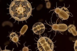
Cryptosporidium Sporulated Oocyst Morphology
- Round shape
- Size: approximately 4-5 micrometers
- Contains 4 sporozoites, crescent-shaped
Cryptosporidiosis Risk Factors: Case-Control Studies in Developed Countries since 1990
- Robertson et al., 2002 (Melbourne):
- Swimming in public pools: OR 2.7
- Families with less than 6 kids and kids with diarrhea: OR 7.4
- Pet ownership: OR 0.6
- Contact with calves: OR 2.9
- Drinking unfiltered water from rivers or lakes: OR 1.5
- Robertson et al., 2002 (Adelaide):
- Swimming in public pools: OR 1.2
- Families with less than 6 kids and kids with diarrhea: OR 8.6
- Pet ownership: OR 0.6
- Contact with calves: OR 5.1
- Drinking unfiltered water from rivers or lakes: OR 3.1
- Roy et al., 2004 (United States):
- Contact with children (2-11 years) with diarrhea: OR 3.0
- Contact with cattle/calves: OR 3.5
- International travel: OR 7.7
- Swimming in freshwater: OR 1.9
- Eating raw vegetables: OR 0.5
- Hunter et al. 2004 (Wales and NW England):
- Travel outside the UK: OR 5.7
- Contact with cryptosporidiosis patients: OR 4.6
- Contact with cattle/buffalo: OR 3.9
- Eating raw vegetables: OR 0.5
- Eating ice cream: OR 0.5
Cyclospora
- Found in humans, monkeys, viper snakes, millipedes, and rodents - 17 species identified
- Cyclospora cayetanensis is the species known to cause disease in humans
Cyclospora cayetanensis
- Causes diarrhea in immunocompetent individuals; usually mild
- More severe in immunocompromised individuals.
- Oocyst shape and staining are similar to C. parvum
- Size: 7.7 - 9.9 micrometers (average 8.6 micrometers)
- Oocysts initially aren't sporulated when excreted in feces, taking at least a week to sporulate
- Sporulated oocysts contain 2 sporocysts, each with 2 sporozoites
- Symptoms:
- Loss of appetite
- Nausea
- Indigestion
- Abdominal pain
- Diarrhea
- Low-grade fever
- Weight loss
- Symptoms can be mild in endemic areas
- Children and elderly may experience more severe symptoms
- The only known host is humans, although oocysts have been found in poultry, ducks, and dogs.
- However, these findings haven't been confirmed through direct infection.
Unsporulated Oocyst of C. cayetanensis
- Round shape
- Size: approximately 8-10 micrometers
- Contains small, round, green granules
- Neon blue autofluorescence with UV illumination
Sporulated Oocysts Morphology
- Sporulated oocyst is round, 6-8 micrometers in size
- Two-layered wall with multiple, small, round, green granules inside
- Under UV light, the oocyst wall is neon blue autofluorescent.
- Modified acid-fast stain causes oocysts to appear red against a blue background, often mottled with darker red spots, or a pink, bubbled appearance
- Sporulated oocysts contain 2 oval sporocysts, each with 2 sporozoites (crescent-shaped)
Cyclospora cayetanensis Life Cycle
- The lifecycle is not fully understood, but is presumed to be similar to C. parvum.
Cyclospora cayetanensis
- Can cause cyclosporiasis, a diarrheal illness.
- Does not cause autoinfection.
- Oocysts released in feces are unsporulated and not immediately infectious.
- Oocysts require 5-6 days to mature and become sporulated in the environment.
- Sporulated oocysts are infectious.
Treatment of Cyclosporiasis
- Trimethoprim/sulfamethoxazole (co-trimoxazole) is the drug of choice for treatment.
Isospora belli
- A protozoan that causes diarrhea in both immunocompromised and immunocompetent individuals.
- Prevalence of infection in HIV patients is 0.2-2%, and 8% in Thailand.
- Oocysts released in feces are unsporulated and not immediately infectious.
- Oocysts need 4-5 days to mature and become sporulated in the environment.
- Oocysts are oval-shaped, measuring 20-30 x 10-20 micrometers.
- They have a thin, smooth, colorless wall with 1-2 round sporoblasts inside.
- Mature oocysts contain 12-14 x 7-9 micrometer sporocysts.
- Each sporocyst contains 4 crescent-shaped sporozoites with a single nucleus.
Isospora belli Characteristics
- Releases immature (unsporulated) oocysts from the intestinal wall.
- Oocysts mature into sporozoites within 4-5 days.
Isospora belli Oocysts
- Oval-shaped
- Approximately 20-33 x 10-19 micrometers in size
- Have a transparent, thin, two-layered wall
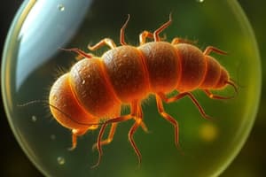
Microspora
- Thick-walled spores contain an infective body called sporoplasm.
- Sporoplasm is surrounded by a coiled, hollow tube called the polar filament.
- The polar filament injects the sporoplasm into the host.
- Microsporidia can infect the intestinal epithelium of AIDS patients.
- They can also invade other epithelium and have been found in immunocompetent patients with diarrhea.
Important Microsporidial Species that Infect Humans
- Enterocytozoon bieneusi
- Encephalitozoon hellem
- Encephalitozoon cuniculi
- Encephalitozoon intestinalis
Microsporidial Species and Infection Prevalence
- Enterocytozoon bieneusi
-
1,000 infections in immunocompromised patients.
- Infections in immunocompetent individuals.
-
Studying That Suits You
Use AI to generate personalized quizzes and flashcards to suit your learning preferences.