Podcast
Questions and Answers
What are the three major stages of prenatal development?
What are the three major stages of prenatal development?
- Pre-embryonic, Embryonic, Fetal (correct)
- Pre-embryonic, Blastocyst, Fetal
- Germinal, Embryonic, Fetal
- Zygote, Embryonic, Fetal
What is gametogenesis primarily responsible for?
What is gametogenesis primarily responsible for?
- Fertilization of the egg
- Development of the embryo
- Formation of the blastocyst
- Creation of gametes (correct)
During which weeks does the pre-embryonic period occur?
During which weeks does the pre-embryonic period occur?
- Weeks 1-3
- Weeks 1-4
- Weeks 1-2 (correct)
- Weeks 2-4
Which process describes the formation of a blastocyst?
Which process describes the formation of a blastocyst?
What structure is formed when the bilaminar embryonic disc develops?
What structure is formed when the bilaminar embryonic disc develops?
Which of the following is NOT an extraembryonic membrane formed during week two?
Which of the following is NOT an extraembryonic membrane formed during week two?
What happens during the process of gastrulation?
What happens during the process of gastrulation?
Which of the following germ layers gives rise to the nervous system?
Which of the following germ layers gives rise to the nervous system?
During which weeks is the embryo most susceptible to teratogenic effects?
During which weeks is the embryo most susceptible to teratogenic effects?
What is the result of meiosis in oogenesis?
What is the result of meiosis in oogenesis?
What does the term 'haploid' refer to in the context of gametes?
What does the term 'haploid' refer to in the context of gametes?
What occurs at the centromere during meiosis?
What occurs at the centromere during meiosis?
Which term describes the earliest stage of human development?
Which term describes the earliest stage of human development?
What is formed from the mesoderm during embryonic development?
What is formed from the mesoderm during embryonic development?
What structure does the paraxial mesoderm primarily develop into?
What structure does the paraxial mesoderm primarily develop into?
What is the primary function of intermediate mesoderm?
What is the primary function of intermediate mesoderm?
Which layer of mesoderm is responsible for forming the intraembryonic coelom?
Which layer of mesoderm is responsible for forming the intraembryonic coelom?
What does the endoderm primarily give rise to?
What does the endoderm primarily give rise to?
What is the primary outcome of craniocaudal folding during embryonic development?
What is the primary outcome of craniocaudal folding during embryonic development?
Which of the following defines transverse folding in embryonic development?
Which of the following defines transverse folding in embryonic development?
During which weeks of prenatal development is the embryo most susceptible to teratogens?
During which weeks of prenatal development is the embryo most susceptible to teratogens?
What structure is formed from the somatic layer of lateral plate mesoderm?
What structure is formed from the somatic layer of lateral plate mesoderm?
Which membranes are formed where ectoderm fuses directly with endoderm?
Which membranes are formed where ectoderm fuses directly with endoderm?
What is the primary role of teratogens in embryonic development?
What is the primary role of teratogens in embryonic development?
How many somites typically develop from days 26 to 32?
How many somites typically develop from days 26 to 32?
From which germ layer do the primary derivatives arise?
From which germ layer do the primary derivatives arise?
What is established as a signaling hub that initiates neurulation?
What is established as a signaling hub that initiates neurulation?
What initiates the release of gonadotropins from the pituitary gland during the ovarian cycle?
What initiates the release of gonadotropins from the pituitary gland during the ovarian cycle?
What structure does the follicle transform into after ovulation?
What structure does the follicle transform into after ovulation?
Which of the following occurs during the cleavage process following fertilization?
Which of the following occurs during the cleavage process following fertilization?
What is the primary outcome of the bilaminar disc formation during implantation?
What is the primary outcome of the bilaminar disc formation during implantation?
What is the role of hCG during the early stages of pregnancy?
What is the role of hCG during the early stages of pregnancy?
Which of the following terms describes the transformation of the embryoblast during implantation?
Which of the following terms describes the transformation of the embryoblast during implantation?
What primarily occurs during the gastrulation process in embryonic development?
What primarily occurs during the gastrulation process in embryonic development?
What is formed from the mesoderm during embryonic development?
What is formed from the mesoderm during embryonic development?
Which embryonic structure is responsible for the future formation of the central nervous system?
Which embryonic structure is responsible for the future formation of the central nervous system?
What represents the main anatomical organization established during gastrulation?
What represents the main anatomical organization established during gastrulation?
What happens to the neural folds during neurulation?
What happens to the neural folds during neurulation?
What are the two layers that differentiate in the trophoblast during implantation?
What are the two layers that differentiate in the trophoblast during implantation?
What primarily occurs with the lacunar network in the developing embryo?
What primarily occurs with the lacunar network in the developing embryo?
In which stage of prenatal development does the formation of the amnionic cavity occur?
In which stage of prenatal development does the formation of the amnionic cavity occur?
Flashcards are hidden until you start studying
Study Notes
Stages of Prenatal Development
- Three stages: Pre-embryonic, Embryonic, Fetal
- Most visible development occurs during the embryonic period
Gametogenesis
- Definition: Germ cells transform into gametes
- Meiosis: Chromosome number halves (46 diploid → 23 haploid)
- Gametes: Sex cells capable of reproduction
- Male: Sperms/Spermatozoa
- Female: Oocytes
Spermatogenesis
- Spermatogonia: Diploid germ cells transforming into primary spermatocytes during puberty
- Meiosis I: Two haploid secondary spermatocytes
- Meiosis II: Four haploid spermatids
- Spermiogenesis: Spermatids elongate and mature into sperms
Oogenesis
- Oogonia: Diploid cells surrounded by a follicular layer
- Meiosis I: Begins before birth, arrests in prophase, and completes monthly beginning at puberty, forming one secondary oocyte and one polar body
- Meiosis II: Begins at ovulation, arrests at metaphase, and completes upon sperm penetration, forming one fertilized oocyte and one polar body
Ovarian Cycle
- Begins at puberty, terminates at menopause
- Hypothalamus: Stimulates the pituitary gland to release gonadotropins
- FSH: Stimulates follicular maturation
- LH: Stimulates estrogen production and ovulation
- Ovulation: Follicle becomes corpus luteum, secreting estrogen and progesterone, thickening the endometrium of the uterus
Pre-embryonic Period
- Week 1: Fertilization, cleavage, blastocyst formation, implantation
- Week 2: Bilaminar disc forms, lacunae form, extraembryonic mesoderm develops, hCG secretion begins
Week 1
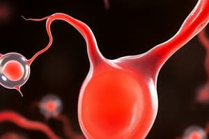
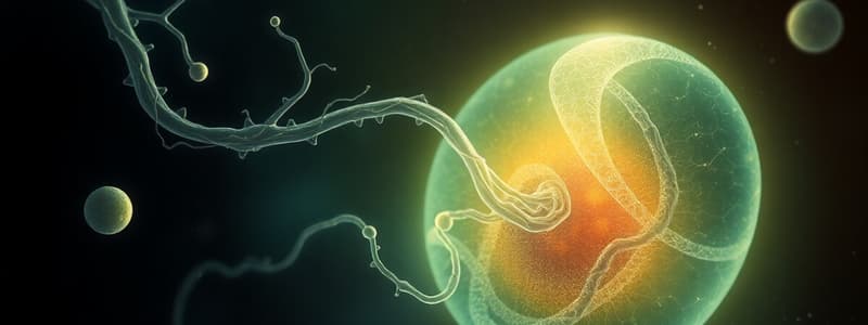
- Fertilization: Union of genetic material, beginning when a capacitated sperm penetrates a secondary oocyte
- Cleavage: Repeated mitotic divisions of the zygote, increasing the number of blastomeres, forming a morula
- Blastocyst Formation: Development of a fluid-filled cavity within the morula, pushing the inner-cell mass to one side, forming the inner cell mass/embryoblast and the trophoblast
- Implantation: Blastocyst embeds in the uterine endometrium, the trophoblast differentiates into two layers: cytotrophoblast and syncytiotrophoblast
Week 2
- Bilaminar Embryonic Disc: Embryoblast differentiates into a bilaminar disc (epiblast and hypoblast)
- Amnionic Cavity: Forms within the epiblast, amnioblasts surround the amniotic cavity forming the amnion
- Primary Umbilical Vesicle: Cells continuous with the hypoblast line the cytotrophoblast forming the primary umbilical vesicle
- Lacunar Network: Syncytiotrophoblast erodes uterine blood vessels forming lacunae, allowing diffusion of nutrients and gases
- Extraembryonic Mesoderm: Cells migrate between the exocoelomic membrane and cytotrophoblast forming spaces, combining to form the future chorionic cavity
- Prechordal Plate: Localized thickening of the hypoblast, an organizing center for head structures
Human Chorionic Gonadotropin (hCG)
- Produced by the syncytiotrophoblast at the end of Week 2
- Maintains the corpus luteum of the ovary, responsible for progesterone and estrogen production
Early Embryonic Period
- Week 3: Gastrulation, neurulation, intraembryonic mesoderm differentiation
- Week 4-8: Organogenesis
Gastrulation
- Bilaminar disc → trilaminar disc (ectoderm, endoderm, mesoderm)
- Establishment of body plan: Cranial-caudal axis, left-right sides
- Begins with the appearance of the primitive streak
Primitive Streak
- Epiblast cells migrate to the median plane
- Primitive node forms, marking the cranial side of the primitive streak
- Invagination of cells creates the primitive groove
Trilaminar Disc
- Ectoderm: Epithelial tissues derived from the epiblast
- Endoderm: Epithelial tissues derived from the hypoblast
- Mesoderm: Mesenchymal or epithelial tissues derived from cells migrating between the ectoderm and endoderm
Notochord Formation
- Migrating mesenchymal cells form the notochord process
- Notochord process binds to the underlying endoderm
- Fused layers degenerate leaving the notochord plate
- Notochord plate folds inward to form the notochord
Neurulation
- Notochord signaling induces the neural plate
- Neural Plate: Thickening of the overlying ectoderm (neuroectoderm)
- Neural Groove: Neural plate invaginates, forming the neural groove and neural folds
- Neural Tube: Neural folds meet in the midline, forming the neural tube
Closure of the Neural Tube
- Begins in the cervical region and occurs bidirectionally
- Complete by the end of Week 4
- Incomplete closure can result in anencephaly or spina bifida
Neural Crest Cells
- Undergo epithelial-to-mesenchymal transition
- Migrate away from the neural folds, forming ganglia, Schwann cells, pharyngeal arches and their derivatives, leptomeninges, melanocytes, odontoblasts, suprarenal medulla
Ectoderm Summary
- Derived from the epiblast
- Neuroectoderm: Thickened region over the notochord that becomes the neural tube and neural crest
- Surface Ectoderm: Becomes the epidermis
Intraembryonic Mesoderm
- Germ layer between the ectoderm and endoderm
- Derived from the epiblast
- Differentiates into three parallel columns:
- Paraxial Mesoderm: Forms somites
- Intermediate Mesoderm: Forms kidneys & gonads
- Lateral Plate Mesoderm: Forms the heart, blood vessels, and lining of body cavities
Paraxial Mesoderm
- Forms paired columns flanking the notochord
- Columns condense and segment into somites around day 22, when the embryo is about 2.5 mm
- Development of 38-39 somites occurs craniocaudally between days 26 and 32
- Somites give rise to:
- Sclerotome: vertebrae, ribs, posterior skull
- Myotome: skeletal muscles of limbs and trunk
- Dermatome: skin over the back and limbs
Intermediate Mesoderm
- Continuous laterally with the paraxial mesoderm
- Precursor to components of the urinary and reproductive systems
- Forms:
- Kidneys and ureters
- Gonads, reproductive ducts, glands
- Uterus
Lateral Plate Mesoderm
- Extends laterally and connects with extraembryonic mesoderm
- Covers the umbilical vesicle and amnion
- Divides into two layers:
- Somatic (parietal) mesoderm
- Splanchnic (visceral) mesoderm
- Transverse folding fuses the right and left sides in the midline
- Intraembryonic coelom forms (future thoracic and abdominopelvic cavities)
Mesoderm Summary
- Derived from the epiblast
- Paraxial mesoderm gives rise to somites
- Intermediate mesoderm forms urogenital structures
- Lateral plate mesoderm splits into somatic and splanchnic layers
Endoderm
- Germ layer surrounding the umbilical vesicle
- Derived from the epiblast
- Forms the lining of the gut tube
- Most internal organs develop from the gut tube
- Divided into three sections:
- Foregut, connected to the oropharyngeal membrane
- Midgut, connected to the secondary umbilical vesicle
- Hindgut, connected to the cloacal membrane
Oropharyngeal & Cloacal Membranes
- Locations where ectoderm directly fuses with endoderm
- Oropharyngeal membrane is at the cranial end:
- Future site of the mouth
- Connects to the foregut
- Cloacal membrane is at the caudal end:
- Future site of the anal and urogenital openings
- Connects to the hindgut
Week 4 Embryonic Development
- Characterized by embryonic folding:
- Neural tube closure
- Conversion of elongated disc into curved cylinder driven by differential growth
Embryonic Folding
- Results in the formation of a curved cylinder from the elongating disc
- Driven by differential growth along the longitudinal axis and lateral parts of the disc
- Craniocaudal and transverse folding occur concurrently
Craniocaudal Folding
- Initiated by rapid growth of the neural tube
- Oropharyngeal and cloacal membranes migrate to the ventral surface
- The endoderm of the umbilical vesicle is internalized, establishing the foregut, midgut, and hindgut
Transverse Folding
- Driven by rapid growth of the somites
- Bilateral edges of the conceptus fuse at the midline
- Splanchnic mesoderm and endoderm fusion forms the gut tube
- Somatic mesoderm and surface ectoderm fusion forms the body wall, except at the vitelline duct
Weeks 3 & 4 Summary
- Gastrulation establishes body planes and generates the three germ layers
- Ectoderm, mesoderm, endoderm
- Notochord acts as a signaling center involved in neurulation
- Neurulation establishes the nervous system and neural crest cells
- Intraembryonic mesoderm differentiates into three paired columns:
- Paraxial, intermediate, lateral plate
- Endoderm forms the lining of the gut tube
- Oropharyngeal and cloacal membranes are formed
- The conceptus transitions from a disc to a tube via folding:
- Craniocaudal folding internalizes thoracoabdominal structures
- Transverse folding forms the gut tube, body wall, and surrounds the conceptus with the amnionic cavity
Teratogens
- Most infant deaths result from birth defects caused by genetic factors, environmental factors, or a combination
- Teratogens are substances, organisms, or physical agents that disrupt normal development
- Consequences of teratogen exposure:
- Slowed growth
- Loss of function
- Malformations
- Death
Studying That Suits You
Use AI to generate personalized quizzes and flashcards to suit your learning preferences.