Podcast
Questions and Answers
Which of the following best describes the primary function of pituicytes within the neurohypophysis?
Which of the following best describes the primary function of pituicytes within the neurohypophysis?
- Transporting hormones from the hypothalamus to the posterior pituitary.
- Acting as a supportive structure for nerve endings from the hypothalamus. (correct)
- Synthesizing ADH and oxytocin.
- Secreting hormones involved in milk ejection and uterine contraction.
The hypothalamus communicates with the neurohypophysis via the:
The hypothalamus communicates with the neurohypophysis via the:
- Corticospinal tract.
- Hypothalamo-hypophyseal portal system.
- Hypothalamo-hypophyseal tracts. (correct)
- Hypothalamo-adrenal axis.
Where are ADH and oxytocin synthesized?
Where are ADH and oxytocin synthesized?
- Adrenal gland
- Anterior pituitary
- Posterior pituitary
- Hypothalamus (correct)
What are the two main hormones synthesized in the magnocellular neurosecretory cells of the hypothalamus?
What are the two main hormones synthesized in the magnocellular neurosecretory cells of the hypothalamus?
Neurophysins are:
Neurophysins are:
Which of the following describes how ADH and oxytocin are transported from their site of synthesis to their site of release?
Which of the following describes how ADH and oxytocin are transported from their site of synthesis to their site of release?
What triggers the release of ADH and oxytocin into the bloodstream from the posterior pituitary?
What triggers the release of ADH and oxytocin into the bloodstream from the posterior pituitary?
ADH and oxytocin are considered neurohormones because:
ADH and oxytocin are considered neurohormones because:
Where are V1 receptors for ADH primarily located, and what is their effect?
Where are V1 receptors for ADH primarily located, and what is their effect?
Which of the following signaling pathways is activated by V2 receptor stimulation?
Which of the following signaling pathways is activated by V2 receptor stimulation?
Which of the following is the primary function of osmoreceptors in regulating ADH secretion?
Which of the following is the primary function of osmoreceptors in regulating ADH secretion?
Which structures are the primary locations of osmoreceptive cells involved in ADH regulation?
Which structures are the primary locations of osmoreceptive cells involved in ADH regulation?
The primary effect of ADH on the kidneys is to:
The primary effect of ADH on the kidneys is to:
The tubular epithelial cells of the late distal tubules and collecting ducts are characterized by:
The tubular epithelial cells of the late distal tubules and collecting ducts are characterized by:
How does ADH increase water permeability in the luminal membrane of tubular cells?
How does ADH increase water permeability in the luminal membrane of tubular cells?
In higher doses, ADH increases arterial blood pressure by:
In higher doses, ADH increases arterial blood pressure by:
Which receptor type mediates the vasoconstrictive effects of ADH?
Which receptor type mediates the vasoconstrictive effects of ADH?
Apart from osmotic stimuli, which of the following directly stimulates ADH secretion?
Apart from osmotic stimuli, which of the following directly stimulates ADH secretion?
How do stretch receptors in the atria and pulmonary vessels influence ADH secretion?
How do stretch receptors in the atria and pulmonary vessels influence ADH secretion?
What effect does atrial natriuretic peptide (ANP) have on ADH secretion?
What effect does atrial natriuretic peptide (ANP) have on ADH secretion?
What is the impact of increased blood volume on ADH secretion?
What is the impact of increased blood volume on ADH secretion?
What condition results from the kidneys' inability to respond to ADH, leading to the excretion of large volumes of dilute urine?
What condition results from the kidneys' inability to respond to ADH, leading to the excretion of large volumes of dilute urine?
Which of the following best describes the mechanism of action of oxytocin on mammary glands?
Which of the following best describes the mechanism of action of oxytocin on mammary glands?
The milk let-down response is caused by which hormone?
The milk let-down response is caused by which hormone?
What is the approximate half-life of oxytocin in the blood?
What is the approximate half-life of oxytocin in the blood?
Sensory input from the nipple during breastfeeding travels to the hypothalamus via:
Sensory input from the nipple during breastfeeding travels to the hypothalamus via:
The primary effect of oxytocin release during childbirth is:
The primary effect of oxytocin release during childbirth is:
Oxytocin receptors are located on the plasma membrane and, upon activation, lead to:
Oxytocin receptors are located on the plasma membrane and, upon activation, lead to:
Which of the following can inhibit oxytocin release?
Which of the following can inhibit oxytocin release?
Which statement is most accurate regarding the roles of oxytocin (OXT) and arginine vasopressin (AVP) in social behaviors?
Which statement is most accurate regarding the roles of oxytocin (OXT) and arginine vasopressin (AVP) in social behaviors?
Flashcards
Pituicytes
Pituicytes
Glia-like cells composing the neurohypophysis, acting as a supporting structure.
Hypothalamo-hypophysial tracts
Hypothalamo-hypophysial tracts
Nerve tracts from the hypothalamus to the neurohypophysis.
ADH and Oxytocine
ADH and Oxytocine
The supraoptic and paraventricular nuclei synthesize these two hormones.
ADH (Antidiuretic Hormone)
ADH (Antidiuretic Hormone)
Signup and view all the flashcards
Oxytocine
Oxytocine
Signup and view all the flashcards
Herring body
Herring body
Signup and view all the flashcards
Supraoptic and paraventricular nuclei
Supraoptic and paraventricular nuclei
Signup and view all the flashcards
Neurophysins
Neurophysins
Signup and view all the flashcards
V1 receptors
V1 receptors
Signup and view all the flashcards
V2 receptors
V2 receptors
Signup and view all the flashcards
V3 receptors
V3 receptors
Signup and view all the flashcards
Osmoreceptors
Osmoreceptors
Signup and view all the flashcards
Circumventricular organs
Circumventricular organs
Signup and view all the flashcards
Angiotensin receptors
Angiotensin receptors
Signup and view all the flashcards
Atrial natriuretic peptide (ANP)
Atrial natriuretic peptide (ANP)
Signup and view all the flashcards
Aldosterone
Aldosterone
Signup and view all the flashcards
Neurohormones
Neurohormones
Signup and view all the flashcards
Diabetes insipidus
Diabetes insipidus
Signup and view all the flashcards
Nephrogenic Diabetes Insipidus
Nephrogenic Diabetes Insipidus
Signup and view all the flashcards
Thirst center
Thirst center
Signup and view all the flashcards
Increased blood volume
Increased blood volume
Signup and view all the flashcards
Decreased blood pressure
Decreased blood pressure
Signup and view all the flashcards
Angiotensin II
Angiotensin II
Signup and view all the flashcards
Hypovolemia
Hypovolemia
Signup and view all the flashcards
Oxytocin (Oxt or OT)
Oxytocin (Oxt or OT)
Signup and view all the flashcards
Oxytocin
Oxytocin
Signup and view all the flashcards
Oxytocin secretion and milk ejection
Oxytocin secretion and milk ejection
Signup and view all the flashcards
Study Notes
Posterior Pituitary Gland Hormones
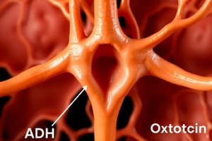
- The posterior pituitary gland releases hormones that are synthesized in the hypothalamus
- Two important hormones synthesized are ADH and oxytocin
Neurohypophysis Composition
- The neurohypophysis is mainly composed of glia-like cells called pituicytes
- Pituicytes act as supporting structures for terminal nerve endings from nerve tracts originating in the magnocellular part of the supraoptic and paraventricular nuclei of the hypothalamus, but do not secrete hormones
- Nerve tracts running from the hypothalamus to the neurohypophysis are called the hyphothalamo-hyphophysial tracts
- These tracts pass to the neurohypophysis through the hypophysial stalk
Hypothalamic Control of Neurohypophysis
- Hypothalamic neurons synthesize oxytocin or antidiuretic hormone (ADH)
- Oxytocin and ADH are transported down the axons of the hypothalamic-hypophyseal tract to the posterior pituitary
- Oxytocin and ADH are stored in axon terminals in the posterior pituitary within Herring bodies
- When hypothalamic neurons fire, action potentials cause oxytocin or ADH to be released into the blood
ADH, OTC, and Neurophysin
- ADH & OTC genes can synthesis similar preprohormones
- OTC and ADH are similar preprohormones of Neurophysin, including:
- Neurophysin I preprooxyphysin
- Neurophysin II prepropressophysin
- Neurophysins are cosecreted with AVP or oxytocin, but have no known hormonal actions
ADH Receptors
- 3 receptor types: V1, V2, and V3
- V1: Found on vessel walls, causes vasoconstriction via IP3/DAG
- V2: Located in the collecting duct of the nephron and stimulate FVIII production, via cAMP
- V3: Stimulate ACTH secretion
- ADH is also known as vasopressin
Osmoreceptors
- Osmoreceptive cells reside in the circumventricular organs, particularly the organum vasculosum of lamina terminalis and subfornical organ, near the third ventricle
- Axons from this region connect with ADH-producing cells in the supraoptic and paraventricular nuclei, stimulating hormone release
Renal Tubules
- The renal tubules are a key site of ADH action, affecting water reabsorption
Effects of ADH
- The main effect is water retention by the kidneys
- Tubular epithelial cells of late distal tubules and collecting ducts have tight junctions and are relatively impermeable to water
- ADH binds to V2 receptors on the basal side of tubular cells, increasing intracellular cAMP
- Cyclic AMP increases membrane permeability on the luminal (apical) side, opening water channels
- A large number of special vesicles inside the cell membrane contain highly water permeable pores called aquaporins
- ADH combines with membrane V2 receptors, activating adenylyl cyclase and forming cAMP inside the tubular cell cytoplasm
- This causes phosphorylation of elements in the special vesicles, leading to their insertion into apical cell membranes for high water permeability
- The process takes 5-10 minutes and reverses in the absence of ADH within another 5-10 minutes
ADH at Higher Doses
- At higher doses, ADH increases arterial blood pressure by constricting smooth muscle of arterioles (vasopressin)
- ADH binds to V1 receptors via the IP3 pathway, increasing intracellular Ca level and constricting smooth muscles of arterioles
- Normal physiological ADH concentrations are below its vasoactive range
- During severe hypovolemic shock, high ADH release contributes to compensatory increase in systemic vascular resistance
Control of ADH Secretion
- Osmotic stimuli: rapid response
- Extracellular volume (ECF): relatively slow response
- Angiotensin II
- Stretch receptors
- Baroreceptors
- Atrial Natriuretic Peptide
Water Intake Regulation
- The thirst center is located in the lateral hypothalamus
- ADH secreting cells are located in supraoptic and paraventricular nuclei
- The lamina terminalis contains two specialized circumventricular organs: the OVLT and the SubFornical Organ (SFO)
- These are located outside the blood-brain barrier
Osmometric Thirst
- The osmoreceptors responsible for osmometric thirst are located in OVLT and SFO neurons, which contain angiotensin receptors
- Neurons in the subfornical organ and organum vasculosum terminalis send their axons to the median preoptic nucleus, which has connections with the supraoptic nucleus (ADH) and lateral hypothalamus
Control By ECF Volume
- Low-pressure receptors in the big veins, atria, and pulmonary vessels detect fullness of the vascular system
- There is an inverse relation between the rate of ADH secretion and the rate of discharge from the stretch receptors
- These receptors are excited by overfilling and send inhibitory signals by the glossopharyngeal nerve to the supraoptic and paraventricular nuclei, inhibiting ADH secretion
- When these receptors are unexcited due to underfilling (decrease in ECF), the supraoptic and paraventricular nuclei are not inhibited and secrete more ADH
Barroreceptors
- Barroreceptors are found in the aortic arch and carotid bodies and detect arterial pressure
- There is a similar inverse relation between the the rate of ADH secretion and the rate of discharge from the barroreceptors
- Decreased stretch of the barroreceptors from low arterial blood pressure causes ADH secretion by the disinhibition of the supraoptic and paraventricular nuclei
- The carotid sinus baroreceptors are innervated by the glossopharyngeal nerve (CN IX).
- The aortic arch baroreceptors are innervated by the vagus nerve (CN X).
- Impulses pass from them via the vagi and glossopharyngeal nerves to the nucleus of the tractus solitarius (NTS).
Atrial Natriuretic Peptide (ANP) & Factors That Regulate ADH
- ANPIn atria and in the brain (BNP) inhibits ADH secretion when there is an increase in the blood volume.
- Factors That Regulate ADH -Stumulation: - Extracellular fluid osmolality increase - Volume decrease - Pressure decrease - Cerebrospinal fluid sodium increase - Angiotensin II - Pain - Nausea and vomiting - Stress - Hypoglycemia - Cytokines - Temperature increase - Senescence - Drugs Nicotine - Opiates - Barbiturates - Sulfonylureas - Antineoplastic agents -Inhibitions: - Extracellular fluid osmolality decrease - Volume increase - Temperature decrease - a-Adrenergic agonists - γ-Aminobutyric acid (GABA) - Ethanol - Cortisol - Thyroid hormone - Atrial natriuretic peptide
ADH Actions
- ADH plays an important role in social behavior, sexual motivation, and bonding in monogamic species
- ADH may have analgesic effects
- ADH has been implicated in memory formation
- ADH released from centrally projecting hypothalamic neurons is involved in aggression
- ADH acts with corticotropin-releasing hormone to modulate the release of corticosteroids from the adrenal gland in response to stress
Diabetes Insipidus
- Two types
- Primary or nephrogenic
- Primary (central): Tumors, posttraumatic, infection
- Nephrogenic DI: The kidneys respond inadequately to normal or even elevated levels of circulating AVP.
- Nephrogenic: ADH/V2 cannot produce cAMP
Oxytocin
- Oxytocin is a peptide hormone and neuropeptide produced in the hypothalamus and released by the posterior pituitary
Oxytocin Actions
- Increases the contractility of contractile myoepithelial cells in mammary glands, resulting in a pressure of 10-20 mmHg
- Causes milk let-down response
- Stimulates uterine contraction
- Circulates freely in blood
- The half-life is 3-5 minutes
Oxytocin Secretion
- Sensory input from nerve endings in the nipple is transmitted to the hypothalamus via thoracic nerves and the spinal cord
- Stimulates neurons in the supraoptic and paraventricular nuclei to release oxytocin from terminals in the posterior lobe
- Neurons can be activated by higher brain centers, such that the mere sight of the baby or hearing it cry can stimulate milk let-down
- Oxytocin secretion and milk ejection are a neuroendocrine reflex
- The normal stimulus is an infant suckling at the breast, with sensory nerves relaying information to the hypothalamus which causes a release in oxytocin
Oxytocin Effects and Control
- Receptors on plasma membrane leads to H-R binding, which increases IP3 and DAG, and increases intracellular Ca++
- Stimulation of milk ejection (milk letdown) and uterine smooth muscle contraction at birth, with the goal of establishing of maternal behavior
- The most important stimulus for release of hypothalamic oxytocin is initiated by physical stimulation of the nipples or teats.
- The act of nursing or suckling is relayed within a few milliseconds to the brain via a spinal reflex arc
- These signals impinge on oxytocin-secreting neurons, leading to release of oxytocin.A number of factors can inhibit oxytocin release, among them acute stress. Both the production of oxytocin and response to oxytocin are modulated by circulating levels of sex steroids.
- The burst of oxytocin released at birth seems to be triggered in part by cervical and vaginal stimulation by the fetus, but also because of abruptly declining concentrations of progesterone.
- Another well-studied effect of steroid hormones is the marked increase in synthesis of uterine (myometrial) oxytocin receptors late in gestation, resulting from increasing concentrations of circulating estrogen.
- The release of oxytocin has various kinds of social interactive behaviours including maternal behaviour and bonding.
- It decreases fear and the sensation of pain and it may promote trust and well-being.
- Oxytocin induces powerful anti-inflammatory and anti-stress eects including lowering of blood pressure, heart rate, and cortisol levels.
- Decreasing the activity in the stress system (the hypothalamo-pituitary-adrenal axis and the sympathetic nervous system), it enhances the activity in the parasympathetic system.
- Low oxytocin levels have been linked to autism and autistic spectrum disorders
Neuropeptides: Oxytocin & Arginine Vasopressin (AVP)
- Neurohypophysis releases antidiuretic hormone (ADH) to the kidneys
- Reabsorbes water, raising blood volume and pressure
- This gets transported along axons from the supraoptic nucleus to neurohypohysis
- Oxytocin(OXT) the Uterus, mammary glands (females), Ductus deferens and prostate gland (males) -Labor contractions, milk ejection -Contractions of ductus deferens and prostate gland -Gets Transported along axons from paraventricular nucleus to neurohypophysis
- Are key mediators of complex social behaviors, including attachment, social recognition and aggression
- Oxytocin (OT) system provides the neurohormonal substrate for parental, romantic, and filial attachment in humans
- AVP has primarily been implicated in male-typical social behaviors, including aggression and pair-bond formation, and mediates anxiogenic effects
- Initial studies in humans suggest behavioral, neural, and endocrine effects of both neuropeptides, similar to those found in animal studies
Studying That Suits You
Use AI to generate personalized quizzes and flashcards to suit your learning preferences.