Podcast
Questions and Answers
What structure delineates the rostral boundary of the pons?
What structure delineates the rostral boundary of the pons?
- Medulla
- Diencephalon
- Midbrain (correct)
- Cerebellum
In which portion of the pons are the descending cortical fibers, pontine nuclei, and transverse fibers primarily located?
In which portion of the pons are the descending cortical fibers, pontine nuclei, and transverse fibers primarily located?
- Caudal portion
- Basilar portion (correct)
- Tegmental portion
- Rostral portion
Damage to the cerebellopontine angle is most likely to directly affect which cranial nerve(s)?
Damage to the cerebellopontine angle is most likely to directly affect which cranial nerve(s)?
- Olfactory nerve (CN I)
- Trochlear nerve (CN IV)
- Vestibulocochlear nerve (CN VIII) (correct)
- Optic nerve (CN II)
In a transverse section of the caudal pons, which of the following is a newly appearing structure?
In a transverse section of the caudal pons, which of the following is a newly appearing structure?
What is represented by the transverse fibers found within the pontine nuclei?
What is represented by the transverse fibers found within the pontine nuclei?
What is the primary neurotransmitter found within the cell bodies of the locus coeruleus, located in the rostral pons?
What is the primary neurotransmitter found within the cell bodies of the locus coeruleus, located in the rostral pons?
Occlusion of which artery, or its branches, could lead to 'locked-in' syndrome?
Occlusion of which artery, or its branches, could lead to 'locked-in' syndrome?
What landmark is found at the caudal boundary of the midbrain?
What landmark is found at the caudal boundary of the midbrain?
Which cranial nerve nuclei are located within the midbrain?
Which cranial nerve nuclei are located within the midbrain?
What is contained within the basis pedunculi of the midbrain?
What is contained within the basis pedunculi of the midbrain?
Which of the following passes through the midbrain?
Which of the following passes through the midbrain?
At the level of the caudal midbrain, the lateral lemniscus axons are synapsing primarily on what structure?
At the level of the caudal midbrain, the lateral lemniscus axons are synapsing primarily on what structure?
What is the function of the enkephalins located within the periaqueductal gray (PAG)?
What is the function of the enkephalins located within the periaqueductal gray (PAG)?
What axons decussate at the level of the caudal midbrain?
What axons decussate at the level of the caudal midbrain?
Which disease results from the death of dopamine-containing cells of the substantia nigra?
Which disease results from the death of dopamine-containing cells of the substantia nigra?
At the level of the rostral midbrain, the axons of the oculomotor nucleus project to innervate how many extraocular muscles?
At the level of the rostral midbrain, the axons of the oculomotor nucleus project to innervate how many extraocular muscles?
The Edinger-Westphal nucleus, a parasympathetic nucleus, is associated with which cranial nerve?
The Edinger-Westphal nucleus, a parasympathetic nucleus, is associated with which cranial nerve?
What is the blood supply source for the midbrain?
What is the blood supply source for the midbrain?
Which of the following functions is NOT influenced by the reticular formation?
Which of the following functions is NOT influenced by the reticular formation?
Where do axons of the oculomotor nuclei exit?
Where do axons of the oculomotor nuclei exit?
Which of the following best describes the anatomical relationship of the pons?
Which of the following best describes the anatomical relationship of the pons?
A patient presents with an acoustic neuroma affecting the cerebellopontine angle. Initially, which cranial nerve is most likely to be affected?
A patient presents with an acoustic neuroma affecting the cerebellopontine angle. Initially, which cranial nerve is most likely to be affected?
Which of the following functions is primarily associated with the basilar pons?
Which of the following functions is primarily associated with the basilar pons?
Transverse fibers in the pons primarily serve which function?
Transverse fibers in the pons primarily serve which function?
Which of the following arteries is the primary blood supply to the pons?
Which of the following arteries is the primary blood supply to the pons?
What is a key function of the reticular formation within the pons and midbrain?
What is a key function of the reticular formation within the pons and midbrain?
Which nuclei gives rise to axons that innervate the ipsilateral muscles of mastication?
Which nuclei gives rise to axons that innervate the ipsilateral muscles of mastication?
What information is relayed by the mesencephalic nucleus and tract found in the mid-level pons?
What information is relayed by the mesencephalic nucleus and tract found in the mid-level pons?
What is the main function of the superior cerebellar peduncle?
What is the main function of the superior cerebellar peduncle?
What functional change would you expect from increased discharge rates of the neurons within the Locus Coeruleus?
What functional change would you expect from increased discharge rates of the neurons within the Locus Coeruleus?
Which structure is located within the midbrain?
Which structure is located within the midbrain?
What are the boundaries of the midbrain relative to other brain structures?
What are the boundaries of the midbrain relative to other brain structures?
Damage to the superior colliculus would most likely affect which function?
Damage to the superior colliculus would most likely affect which function?
The axons from which cranial nerve exit the brainstem between the two cerebral peduncles?
The axons from which cranial nerve exit the brainstem between the two cerebral peduncles?
A lesion in the periaqueductal gray (PAG) of the midbrain would most likely affect:
A lesion in the periaqueductal gray (PAG) of the midbrain would most likely affect:
Which axonal tract decussates at the level of the caudal midbrain allowing for cerebellar influence on the contralateral cortex?
Which axonal tract decussates at the level of the caudal midbrain allowing for cerebellar influence on the contralateral cortex?
What is the primary function of the red nucleus?
What is the primary function of the red nucleus?
What part of the midbrain contains dopamine producing cells that when they die result in Parkinson's disease?
What part of the midbrain contains dopamine producing cells that when they die result in Parkinson's disease?
Which arteries supply blood to the midbrain?
Which arteries supply blood to the midbrain?
What is the name for axons contained within the middle 3/5 of each cerebral peduncle?
What is the name for axons contained within the middle 3/5 of each cerebral peduncle?
Flashcards
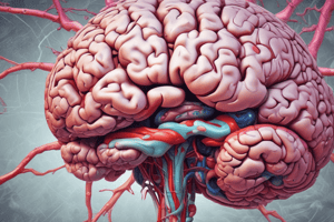
What is the Pons?
What is the Pons?
Middle segment of the brainstem, located between the midbrain and medulla.
What is the basilar pons?
What is the basilar pons?
The pons contains descending cortical fibers, pontine nuclei, and transverse fibers that enter the contralateral cerebellum.
What is the tegmental portion of the pons?
What is the tegmental portion of the pons?
Sensory and motor nuclei for cranial nerves V, VI, VII, and VIII.
What is the reticular formation?
What is the reticular formation?
Signup and view all the flashcards
What is the Abducens nucleus?
What is the Abducens nucleus?
Signup and view all the flashcards
What is the facial nucleus?
What is the facial nucleus?
Signup and view all the flashcards
What is the Mesencephalic nucleus and tract?
What is the Mesencephalic nucleus and tract?
Signup and view all the flashcards
What are Pontocerebellar fibers?
What are Pontocerebellar fibers?
Signup and view all the flashcards
What is the Locus Coeruleus?
What is the Locus Coeruleus?
Signup and view all the flashcards
What is Basilar artery stroke?
What is Basilar artery stroke?
Signup and view all the flashcards
What is the midbrain?
What is the midbrain?
Signup and view all the flashcards
What are the three parts of the midbrain?
What are the three parts of the midbrain?
Signup and view all the flashcards
What is the tectum?
What is the tectum?
Signup and view all the flashcards
Which cranial nerve nuclei are located in the midbrain?
Which cranial nerve nuclei are located in the midbrain?
Signup and view all the flashcards
What is the Lateral Lemniscus?
What is the Lateral Lemniscus?
Signup and view all the flashcards
What is the substantia nigra?
What is the substantia nigra?
Signup and view all the flashcards
What is the inferior colliculus?
What is the inferior colliculus?
Signup and view all the flashcards
What is the cerebral aqueduct?
What is the cerebral aqueduct?
Signup and view all the flashcards
What is the Periaqueductal gray?
What is the Periaqueductal gray?
Signup and view all the flashcards
What are the superior cerebellar and posterior cerebral arteries?
What are the superior cerebellar and posterior cerebral arteries?
Signup and view all the flashcards
Acoustic neuromas
Acoustic neuromas
Signup and view all the flashcards
Clinical significance of cerebellopontine angle
Clinical significance of cerebellopontine angle
Signup and view all the flashcards
Cerebellar Peduncles
Cerebellar Peduncles
Signup and view all the flashcards
Corticospinal tract (Pons)
Corticospinal tract (Pons)
Signup and view all the flashcards
Medial Lemniscus (Pons)
Medial Lemniscus (Pons)
Signup and view all the flashcards
Pontine Nuclei
Pontine Nuclei
Signup and view all the flashcards
Middle Cerebellar Peduncle
Middle Cerebellar Peduncle
Signup and view all the flashcards
Lateral Lemniscus (pons)
Lateral Lemniscus (pons)
Signup and view all the flashcards
Trigeminal motor nucleus
Trigeminal motor nucleus
Signup and view all the flashcards
Principal Sensory Nucleus
Principal Sensory Nucleus
Signup and view all the flashcards
Superior Cerebellar Peduncle
Superior Cerebellar Peduncle
Signup and view all the flashcards
Locked-in syndrome
Locked-in syndrome
Signup and view all the flashcards
ASCT
ASCT
Signup and view all the flashcards
ALS
ALS
Signup and view all the flashcards
Trochlear nucleus
Trochlear nucleus
Signup and view all the flashcards
Decussation of the Superior Cerebellar Peduncles
Decussation of the Superior Cerebellar Peduncles
Signup and view all the flashcards
Oculomotor Nucleus
Oculomotor Nucleus
Signup and view all the flashcards
Red Nucleus
Red Nucleus
Signup and view all the flashcards
Study Notes
- The pons and midbrain make up the brainstem
- Pons is studied in three sections: general info, external anatomy, internal anatomy, and blood supply
- Midbrain is studied based on general information, external anatomy, internal anatomy, and blood supply
Learning Objectives
- Describe the boundaries/landmarks distinguishing the medulla, pons, and midbrain
- Identify external anatomical landmarks/structures of the pons and medulla on images
- Identify the three major levels of the pons and their associated nuclei and tracts
- Identify the two major levels of the midbrain and their associated nuclei and tracts
- Identify structures at the midbrain to diencephalon transition zone
- Describe the relationship between the ventricular system and brainstem, noting which part is associated with the midbrain and pons
- Describe and identify the major arteries that supply the pons and midbrain
- Describe signs/symptoms from brainstem lesion images affecting ALS, PC/ML, or voluntary motor pathways
Pons General Information
- The pons is the middle segment of the brainstem
- Rostral boundary - midbrain
- Caudal boundary - medulla
- Posterior - 4th ventricle and cerebellum
- The word "pons" translates to "bridge" in Latin
- Early anatomists saw the pons as a bridge connecting to the cerebellum
- The pons and cerebellum together are called the metencephalon
- The basilar section contains descending cortical fibers, pontine nuclei, and transverse fibers that enter the contralateral cerebellum
- Cortical fibers innervate neurons in the pontine nuclei, which project to the cerebellum as transverse fibers
- The tegmental portion contains sensory and motor nuclei
- Cranial nerves in tegmental portion include V, VI, VII, and VIII
Pons External Anatomy
- Anterior view shows the basilar pons and cranial nerve VI
- Lateral aspect features the middle cerebellar peduncle and cranial nerves V, VII, and VIII
- Posterior view (with cerebellum removed) reveals middle and superior cerebellar peduncles, and the facial colliculus
- Cerebellopontine angle is clinically significant; acoustic neuromas can grow here affecting CN VIII, and then CN VII
Internal Anatomy of the Pons
- Pons has three major sections that depict various nuclei and tracts: caudal, mid, and rostral
- Caudal Pons: Features include the:
- Medial lemniscus starts to bend laterally towards the PC/ML pathway
- ALS (anterolateral system)
- ASCT (anterior spinocerebellar tract)
- inferior cerebellar peduncle
- spinal trigeminal nucleus and tract
- medial longitudinal fasciculus (MLF)
- medial and inferior vestibular nuclei
- corticospinal tract
- Caudal Pons: New structures in this section include the:
- Abducens nucleus contains cell bodies of CN VI, which innervate the ipsilateral lateral rectus extraocular muscle
- Facial nucleus which cell bodies will innervate muscles of facial expressions
- Middle cerebellar peduncle which axons will enter into the cerebellum
- Pontine nuclei gives rise to pontocerebellar fibers that project to the cerebellum via the middle cerebellar peduncle
- Mid-level Pons: Previous structures include the:
- MLF
- middle cerebellar peduncle
- medial Lemniscus as part of the posterior columns/medial lemniscus pathway
- ALS
- pontine nuclei
- CST
- Mid-level Pons: New structures include the:
- Lateral Lemniscus as part of the auditory pathway
- Trigeminal motor nucleus - Cell bodies whose axons will innervate the ipsilateral muscles of mastication
- Principal sensory nucleus - Cell bodies receiving tactile, vibratory sensations from the ipsilateral face
- Mesencephalic nucleus and tract - pseudounipolar neurons relaying ipsilateral proprioceptive information from the mastication muscles
- Transverse fibers in the pontine nuclei represent pontocerebellar fibers - axons from pontine nuclei traveling to the cerebellum
- Rostral Pons: Previous structures include the:
- Mesencephalic nucleus and tract
- MLF
- Trigmenial motor nucleus
- Principal Sensory nucleus
- ALS
- Medial Lemniscus
- Lateral Lemniscus
- Pontine nuclei
- CST
- Middle cerebellar peduncle
- Rostral Pons: New structures include the:
- Superior Cerebellar Peduncle sends axons to and from the cerebellum
- Locus Coeruleus contains cell bodies with a smokey blue appearance that contain Noradrenaline/Norepinephrine, and their axons project throughout the CNS
- Cell bodies have low discharge rates during sleep and high discharge rates during stress
Reticular Formation
- Reticular formation spans the brainstem
- Network of nuclei and neurons (reticular & raphe nuclei without distinct boundaries)
- Name from "reticulum," Latin for "little net"
- Functions as a major integration and relay center for crucial neural pathways essential for survival and coordinating various functions
- Influences control of heart and respiratory rate, pain modulation, habituation, coordination of somatic motor movements, sleep-wake cycles, circadian rhythm, consciousness, and arousal
- Ventral lateral reticular areas aid in controlling heart rate and respiration
Cranial Nerve Nuclei in Pons
- Nuclei of CN V (partial), VI, VII & VIII (partial) can be found here
- Superior and inferior salivatory nuclei (of IX) are too small to identify in the lab
Blood Supply to the Pons
- Supplied by branches of the basilar artery
- Basilar artery stroke: due to blockage of the basilar artery/branches, can result in "locked-in" syndrome with total body and face paralysis, some eye movements and pupillary reflex may be spared
- Large strokes damaging nuclei of the ascending reticular activating system (ARAS) bilaterally can lead to coma or death
Midbrain General Information
- Rostral Boundary: Diencephalon
- Caudal Boundary: Pons
- Cerebral aqueduct runs through it
- Cranial nerve nuclei III & IV are located here
- Midbrain has a basis pedunculi (anteriorly), a tegmentum (middle), and a tectum (posteriorly)
- Basis pedunculi contains the substantia nigra and cerebral peduncles (also called the crus cerebri)
- Tectum contains the superior and inferior colliculi, also known as the quadrigeminal plate
Cranial Nerve Nuclei in Midbrain
- Nuclei of CN III and IV can be found here
- Parts of the nuclei for CN V can also be found in the midbrain, but are too small to identify in lab.
Internal Anatomy of the Midbrain
- Midbrain has two major transverse sections: caudal, and rostral
- Caudal Midbrain previous structures
- Lateral lemniscus (these axons synapse on the inferior colliculus, part of auditory pathway)
- MLF
- ALS
- Medial Lemniscus
- Lateral lemniscus (these axons synapse on the inferior colliculus, part of auditory pathway)
- Caudal Midbrain new structures
- Inferior colliculus - part of auditory pathway
- Cerebral aqueduct - part of ventricular system
- Periaqueductal gray - enkephalins present to block pain transmission
- Trochlear nucleus (CN IV) - cell bodies innervate superior oblique muscle
- Decussation of superior cerebellar peduncles cross here, ascend to cortex or synapse in red nucleus
- Substantia nigra - part of basal nuclei (ganglia), contains dopamine, death of these cells results in Parkinson's disease
- Cerebral peduncles contains axons from many sources; middle 3/5 contains corticospinal tracts which are axons from upper motor neurons descending to the spinal cord
- Rostral Midbrain previous structures:
- Cerebral Aqueduct
- Periaqueductal Gray
- ALS
- Medial Lemniscus
- Substantia Nigra
- Cerebral Peduncles
- Rostral Midbrain new structures:
- Superior colliculus - visual system
- Oculomotor nucleus (CN III) - projects axons to innervate 4 extraocular muscles
- Edinger-Westphal nucleus - parasympathetic nucleus to CN III; contains preganglionic parasympathetic cell bodies whose axons will synapse on the ciliary ganglion and cause pupillary constriction
- Axons of oculomotor nuclei exit between the two cerebral peduncles (in the interpeduncular fossa), and are important when discussing lesions
- Red nucleus projects as rubrospinal tract, influencing lower motor neurons in spinal cord
Blood Supply to the Midbrain
- Branches of the superior cerebellar artery and posterior cerebral artery supply the midbrain
Studying That Suits You
Use AI to generate personalized quizzes and flashcards to suit your learning preferences.