Podcast
Questions and Answers
What is the reference point for measuring the foot progression angle?
What is the reference point for measuring the foot progression angle?
- The angle between the heel and the first metatarsal
- The bisection of the calcaneus and the line of progression
- The angle of the toes relative to the midline of the body
- The bisection of the foot from heel to the second intermetatarsal space and the line of progression (correct)
A positive foot progression angle value indicates which condition?
A positive foot progression angle value indicates which condition?
- Normal gait
- Out-toeing (correct)
- Equinus deformity
- In-toeing
In a newborn, the head and neck of the femur are typically rotated laterally how many degrees from the frontal plane?
In a newborn, the head and neck of the femur are typically rotated laterally how many degrees from the frontal plane?
- 45 degrees
- 60 degrees (correct)
- 30 degrees
- 90 degrees
At what age does tibial torsion typically reach adult values?
At what age does tibial torsion typically reach adult values?
Which of the following is described as the most common cause of in-toeing gait in infants?
Which of the following is described as the most common cause of in-toeing gait in infants?
For a child aged 1-2 years, what is the most common cause of in-toeing gait?
For a child aged 1-2 years, what is the most common cause of in-toeing gait?
In children older than 3 years, increased femoral anteversion is often associated with in-toeing gait. What factor contributes significantly to this condition?
In children older than 3 years, increased femoral anteversion is often associated with in-toeing gait. What factor contributes significantly to this condition?
According to POSNA guidelines for simple in-toeing, which of the following interventions is typically NOT recommended for children under 8 years old?
According to POSNA guidelines for simple in-toeing, which of the following interventions is typically NOT recommended for children under 8 years old?
Which of the following is NOT a typical symptom associated with in-toeing?
Which of the following is NOT a typical symptom associated with in-toeing?
Retained in-toeing gait into adulthood can potentially lead to which of the following musculoskeletal issues?
Retained in-toeing gait into adulthood can potentially lead to which of the following musculoskeletal issues?
In newborns, how is the tibia described in the transverse plane?
In newborns, how is the tibia described in the transverse plane?
What is identified as the most common cause of in-toe gait during the first three years of life?
What is identified as the most common cause of in-toe gait during the first three years of life?
What physical examination finding is characteristic of internal tibial torsion when observing a child sitting?
What physical examination finding is characteristic of internal tibial torsion when observing a child sitting?
At what age range is internal tibial torsion most commonly noticed by parents?
At what age range is internal tibial torsion most commonly noticed by parents?
Laterally faced hips can mask the appearance of which condition?
Laterally faced hips can mask the appearance of which condition?
In pseudobowing associated with internal tibial torsion, what will an X-ray of the tibia typically reveal?
In pseudobowing associated with internal tibial torsion, what will an X-ray of the tibia typically reveal?
What is the typical malleolar position at birth?
What is the typical malleolar position at birth?
At 6 years old, what is the approximate adult value for malleolar position in degrees?
At 6 years old, what is the approximate adult value for malleolar position in degrees?
In a normal child, how should the medial malleolus be positioned relative to the lateral malleolus?
In a normal child, how should the medial malleolus be positioned relative to the lateral malleolus?
If the medial malleolus is positioned even with or posterior to the lateral malleolus, what condition might this indicate?
If the medial malleolus is positioned even with or posterior to the lateral malleolus, what condition might this indicate?
In the thigh-foot angle assessment for tibial torsion, in a normal alignment, how should the foot be positioned relative to the thigh?
In the thigh-foot angle assessment for tibial torsion, in a normal alignment, how should the foot be positioned relative to the thigh?
For internal tibial torsion, when using night time splinting, what is the initial setting for out-toe?
For internal tibial torsion, when using night time splinting, what is the initial setting for out-toe?
In night time splinting for internal tibial torsion, by how much should the out-toe setting be increased monthly, up to a maximum?
In night time splinting for internal tibial torsion, by how much should the out-toe setting be increased monthly, up to a maximum?
What is the normal femoral version range in newborns?
What is the normal femoral version range in newborns?
What is the normal femoral version range in adults?
What is the normal femoral version range in adults?
Medial femoral torsion is also known as:
Medial femoral torsion is also known as:
Squinting patellae and 'W' sitting are clinical signs associated with:
Squinting patellae and 'W' sitting are clinical signs associated with:
In medial femoral torsion, if a patient lays prone and their legs flop outward and barely cross over, this suggests:
In medial femoral torsion, if a patient lays prone and their legs flop outward and barely cross over, this suggests:
Why might there be no apparent in-toeing from the hip in a child younger than 3 years old with medial femoral torsion?
Why might there be no apparent in-toeing from the hip in a child younger than 3 years old with medial femoral torsion?
Which imaging modality is considered the best for evaluating femoral anteversion, although it does not capture dynamic motion?
Which imaging modality is considered the best for evaluating femoral anteversion, although it does not capture dynamic motion?
At what age is the greatest change in femoral anteversion angle typically observed?
At what age is the greatest change in femoral anteversion angle typically observed?
According to the Netters method for measuring femoral anteversion, what position should the patient be in?
According to the Netters method for measuring femoral anteversion, what position should the patient be in?
Femoral anteversion/torsion typically presents at what age and peaks around which age?
Femoral anteversion/torsion typically presents at what age and peaks around which age?
In infants, what is the typical degree of lateral femoral rotation?
In infants, what is the typical degree of lateral femoral rotation?
In children aged 1-4 years, what is the typical range for medial femoral rotation?
In children aged 1-4 years, what is the typical range for medial femoral rotation?
What clinical finding suggests tight hamstrings as a cause of intoe when assessing femoral rotation?
What clinical finding suggests tight hamstrings as a cause of intoe when assessing femoral rotation?
By what age should a child's knees point straight ahead during gait?
By what age should a child's knees point straight ahead during gait?
What is the approximate decrease in femoral twist from birth to age 4?
What is the approximate decrease in femoral twist from birth to age 4?
What hip flexion angle should be achieved to accurately measure the popliteal angle for hamstring flexibility?
What hip flexion angle should be achieved to accurately measure the popliteal angle for hamstring flexibility?
What is the normal popliteal angle range for children aged 5 years and older?
What is the normal popliteal angle range for children aged 5 years and older?
A 6-year-old female presents with 75° medial and 5° lateral femoral rotation. Femoral torsion is 25° internal and tibial torsion is 22°. Bleck’s test is positive with the 2nd digit. What is the most likely cause of her intoe?
A 6-year-old female presents with 75° medial and 5° lateral femoral rotation. Femoral torsion is 25° internal and tibial torsion is 22°. Bleck’s test is positive with the 2nd digit. What is the most likely cause of her intoe?
A 2.5-year-old female has lateral > medial femoral rotation, Bleck's test positive for the 3rd digit, and -5° bilateral tibial torsion. What is the cause of her intoe?
A 2.5-year-old female has lateral > medial femoral rotation, Bleck's test positive for the 3rd digit, and -5° bilateral tibial torsion. What is the cause of her intoe?
At what foot progression angle is out-toe defined?
At what foot progression angle is out-toe defined?
Should an early walker with out-toe be immediately referred for surgery?
Should an early walker with out-toe be immediately referred for surgery?
An obese 12-year-old male presents with out-toe, a limp/waddle, and groin pain for 6 months. What condition should be suspected?
An obese 12-year-old male presents with out-toe, a limp/waddle, and groin pain for 6 months. What condition should be suspected?
Which of the following is NOT a risk factor for causing out-toeing gait in infancy?
Which of the following is NOT a risk factor for causing out-toeing gait in infancy?
Flashcards
Foot progression angle
Foot progression angle
Angle between foot bisection and progression line. Positive = out-toe, Negative = in-toe
Intoe gait pattern
Intoe gait pattern
Internal rotation of the foot's long axis relative to the line of progression.
Common causes of in-toeing
Common causes of in-toeing
Combination of metatarsus adductus and clubfoot in infancy, internal tibial torsion (1-2 y.o), femoral anteversion (>3 y.o)
Simple in-toeing
Simple in-toeing
Signup and view all the flashcards
Evaluate in-toeing deformity
Evaluate in-toeing deformity
Signup and view all the flashcards
Intoe symptoms
Intoe symptoms
Signup and view all the flashcards
Sequelae of retained intoe gait
Sequelae of retained intoe gait
Signup and view all the flashcards
Internal tibial torsion
Internal tibial torsion
Signup and view all the flashcards
Internal tibial torsion: parental signs
Internal tibial torsion: parental signs
Signup and view all the flashcards
Compensation for ITT.
Compensation for ITT.
Signup and view all the flashcards
Knee direction
Knee direction
Signup and view all the flashcards
ITT: foot to thigh
ITT: foot to thigh
Signup and view all the flashcards
ITT Treatment
ITT Treatment
Signup and view all the flashcards
Femoral Version
Femoral Version
Signup and view all the flashcards
Femoral version angle (normal)
Femoral version angle (normal)
Signup and view all the flashcards
Abnormally increased version
Abnormally increased version
Signup and view all the flashcards
Increased femoral torsion Treatment
Increased femoral torsion Treatment
Signup and view all the flashcards
Femoral anteversion
Femoral anteversion
Signup and view all the flashcards
Medial femoral rotation rule
Medial femoral rotation rule
Signup and view all the flashcards
tight hamstrings
tight hamstrings
Signup and view all the flashcards
Femoral antetorsion
Femoral antetorsion
Signup and view all the flashcards
SCFE
SCFE
Signup and view all the flashcards
Causes of out-toeing gait
Causes of out-toeing gait
Signup and view all the flashcards
Out Toeing
Out Toeing
Signup and view all the flashcards
Femoral Retroversion
Femoral Retroversion
Signup and view all the flashcards
Femoral retroversion = ?
Femoral retroversion = ?
Signup and view all the flashcards
Tight hip ligaments test
Tight hip ligaments test
Signup and view all the flashcards
Knock knees: progression
Knock knees: progression
Signup and view all the flashcards
Blounts disease
Blounts disease
Signup and view all the flashcards
Blount's proximal beak knee
Blount's proximal beak knee
Signup and view all the flashcards
Rickets
Rickets
Signup and view all the flashcards
Newborn norm
Newborn norm
Signup and view all the flashcards
Child muscle strength
Child muscle strength
Signup and view all the flashcards
Gowers sign
Gowers sign
Signup and view all the flashcards
Normal STJ
Normal STJ
Signup and view all the flashcards
Tibial measurement rule
Tibial measurement rule
Signup and view all the flashcards
Stance relaxed assessment
Stance relaxed assessment
Signup and view all the flashcards
Lateral x-ray assess
Lateral x-ray assess
Signup and view all the flashcards
Plane angles Talar
Plane angles Talar
Signup and view all the flashcards
Oblique views evaluate
Oblique views evaluate
Signup and view all the flashcards
Study Notes
- Podopeds is the final word in podiatric medicine.
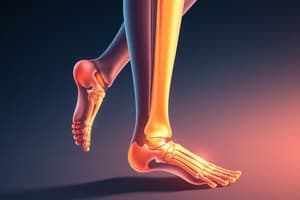
Foot Progression Angle (Angle of Gait)
- This angle is determined by the bisection of the foot from heel to the second intermetatarsal space, relative to the line of progression.
- A positive value, indicating out-toeing, exceeds 20°.
- A negative value, indicating in-toeing, is greater than -20°.
Knee Position in Newborns
- The head and neck of the femur are laterally angled at 60° from the frontal plane.
- There is a 30° medial twist of the femur.
- The knee position is 30° from the frontal plane, calculated as 60° - 30° = 30°.
- Inward twist is masked by outward rotation at the hip, making outward influence greater than inward influence.
Tibial Torsion Degrees
- At birth, tibial torsion is 0°.
- By age 6, it reaches an adult value of 18-23°.
Intoe
- Intoe is an internal rotation of the foot's long axis relative to the line of progression.
- Many intoe deformities resolves spontaneously.
- The genetic component of in-toeing does not self-correct/spontaneously.
- Cerebral palsy is often a causative etiology of in-toeing.
- Common causes of in-toeing gait is a combination of factors:
- Metatarsus adductus and clubfoot (TEV) in infancy.
- Internal tibial torsion for 1-2 year olds.
- Femoral anteversion for those older than 3 years.
- Spasticity of medial hip rotators and adductors, as seen in cerebral palsy (often with toe walking).
- The most common cause in infants is metatarsus adductus while internal tibial torsion is most common at age 1-2, and increased femoral anteversion is most common after age 3.
- Severe cases results from complex causes so consider all options.
- POSNA recommends no radiographs, surgery, or bracing for simple in-toeing in children under 8.
- Most untwisting of the femur happens before 9 years old.
- Check hip rotations, femoral torsion, tibial torsion, foot deformity, and foot progression angle to evaluate in-toeing deformity.
- Symptoms of intoe includes tripping, clumsiness, fatigue-like pains, especially in 3-year-old males.
- Sequelae of retained intoe gait into adulthood includes patellofemoral pathology, abnormal pronation, patellar instability, and lever arm dysfunction.
Newborn Tibia
- At birth, the tibia is relatively straight without twisting.
- Opposite to the femur, the tibia starts straight and then twists.
Internal Tibial Torsion
- Most common cause of in-toe gait during the first 3 years of life.
- Intrauterine positioning leads the rotation of the distal leg on the thigh at the knee joint, accompanied by tilting of the fibula about the tibia.
- Feet point inward toward each other when sitting on their parent's lap.
- Often noticed by parents around ages 6-18, presenting as a complaint of bowed leg or pigeon-toed gait, which compensates resulting in:
- Increased lateral femoral rotation
- Compensatory foot pronation
- Laterally faced hips may mask the deformity, making ITT or medial femoral torsion less noticeable.
- Tibial deformity results in straight or outward knees, while femoral or hip deformity results in inward knees.
- ITT can give the appearance of bowing, known as pseudobowing, but x-rays show a normal tibia, it presents as an illusion from the bulky calf and disappears when the leg is turned outward.
Malleolar Position
- At birth, the malleolar position is at -5°.
- By age 6 (adult value), it is 13-18°, which is 5° less than tibial torsion.
- Medial malleolus should be anterior to the lateral malleolus in a normal child.
- If the medial malleolus is even with or posterior to the lateral malleolus, suspect ITT.
- Evaluate tibial torsion by having the patient lay on their stomach with knees flexed, comparing the foot's long axis to the thigh.
- Normal: STJ is neutral, and the foot is lateral to the thigh.
- ITT: the foot is medial to the thigh.
Treatment for ITT
- Use above knee casting in preambulatory children.
- Implement night time splinting for 12-15 months, where it is more beneficial for those less than a year old, as older children exceeding 2 years are less likely to respond to the splint.
- Initial setting: 20-25° out-toe + add another 5° each month until a maximum of 45° is reached.
- Keep the unaffected side at the initial 20° setting in unilateral ITT cases.
- Wheaton bracing is used for unilateral internal tibial torsion.
- Use pelvic band and derotation straps usually from 5-6 y.o (after toilet training) for severe intoe with tripping and falling.
- Dr. Ressesque prefers the Dobbs brace as it is extremely flexible and the width should be shoulder-width.
Femoral Version
- Version is the normal twisting of a long bone on its anatomic longitudinal axis.
- Normal angular difference exists between the transcondylar axis of the knee and the head-neck axis of the femur.
- A higher angle leads to intoe.
- Normal femoral version:
- Newborn = 30-50°
- Adults = 10-15°
- A version that is abnormally increased is more than 2 standard deviations from the norm for a given age and is termed medial femoral torsion or increased femoral anteversion.
- Squinting patella and "W" sitting signs can be present with medial femoral torsion.
- Medial femoral torsion typically presents as 80° medial, 20° lateral.
- True bone problems maintain the same angle whether flexed or extended.
- Legs flop outward and barely cross over when attempting to lay on their stomach with legs go.
- The lateral soft tissue contracture masks the internal twist that there is no appearance of an intoe from the hip in a child less than 3 years old when the medial femoral torsion is high.
- High medial twist of the femur in newborns is masked due to the lateral rotation contracture of the intrauterine position.
- Reduction is achieved through growth, reduced hip flexor tightness, and lateral hip rotation.
Treatment
- Activities such as ballet, martial arts, and biking is recommended with less flexible shoes.
- Gait plates serve to improve the angle of gait and requires > 25° lateral hip rotation, working at the push-off phase and must be used for 3.5-4.5 y.o. with flexible shoe/sneakers.
- Denis Browne bar is not indicated for femoral torsion treatment, as it will create problems as the bar is for tibial torsion.
Femoral Anteversion
- Femoral torsion is pathology while femoral version is normal.
- Femoral anteversion measures the angle between the head and neck axis of the femur and the frontal plane of the body.
- Anteversion of the head and neck results in in-toeing and is best evaluated with CT scans but it won't count for dynamic motion.
- With medial hip rotation measured to be > 70°, the angles at birth are 37°, which changes to 12° at 14-15 y.o.
- The most improvement to be expected is at 9 years old, which is why osteotomies are performed then.
- Netters method is patient prone with the knee flexed 90°.
- Palpate the greater trochanter with one hand while internally rotating the hip with the other; the neck is parallel to the table at the point of maximum greater trochanter prominence.
Femoral Anteversion/Torsion
- Typically presents as age 3-4 y.o, peaking around 5-6 y.o, and often symmetric, it is commonly seen more in females.
Femoral Antetorsion
- Measures osseous twist between upper and lower ends, measuring between the head & neck vs transcondylar axis of femur.
Lateral and Medial Femoral Rotations
- Infants = 60° lateral, 0-30° medial
- 1-4 y.o = 40-45° lateral, 35-45° medial
- Medial should never be larger than lateral.
- Tight hamstrings can cause intoe if lateral/medial femoral rotation is affected differently in different positions (hip/knee flexed or extended).
- Soft tissue influences the amount of medial & lateral femoral rotation changes when the hip is flexed versus extended.
- 40° of medial rotation & 45° of lateral rotation as hip & knees are extended vs. 55° of medial rotation & 10° of lateral rotation as hip & knees are flexed indicates a tight medial hamstring that limits lateral femoral rotation.
- In medial femoral rotation (anteversion), the child is prone, have their legs flop to the sides of the chair, which rotates the thighs and femurs inward.
- In lateral femoral rotation, the child is put in the prone position to cross the tibia over the midline.
- Knees point straight by age 4 years old.
- The twist in the femur decreases (derotates) to -15° (from the previous 30°) inward, the angle of the head & neck to the frontal plane decreases to +15° (from the previous 60°).
- There is +15 opposed by -15 = 0° in the frontal plane, which is why the knees point straight.
- Hamstring flexibility measurement is assessed with the 70° of hip flexion needed for a straight leg raise.
Popliteal Angle
- The superior method to test hamstring flexibility
- Flex the hip 90° then extend the knee until resistance is met - Measure the acute angle between the lower leg and the imaginary line extending up from the flexed femur.
- Normal popliteal angle:
- Birth-2 y.o = 0-6°
- Greater than 5 y.o = 0-25°
Torsional Profiles
- A child has intoe, despite a normal torsional profile, possibly ligamentous laxity.
- With torsional profiles in 6-year-old females with 75° medial & 5° lateral femoral rotation w/ hip flexed & extended at a femoral torsion of 25° internal, tibial torsion of 22°, and Bleck's test showing 2nd digit means the Femoral anteversion/torsion
- This outcome can be determined by the difference in medial & lateral torsion, and also the femoral torsion is elevated.
- In 2.5-year-old females, presented with lateral > medial femoral rotation with hip flexed and extended, tibial torsion is -5° bilateral and Bleck's test shows 3rd digit means met adductus and ITT.
Out-Toe
- Foot progression angle is measured to be > 20°.
- Early walkers with out-toe does not need surgery; it is normal.
- Obese, 12-year-old male who presents with a limp or waddle with out-toe and groin pain.
- Suspect slipped capital femoral epiphysis (SCFE).
Risks of Gaits
- Infancy for calcaneovalgus (left), convex pes valgus (right).
- Childhood for contracture of triceps surae, causing pes valgus; tarsal coalition with peroneal spasm.
- All age groups for neuromuscular equinovalgus (Cerebral Palsy).
Hip Deformities
- Tightness of lateral hip rotators (infancy)
- Developmental dysplasia (when a child begins to walk)
- Lateral femoral torsion aka retrotorsion (pre-adolescence & adolescence)
- Neuromuscular disorders (spina bifida) (all age groups)
Convex Pes Valgus
- Less common but much more difficult congenital flatfoot deformity to treat.
- Identified as rocker-bottom foot primarily caused by dislocation of TN joint.
- Treatment is surgery.
Calcaneovalgus
- Congenital deformity: foot is dorsiflexed and abducted.
- Also referred as "Up & Out foot" in addition to a congenital clubfoot and flatfoot deformity.
- Diagnostic of fixed dorsiflexion, a valgus heel, tight anterior and lateral structures, low arch and prominent medial talus.
- Patients say "My baby always holds her foot out”.
- Etiology accounts for uterus wall pressure during pregnancy seen among primigravida with tight uterus.
Slipped Capital Femoral Epiphysis (SCFE)
- Associated with lateral femoral torsion aka retrotorsion
- 12 y.o male presents with a right-sided limp, out-toe, right flatfoot and right groin pain and limited internal rotation, abduction, and flexion of the right hip.
- Adolescence (boys 13-16; girls 11-14) often occur.
- Pain referred to groin or medial knee causing treatment limitations of internal rotation, abduction, & flexion, which call for surgery.
Femoral Retroversion
- Occurs when the head & neck of the femur are directed posteriorly from the transcondylar axis when the axis is on the frontal plane.
- There is not enough medial twist in the bone with 10° of torsion.
- Say "OUT TOE” if a provider says femoral retroversion.
- Can also be called external femoral torsion or lateral femoral torsion.
Examples
- If there is 90° lateral & 10° medial femoral rotation whether the hip is flexed or extended, the condition is likely Femoral retroversion instead of soft tissue influence.
- The angle doesn't change and indicates a bone problem versus cases with 45° of medial femoral rotation & 50° of lateral femoral rotation as hip is flexed; 45° of medial femoral rotation & 20° of lateral femoral rotation when the hip is extended indicates soft tissue influence
- In situations with decreased lateral rotation, influenced due to tightness of hip ligaments.
- In example: limited medial femoral as the hip is flexed & the knee is extended
- What condition accounts for 35° medial rotation & 45° lateral rotation hip & knee extended and 10° medial rotation & 45° lateral rotation hip flexed & knee extended? This is characteristic of out-toe due d/t tight lateral hamstrings.
Knock Knees
- Includes genu varum and valgum in childhood
- Progresses as birth - 1.5 years = genu varum
- 1.5 - 3 years = straight
- 3 - 6 years = genu valgum
Genu Varum and Valgum
- Genu Varum (bowed legged) causes asymmetrical abnormalities that require treatment.
- Symmetrical cases are likely normal and will not require treatment.
- Associated with Blount’s disease, rickets, achondroplasia
- Blount’s disease (tibia vara) causes growth disturbances of the medial aspect of the proximal tibia, form a "beaking."
- Bowing of the distal tibia is normal physiologic bowing, though presentation can can present as genu varum, while result of medial angulation in the metaphyseal which causes an metaphyseal-diaphyseal angle > 11.
- Seen among early walkers, short statured, overweight, black female, has associated ITT, the bow being in the proximal ⅓ of the tibia and can be classified by Langenskiöld classification
- To manage the disorder:
- plan for a child than 4-year-old w/ langenskiold of I or II; use a brace
- plan for a child than 5 y.o w/ langenskiold of III or VI; use surgical osteotomy
- A "cover up test" is a technique in which the child is placed on back, covering middle ⅓ of tibia with hand and evaluating valgus/varus relationship between tibia and thigh Used to differentiate physiologic genu varum from Blounts disease, used as Juveniles & infantile bilaterally, with these specific results:
- negative valgus, there is physiologic or pseudobowing
- positive varus Blounts disease results
Rickets
- Vitamin D deficiency -> weak bones, multiple problems, and possibility of tibial varum
- Congenital tibial angulation causes Group 1 disease:
- Angulation = anterolateral bowing of tibia combined with neurofibromatosis
- Congenital tibial angulation causes Group 2 disease:
- Angulation = anterior bowing of tibia with fibular Deficiency that is related to fibular hemimelia and short leg
- Congenital tibial angulation causes Group 3 disease:
- Angulation = posteromedial bowing of tibia that is related to calcaneovalgus
Genu Valgum
- Refer a bowlegged child to a pediatric orthopedist when greater than 3 y.o and in cases of knock knees,
refer them when greater than 7 due to causes including:
- unilateral case
- intermalleolar distance greater than 3.5-inches
- trauma causing injury
- congenital cause stemming from short fibula
- those caused by physiological causes and sources of infection
- Ollier's disease being unilateral
- lumps/bumps all over the body on the feet & legs in cases of bone dysplasia (multiple enchondromatosis)
- Renal osteodystrophy also relates to:
- bone disease in kids with kidney disease and with kidneys failing to maintain the balances of Ca and phophorus in bone
- lumps/bumps all over the body on the feet & legs in cases of bone dysplasia (multiple enchondromatosis)
- the disorder can indicate ligamentous laxity, or down syndrome
Symptoms and Diseases related to Valgum
- Symptoms include pain & limping
- Associated diseases relates to Multiple Enchondromatosis (Ollier's Disease) that are benign bone tumors called enchondromas all over renal resulting in osteodystrophy or cases of ligamentous laxity and down syndrome
Causes of Violence
- Includes acts of abuse against children and their bodies (whether through sex or physical harm)
- Another factor relates to neglect of the child with failure to provide or meet basic needs.
- It is expected, with SCR and a mandated report, that calls be made/reports to be given regarding the child's condition being impaired or if the child is at imminent danger. A written report is therefore required, and, when reporting to the SCR, reasonable cause is needed, which does not necessitate proof of condition
- Emergency cases are turned over to police to resolve
Pharmacology
- Ensure that you are giving an antibiotic for the right reason.
- overprescription can cause the child to build resistance and intolerance to the medication
- to treat infection, the right method follows narrow spectrum penicillins that are also the most common abx used in these pediatric cases:
- Penecillin that is penicillinase ressistant (NOT MRSA)
- Amoxacillin
- 1st generation cephalosporin like Cephalexin (kefflex)- covers gram positives and negative
- Sulfonamides
- Nitrofuratoin
- Dicloxacillin
Drug Combinations to Note:
- Oral penicillin can be used as treatment for staph aureus
- Penecillin is also the penicillin of choice
- For indicated conditions that need Group A beta hemolytic strep for treatment:
- Amoxicillin, Penicillin (for strep, ear, and sinus infections), & Ampicillin
Medical Considerations
- Contraindications exist for sulfa allergy patients.
- Clindamycin is the drug of choice for patient with MRSA infection that is indicated for soft tissue infections with penicillin allergy
- Tetracycline must be avoided for treatment on patients if they are younger than 8, as they permanently stain the teeth and Fluoroquinolones
- Must be avoided on pediatric patients as concerns are with increased pneumococcal resistance
- In adults: cartilage and joint toxicity
- foot abscess without signs, always perform incision and drain — Use Mupirocin (bactroban)
- for topical skin infections like impetigo and MRSA
- but must take care if treatment and remedy for topical are bacterial with possible presence of staph
- or if the area of infection is commonly found at the face and and face excluding palms and soles
- for burns: Silvadene first line therapy will Tylenol (acetaminophen)
Medical Prohibitions
- Aspirin can't be prescribed to kids as this can worsen the flu and potentially cause Reye's Syndrome.
- When a wound has been bit and is infected, one shouldn't prescribe antibiotics or give cephalosporins in allergy cases.
Drug Applications
- Ampicillin and subactam are prescribed to infected bitten wounds. Clindamycin for patients with staphylococcal soft tissue infection and true allergy to penicillin
- A 9 year old with an abscess doesn't require oral medication (incision 7 drainage is enough)
Disease Applications
- 70% of infections with skin are result of S. Aureus.
- the preferred and first method for treatment is by prescription penicillins like flucloxacillin and dicloxacillin — take Cephalexin/Erythromicin if alternative but use risks of developing resistance to multiple antibiotics.
- Antihistamines help reduce inflammation caused by the drugs.
- can also use topical diphenhydramine
- Promethazine in a 2 y.o max doses would be 25mg
Method To Prevent Damage
- ointments for dry locations; powders if the area is known to be wet
- powders absorb better
- solutions for interdigital lesions with great drying abilities
- lotions are more watered
- steroids used topically shouldnt be for long durations, as atrophy, red marks, adrenal effects, stretch marks can occur reduce steroid dosage -example: hydrocortisone 1%
- use topical antifungals like Lamasil that are great. However, ketoconazole use should be avoided due being prone to liver toxicity. Onychoymcodis must be treated in combination to work with an effective agent.
Dosages
- (West nomogram) - use body surface to calculate medication needed, as it accurate
- dosage in common use for medication:
- 2.2 lbs → 1 kg
- 5 mL → 1 teaspoon
- 15 mL → 1 tablespoon
- When having issues with pill swallowing, 44 lb. female w/ infected ingrown toenail in need prescribed cephalexin, the female will need 250 500 750 mg tablets AND 125 mg/0.5 mL or 250 mg/5 mL, with 25 mg/kg/day divided every 12 hrs
Prescribing Medicine
- use these method to get it correct, remember that in oral suspensions, 250mgs/5 mL and that
- 44 over 2.2- that equals 20kg
- 2, 20 from multiply to 25 to achieve 500mg dosage
- 500 divide in 2 so dose
- 250 should result in 5mL in 1 tsp
- to ensure correct medicine, the message here needs to say "oral suspension of 250 over 5. give 100 MLS with a sig of 1 tsp for 12 hrs for 10 days
More Examples
55 lb. female, you should use Medicine D in liquid suspension of 200 mgs/5mL, with a rate of 25 mgs/kg/day and the requirement to prescribe every 12 hrs for 10 days
- here you need calculate, 55lb in 25 kg
- 25 times 25 = 6.5 mg dose in one usage
-
- 5 divided in 2.0 results in 3.5 To account for medicine "medication B” over 7 days " if a medical director needs to prescribe 3 tsps with meals in a 15ml volume and 15ml over 7 days, with 25mds/kg/day, what will the medication be? Its 125/25lbs.
Precautions
Maximum dose size of lidocaine is calculated by 5md/kg/dose and can't be overcome by a person, otherwise the 300 md dose should be the most in that scenario
Verrucae and Similar Conditions
To effectively treat common conditions and warts, there are number of methods. It is to stimulate the immune system more than eliminate the problem:
- Line of defense- salicyclic acid 70-80% - stimulate immune system, apply aldara after
- cryotherapy more so the alicylic acid because it can have a hurt
- FDA has approved cimetidine for treating warts, as it is immunomodulatory If these attempts have fail, consider squaric acid
- There are also vaccines but should first focus on treating underlying concerns
Onchymycosis
A highly common infection in the nails that is caused by the T. rubrum fungus, this can be treated (in specific context such as pt and those under Down Syndrome or immunocompromised ). the treatment options: terbinafine or Itraconazole. DO consider if a child is more than 40kg
Drug Contraindications
Griseofulvin (treats ringworm), instead using Terbinafine that safer and well more Onychyrocryptosis is an ingrown toenail. And is cause often by the nail. The treatment is by partial avulsion where the lido block step won't be needed in babies, there isn't any trauma is required in this step
Tinea Infections
Common forms of skin fungul inflammation
- Tinea corporis & and capitis occurs often among children
- Tinea cruries and Tinea Pedias and tinea unguinum happen during the adolescents
- Tineas like mentagrophyes - can be seen, and there are blisters containing fluid in the plantar foot distal
More Considerations
- Skin rash steroid needs to help with tinea
- topical turbinafine is mild tinea infections needs to occur
- Nystatin cannot be prescribed in these cases due liver concerns
Treatment Methods and Practioner Tactics
- When determining mimic of onchomycoses, consider the potential of
- (seen during sport): Dystrophic nails
- that produces a pitside: Psoriasis
- Trachyichia
- Atopic dermatitis exists in kids with allergies - in a variety of forms: contact to facial surfaces, skin at the hallux
- Juvenile planar dermatoses affects those young and creates a look that is glaze
Piezogeneic
- Can happen by bump from fat near foot. Need surgical interventions or touch these as they all improve on your own
• Growing pains
- Usually happens while asleep at night , but cant be localised vs severe pains the usual treatment must be to focus steroid and make some modification -the calf may be a trigger
Bone Structures and Other Conditions
- Occurance when pain is near tibia - related with exercise.
- Other related issues can lead into musculoskeletal issues because of leg, with Osgood
- Schlatter which results in pain to the bone or over tubercle.
- There could also conditions, in this case such for running women. Is discold meniscus that lead to the catching and stopping of the legs which creates to the locking up.
- All and any of these can relate to Amps (amplified musculokeletal pain syndrome). Tenderness can occur in joints
Juvenile Idiopathic Arthritis (JIA)
- occurs for several weks
- This causes severe heel pain needing NSAIDS, associated anterior Uveitis
- oligarioarthritis cause swollen joints mostly in those affected by JIA
Tests and Methods to Help
- Check if lab results are normal
- Look for Micrognathia with the two differentiating - RF (-) = MG - RF(+) = Nodules
- Check for diagnosis via clinics, not the lab. This might indicate knee problems, which are managed by NSAIDs of all kinds
Calcaneal Apophysitis and Sever Disease
- Sever Disease can also cause some similar heel problems • These results from squeezes from side, causing movement.
- All tests results must have some scientific reason to exist or be known.
Bones
- The enthesophathy in the body can often tested to be B25
- there must be a scan test to review where and what in the bone
Osteoporosis
- caused by achilles and plantar fasciitis Test and identify the deformity - and check for swollen toe
Additional Information
- Tests with show and confirm and what side and how the bone will respond 15 or 16 and the like. B
- Be aware of what is seen: the joint ,talus,bones
- Are there abnormalities - The goal is review the parts of feet
- Calcaneous (is that a normal view)
- Are there abnormalities - The goal is review the parts of feet
- Flat with what we see when reviewing images
Test Method
- Always review the legs. Where they are and how, what part do we evaluate?
Calcaneus Fractures
- In X-ray
- It means something bad happen
- Does the patient have any bone
- Is bone fluid or has cyst which must have medical operations
Rare Deformities
- Look for Z foot and how many components and what that may involve (rare)
- Pes Planius
- The test the deformity with blocks.
- Evaluate what may need the treatment
Metatarsias
Be clear for 4th with shortening. The medial deviation with halux
Bone Identifiers
- Know the bones at birth seen by x-ray
- Bones do ossy or come together - do they do in the right order or they'll come together like what happens in an army where every comes in formation and a team.
- What does that indicate about an anomaly caused by or that may cause the condition we are seeing
- That helps diagnose many patients, especially for 5th M and to identify it away for fractals by showing how it may show in x ray (how transverse and the apophy)
Treatment, Symptoms and More Related Conditions
-
the treatment plan should be evaluated and analyzed in way that shows good relationship
-
Is joint unusal to point in what condition we are seeing or that the patient may have and what the results are from
-
Check if some had cavus foot is always concern
-
the treatment plan should be evaluated and analyzed in way that shows good relationship
-
Be sure to keep the process simple not complex
- It's meant to help, so keep it to that standard
the treatment plan should be evaluated and analyzed in way that shows good range relationsh
-
Be aware can or May is -What do results mean
-
Toes can be cause by : Cerebral Pulsy with :
-
Briskers lower than upper , exensor babinski
-
History . They were early and low when birth
-
Dystrophy-
- Trouble going up and going.
- Absent of test and history they stop normal and slow progress • CMT symptoms similar to cavus and abnormal patterns -Tether :
- Cord with bladder functions and order to help
-
And must what we can fix all these causes
-
Must treat them by treating what there'll be and is always better For neuro cases must refer to the right Dr.
Studying That Suits You
Use AI to generate personalized quizzes and flashcards to suit your learning preferences.