Podcast
Questions and Answers
Which of the following diagnostic findings is commonly associated with asbestosis?
Which of the following diagnostic findings is commonly associated with asbestosis?
- Diffuse reticulonodular infiltrate at bases on chest X-ray (correct)
- Normal pulmonary function tests
- Elevated forced expiratory volume in 1 second (FEV1)
- Absence of pleural plaques on CT scan
Silicosis is caused by the inhalation of asbestos fibers.
Silicosis is caused by the inhalation of asbestos fibers.
False (B)
What is the most common cause of cor pulmonale?
What is the most common cause of cor pulmonale?
COPD
In coal workers pneumoconiosis, simple cases often present with small upper lobe ______ opacities.
In coal workers pneumoconiosis, simple cases often present with small upper lobe ______ opacities.
Match the following types of hypersensitivity pneumonitis with their associated cause:
Match the following types of hypersensitivity pneumonitis with their associated cause:
A patient presents with fever, chills, cough, chest tightness, and dyspnea. Auscultation reveals diffuse inspiratory crackles but no wheezing. Which condition is most likely?
A patient presents with fever, chills, cough, chest tightness, and dyspnea. Auscultation reveals diffuse inspiratory crackles but no wheezing. Which condition is most likely?
Vaping-associated lung injury is definitively diagnosed through a specific biomarker test.
Vaping-associated lung injury is definitively diagnosed through a specific biomarker test.
What is the primary symptom of pleuritis?
What is the primary symptom of pleuritis?
Fluid accumulation in the pleural space is known as a pleural ______.
Fluid accumulation in the pleural space is known as a pleural ______.
Match the following characteristics with the correct type of pleural fluid.
Match the following characteristics with the correct type of pleural fluid.
Which of the following is the initial treatment approach for a patient diagnosed with small primary spontaneous pneumothorax?
Which of the following is the initial treatment approach for a patient diagnosed with small primary spontaneous pneumothorax?
Cystic fibrosis is an autosomal dominant disorder.
Cystic fibrosis is an autosomal dominant disorder.
What is the genetic abnormality responsible for Cystic Fibrosis?
What is the genetic abnormality responsible for Cystic Fibrosis?
Solitary pulmonary nodules are considered benign if they are isolated, round, opacity, and less than ______ cm.
Solitary pulmonary nodules are considered benign if they are isolated, round, opacity, and less than ______ cm.
Match the following characteristics to their respective classification as either a benign or malignant solitary pulmonary nodule (SPN):
Match the following characteristics to their respective classification as either a benign or malignant solitary pulmonary nodule (SPN):
Which of the following is the most common type of lung cancer?
Which of the following is the most common type of lung cancer?
Small cell lung cancer (SCLC) is mainly caused by peripheral masses.
Small cell lung cancer (SCLC) is mainly caused by peripheral masses.
What syndrome can occur if a lung tumor is located in the apex of the lung compressing nerves and blood vessels?
What syndrome can occur if a lung tumor is located in the apex of the lung compressing nerves and blood vessels?
Unlike typical carcinoid tumors, _______carcinoid tumors are intermediate grade that grow a little faster.
Unlike typical carcinoid tumors, _______carcinoid tumors are intermediate grade that grow a little faster.
Match the following ARDS criteria to their description:
Match the following ARDS criteria to their description:
Flashcards
Pneumoconiosis
Pneumoconiosis
Chronic fibrotic interstitial lung disease caused by dust inhalation, usually from workplace exposures, leading to irreversible lung damage.
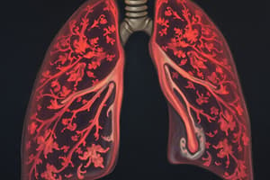
Asbestosis
Asbestosis
Lung disease caused by asbestos exposure, showing restrictive patterns on pulmonary function tests and reticulonodular infiltrates on chest X-rays.
Silicosis
Silicosis
Lung disease resulting from crystalline silica inhalation, presenting with nodules in upper lung fields on CXR and potentially progressing to restrictive lung patterns.
Coal Workers Pneumoconiosis
Coal Workers Pneumoconiosis
Signup and view all the flashcards
Hypersensitivity Pneumonitis
Hypersensitivity Pneumonitis
Signup and view all the flashcards
Vaping-Associated Lung Injury
Vaping-Associated Lung Injury
Signup and view all the flashcards
Pleuritis/Pleurisy
Pleuritis/Pleurisy
Signup and view all the flashcards
Costochondritis
Costochondritis
Signup and view all the flashcards
Pneumothorax
Pneumothorax
Signup and view all the flashcards
Cystic Fibrosis
Cystic Fibrosis
Signup and view all the flashcards
Adenocarcinoma(50%)
Adenocarcinoma(50%)
Signup and view all the flashcards
ARDS
ARDS
Signup and view all the flashcards
Foreign Body Aspiration
Foreign Body Aspiration
Signup and view all the flashcards
Pulmonary Embolism
Pulmonary Embolism
Signup and view all the flashcards
Pulmonary Hypertension
Pulmonary Hypertension
Signup and view all the flashcards
Study Notes
Pneumoconiosis
- Chronic fibrotic interstitial lung disease caused by dust inhalation
- Exposures commonly occur from workplace settings
- Irreversible lung damage results from this condition
Asbestosis
- Directly linked to workplace exposure, with the highest exposure dose occurring directly at work
- Bystander exposure and general community exposure are also risk factors, and it is most common in the general community
- There is progressive worsening of dyspnea as a presentation
- Other presentations: Nonproductive cough, late chest from right-sided heart failure from pulmonary hypertension
- Bilateral inspiratory crackles can be auscultated during a physical exam, some patients will have clubbing and reduced chest expansion
- Later stages of cor pulmonale can cause pedal edema, JVD, and right ventricular heave
- Pulmonary Function Tests will reveal a restrictive pattern for diagnosis
- Chest X-ray shows diffuse reticulonodular infiltrate at bases and shaggy heart borders
- Ground glass opacities, diffuse interstitial fibrosis, pleural thickening, and calcified pleural plaques are revealed via CT imaging
- There is no specific differential diagnosis
- Treatment is supportive, which includes removing the exposure while prevention is best
- Prognosis is chronic with complications that require patient education
Silicosis
- Caused by quartz dust (crystalline silica)
- Considered the world's most prevalent occupational lung disease
- Three phases: acute develops in less than 5 years after exposure, accelerated develops in 5-10 years after, and chronic develops, 10-30 years after exposure
- Progressive dyspnea and nonproductive cough are presentations, lymphadenopathy may also be present
- CXR reveals nodules in the middle and upper lungs plus enlargement of hilar and mediastinal lymph nodes
- PFTs indicate a restrictive pattern
- High resolution CT may be necessary for certain diagnoses
- Support and lung transplant for end-stage acute silicosis are support treatments, primary is prevention
- Prognosis is chronic
- Complications: susceptibility to mycobacterial disease, autoimmune disease, and bronchogenic carcinoma, patient education is necessary
Coal Worker's Pneumoconiosis
- Coal dust inhalation leads to scarring and fibrosis
- Cumulative dust exposure is the main risk factor
- Simple causes upper lobe nodular opacities, while other symptoms might be asymptomatic
- It presents with progressive dyspnea, a productive cough and black sputum production
- CXR and CT will reveal small, acute, rounded, nodular opacities, while complicated cases will show large conglomerate masses
- Black pigmented macrophages are apparent via BAL
- Prevention is key, including respiratory masks and limiting mine dust exposure
- Removal of exposure, smoking cessation, supportive care, and COPD treatment are also part of typical treatment
Hypersensitivity Pneumonitis
- Interstitial lung disease with complex immunological reaction of lung parenchyma due to repetitive inhalation of organic sensitized allergen
- Combo of type III and IV reactions
- Farmer's lung is caused from moldy grain, hay, silage fungi (aspergillus)
- Bird fanciers lung is related to bird proteins like parakeets, pigeons, chickens, turkeys and duck protein
- Hot tub lung: mycobacterium avium complex from contaminated mist and mold ceilings around hot tubs
- Humidifier lung: aureobasidium species, contaminated water in humidifier or AC
- Symptoms: fever, chills, cough, chest tightness, dyspnea, tachypnea, diffuse inspiratory crackles, absence of wheezing in acute
- Symptoms for chronic cases: few physical findings, exertional dyspnea, productive cough, cor pulmonale
- Subacute symptoms happen in between days and weeks
- Requires high index of suspicion
- Clues in history!
- CXR normal in acute, reticular or nodular opacities in chronic
- CT acute: upper and middle lobe patchy ground glass or nodular opacities "head cheese sign" Chronic: reticular opacities and honeycombing
- PFT: advanced restrictive pattern
- Prognosis: if caught early acute is reversible, and chronic is not reversible
- Acute: prednisone 60mg daily for 1-2 weeks then taper down (relieves initial symptoms)
- Chronic: prednisone 30-40mg daily longer course, taper maybe
Vaping Associated Lung Injury
- Acute or subacute respiratory illness characterized by a spectrum of clinicopathologic findings mimicking various pulmonary disease
- People with a median age of 21 are most at risk
- Symptoms: cough, SOB, chest pain, may include Gl upset, fever, chills
- Diagnostic criteria: Use of e-cig within 90 days, pulmonary infiltrates on CXR or CT, and absence of any other possible etiology
- CXR: hazy bilateral opacities with central and peripheral sparing
- CT: diffuse bilateral ground glass opacities
- Diagnostics of exclusion
- Supportive treatments such as O2 or steroids are sometimes needed
- Patient education is necessary
Pleuritis/Pleurisy
- Viral infections are the main cause
- Other causes: bacterial infections, inflammatory disorders, cardiac conditions, pulmonary conditions, idiopathic
- Pleuritic chest pain (severe, sharp pain that worsens with breathing) and a pleural friction rub (high pitched breath sound) are the presentations
- NSAIDS are the recommended treatments
Costochondritis
- Inflammation of costochondral or constosternal joints
- Typically affects rib 2-5
- Recent respiratory infection with coughing, strenuous exercise and physical activity are risk factors
- Presentation: Sharp chest wall pain or pressure like, worsens with deep breath, coughing, lying on affected side
- Pain relievers such as NSAIDS, heating pad, and avoiding strenuous activity are treatments
- Self limiting conditions, but should be distinguished from other conditions
Pleural Effusion
- Abnormal accumulation of fluid in pleural space
- Compresses the lungs and impairs expansion
- Thoracentesis is diagnostic and therapeutic
- Light's Criteria: used to classify fluid as transudative or exudative
- Exam:
- Dullness to percussion over effusion
- Decreased breath sounds over effusion
- Decreased tactile fremitus
- Positive egophony at upper border of effusion
- Decreased bronchophony over effusion
- Peripheral edema and JVD
- Evaluation:
- Upper CXR: blunting of costophrenic angles if 75mL of fluid or more
- Blunting of lateral angles requires more than 200mL
- Lateral CXR: can visualize even smaller amounts of fluid
- Ultrasound can detect as little as 5mL of fluid (hypoechoic or anechoic)
- Serum labs: CBC, CMP, Lipase, Cardiac enzymes, LDH and BNP
- Fluid analysis: pH, protein, LDH, glucose, cell count, gram stain and culture are analyzed
- Light's Criteria is exudative:
- Protein/serum ratio greater than 0.5
- LDH/serum ratio greater than or equal to 0.6
- Pleural LDH greater 2/3 upper limit of normal
- Treatment: Thoracentesis is for all patients with more than 10mm pleural fluid on imaging that is new (ORDER CXR AFTER PROCEDURE) Small bore drains, 10-14 gauge effective as larger drains and more comfortable
- Do not train more than 1500mL
Pneumothorax
- Collection of air outside the lung but within the pleural cavity
- Applies pressure on the ling causing it to collapse
- By trauma or rupture of visceral pleura
- Simple, tension, open: traumatic or atraumatic: primary or secondary
- Risk factors:
- Smoking, tall thin body habitus, marfan syndrome, men, 20-30 years old
- Causes of latrogenic:
- Lung biopsy and central line insertion
- Causes of traumatic:
- Penetration or blunt trauma, rib fracture and diving
- Tension is caused by:
- Barotrauma due to positive pressure ventilation
- Evaluation:
- CXR
- Ultrasound seen as superior
- E-FAST trauma situation
- If unstable, don't image, needle decompression immediately
- Treatment:
- Small primary spontaneous: supplemental 02, repeat CXR 4 hours, outpatient follow up
- LArge primary spontaneous: catheter aspiration then Heimlich valve if ambulatory, tube thoracostomy (4/5 rib) to water seal with suction if hospitalized
- Secondary spontaneous: chest tube with continuous suction, CXR to evaluate -If CXR shows full lung expansion, so suction and observe -If CXR shows no lung expansion, consult surgery
- Tension: immediately with needle thoracostomy 14 or 16 gauge needle catheter through chest wall in 2nd intercostal space at midclavicular line
- Open sucking chest wounds: 3 sided dressing util tube thoracostomy and chest wall defect repair
- VATS: indicated after second occurrence of primary, any bilateral, or failure tube thoracostomy for first episode of primary or any episode of secondary
- Stable criteria:
- RR less than 24
- HR less than 120 but greater than 60
- Normal BP
- 02 greater than 90%
- Whole sentences
Cystic Fibrosis
- It is the most common chronic lung disease is young adults
- Autosomal recessive disorder: abnormalities in CFTR protein that results in altered chloride transport and water flux
- Newborns: meconium too thick, ileus
- Early childhood: pancreatic insufficiency because thick secretions block duct leading to proteins ad fats unable to be absorbed
- Later childhood: recurrent ling infections, respiratory failure that ad lead to death
- Symptoms: productive pough, decreases exercise tolerance, hemoptysis, chronic rhinosnusitis, steatorrhea, diarrhea, abdominal pain
Newborn Diagnosis
- Screening: detects pancreatic enzyme IRT
- Sweat chloride test: historical gold standard (elevated sodium and chloride)
- Genetic testing for CFTR mutation
Adult Diagnosis
- PFTs: obstructive pattern
- Chest CT: peribronchial cuffing, mucous plugging, increased interstitial markings, small rounded periaheral opacities, focal atelectasis
- genetic testing for mutation
- Symptomatic treatment include CFTR modulators (only for certain mutations), airway clearance techniques, inhaled bronchodilators, abx, pancreatic enzyme replacement
- Only definitive is lung transplant
- Keep up to date on vaccines
Mediastinal Masses
-
Anterior compartment: sternum anteriorly and surface of great vessels and pericardium posteriorly
- Thymonma, teratoma, thyroid lesions, lymphoma and fibroma
-
Middle compartment: anterior pericardium to anterior surface of thoracic spine
- Lymphadenopathy, pulm artery enlargement, aneurysm of aorta, developemental cyst
-
Posterior: Paraverterbral -Hital hernia, esophageal tumor, meningocele, neurogenic tumor, and thoracic spine disease
Solitary Pulmonary Nodule
- Isolated, round, opacitu, less than 3 cm NOT associated with infiltrate, atelectasis and adenopathy
- Mainly asymptomatic
- Benigns are moslty infectious granulomas
- Malignancy likelihood increases with age, smoking and prior malignancy -The bigger the nodule higher the risk of malignancy
Benign vs Malignant Nodules
- Benign:
- Stability in size compared to prior imaging
- Smooth well defined edge
- Dense calcifications
- Rapid doubling, less than 30 days is an infection
- Malignant:
- Spiculated margins, peripheral halo
- III defined margins or lobular apearance
- Sparse or stippled calcification
- Cavitory lesion with thick walls more than 16mm
Lung Cancer
- Leading cause of cancer deaths
- Cigarette smoke is primary cause and other risk factors are second hand smoke, radon, asbestos, diesel exhaust, ionizing radiation, metal and family history
- Median age if 71 with 25% 5 years survival rate
Type of Lung Cancer
- Five categories, mainly broken into small cell and non small cell
- Origin
- Adenocarcinoma 50%- mucus glands or epithelial cells hear terminal bronchiole
- Characterics
- Forms peripheral nodules/masses
- Location- peripheral -Squamous Cell Carcinoma (SCC)
- Origin- bronchial epithelium
- Characteristics
- Intraluminal masses, presents with hemoptysis, central -Small Cell Carcinoma
- Origin
- Bronchial origin
- Characteristics
- Causes narrowing bronchus without discrete mass and aggressive to lung
- Adenocarcinoma
- Origin
- Alveoli to lung tumor, spreads along alveoli spreading
- Large Cell Carcinoma
- Origin
- Undifferentiated large cells and aggressive, rapid doubling Times
- Symptoms-New cough, anorexia, weight loss, dyspnea, hemoptysis, pain
- Metastasis,Localized spread: PNA, pleural effusion, hoarseness, SCV, Horner syndrome, pain
- Pancoast: tumor in apices compress , invades nerves & Blood vessels, Brain- Nausea & Vomiting, Seizures , dizziness, Liver/Asthenia, Bones: Bone pain, Adrenal glands
Lung Treatment
- Surgery, chemo (Cisplatin & Etoposide)
- Limited- (20-40%) 2 yr rate, extensive 5% 2 year/IB/II
- Resection, Neoadjuvant therapy (Chemo or Radiotherapy)
- Stage IIIA/ Operable- Resection, chemotherapy,IIIB- concurrent radiation, immunotherapy
- SCLC :Limit tumor in HEMITHORAX
- NSCLC extensive tumor is beyond hemothorax
Bronchial Carcinoid Tumors
- Neuroendocrine tumors of Lung, Low MALIGNANCY and more
- pink or purple growths in central bronchi & Hemoptysis Surgical excision recommended
Mesothelioma
- Aggressive cancer of pleural membranes or peritoneum
- Associated with asbestos exposure
- idious onset dyspnea, nonpleuritic chest pain, weight loss, dullness to percussion, diminished breath sounds, clubbing
- VATS biopsy to obtain specimen for histology and Pleural fluid analysis shows exudative fluid and hemorrhagic NOT-Good Prognosis7-18month
ARDS
- Acute respiratory distress syndrome
- Acute/onset
- Bilateral infiltrates or no cardiac origin
- Hypoxemia, increased RR, progressive symptoms
- Causes alveolar aspiration or damage
- Progsive SX include Oxygen treatment and Ventilation,
Foreign Body Aspiration
- Upper Air Obstruction, Dysphagia, Stidor , Coughing. LOC, medication related Tx/ Remove it quickly as possible
Pulmonary Embolism:
-
obstruction of Pulmanry / Branch. Trhombus - tumor - air - fat
-
RISK FACTOR - INCREASE Clottiing- factor v leidine/immobilization . Malignancy - obsity - pregnancy - OC
-
Increased Respiration & Heart Rate, Sudden
-
Test/ d- dimer -CUA - VIO Scan - Well >2 intermediate and over 6 at risk with CT &EKG
Thrombolytic therapy / AnicOag
Pulmonary Hypertension
Pulmonary HTN: Aloration In Structure & fan. Ntricle of H Heart Cased Primary disorder- results from lung problems Etiology: increased pulm artery + vascular Resistance.
- Pulm Art HTN
- Lung disease (COPD)
- Chrnoic PE rare .Treat underlying (vasodilat / Diuretics/ fluid management)
(MAP - 25 - Loud P2 (high pulmonary pressure) *sign is Right heart pressure and Strain shown on EKG
Test= (ECHO & Catheter).
Studying That Suits You
Use AI to generate personalized quizzes and flashcards to suit your learning preferences.