Podcast
Questions and Answers
Which of the following statements accurately describes the functional role of the placenta?
Which of the following statements accurately describes the functional role of the placenta?
- It serves as the primary site for blood cell production in the developing fetus.
- It is responsible for the exchange of nutrients and waste products between the mother and the fetus. (correct)
- It solely functions to produce hormones that maintain the uterine lining during pregnancy.
- It provides a protective barrier against all pathogens, ensuring a sterile environment for fetal development.
What is the key distinction between an embryo and a fetus in human development?
What is the key distinction between an embryo and a fetus in human development?
- An embryo is defined as the developing human from fertilization until the end of the first week, while a fetus is from the second week until birth.
- The terms are clinically interchangeable, referring to the developing human at any point during gestation.
- An embryo is the term for the developing human up to the end of the ninth week post-fertilization, after which it is called a fetus. (correct)
- A fetus refers to the developing human only after it has a fully formed organ systems, typically after the first trimester.
Why is the placenta essential for human embryonic development, unlike in avian development?
Why is the placenta essential for human embryonic development, unlike in avian development?
- Avian eggs provide a complete source of nutrients via the egg yolk, whereas human embryos rely on the mother for nutrition and waste removal. (correct)
- Human embryos develop externally and require a placenta for protection from the elements.
- Human embryos have a large yolk sac that provides all the necessary nutrients, but they still need a placenta for waste disposal.
- Avian embryos require a placenta for gas exchange but not for nutrition.
Which of the following best describes the roles of the inner cell mass (embryoblast) and outer cell mass (trophoblast) in early placental development?
Which of the following best describes the roles of the inner cell mass (embryoblast) and outer cell mass (trophoblast) in early placental development?
During implantation, what is the initial mechanism by which the embryo loosely attaches to the uterine wall?
During implantation, what is the initial mechanism by which the embryo loosely attaches to the uterine wall?
What is the decidual reaction in the context of embryo implantation?
What is the decidual reaction in the context of embryo implantation?
How do cytotrophoblast and syncytiotrophoblast differ in structure and function?
How do cytotrophoblast and syncytiotrophoblast differ in structure and function?
What is the significance of the lacunar network formed by the syncytiotrophoblast?
What is the significance of the lacunar network formed by the syncytiotrophoblast?
During the second week of development, how do nutrients and waste materials transfer between the mother and the embryo?
During the second week of development, how do nutrients and waste materials transfer between the mother and the embryo?
Which part of the decidua directly contributes to the maternal portion of the placenta?
Which part of the decidua directly contributes to the maternal portion of the placenta?
What is the sequence of the villous development?
What is the sequence of the villous development?
What structural feature distinguishes a tertiary villus from a primary or secondary villus?
What structural feature distinguishes a tertiary villus from a primary or secondary villus?
Which of the following accurately contrasts the chorionic frondosum and chorionic laeve?
Which of the following accurately contrasts the chorionic frondosum and chorionic laeve?
How are the chorionic vessels connected to the developing vasculature of the embryo?
How are the chorionic vessels connected to the developing vasculature of the embryo?
What is the role of the allantois in the formation of the umbilical cord?
What is the role of the allantois in the formation of the umbilical cord?
What happens to the yolk sac and vitelline duct as the embryo develops?
What happens to the yolk sac and vitelline duct as the embryo develops?
How is the amniotic cavity related to the chorion as pregnancy progresses?
How is the amniotic cavity related to the chorion as pregnancy progresses?
What leads to the degeneration and eventual loss of the decidua capsularis?
What leads to the degeneration and eventual loss of the decidua capsularis?
Initially, where does the embryo grow within the uterus, and how does this change later in development?
Initially, where does the embryo grow within the uterus, and how does this change later in development?
Which of the following describes the ultimate fate of the decidua capsularis during pregnancy?
Which of the following describes the ultimate fate of the decidua capsularis during pregnancy?
Flashcards
Placenta
Placenta
A temporary vascular organ facilitating nutrient and waste exchange between mother and fetus.
Morula Stage
Morula Stage
Outer cells arrange for nutrition; inner cells form the embryo.
Decidua
Decidua
The outermost layer of the endometrium after uterine cells undergo decidual reaction.
Cytotrophoblast
Cytotrophoblast
Signup and view all the flashcards
Syncytiotrophoblast
Syncytiotrophoblast
Signup and view all the flashcards
Lacunar Network
Lacunar Network
Signup and view all the flashcards
Decidua Capsularis
Decidua Capsularis
Signup and view all the flashcards
Decidua Basalis
Decidua Basalis
Signup and view all the flashcards
Decidua Parietalis
Decidua Parietalis
Signup and view all the flashcards
Primary Villi
Primary Villi
Signup and view all the flashcards
Secondary Villi
Secondary Villi
Signup and view all the flashcards
Tertiary Villi
Tertiary Villi
Signup and view all the flashcards
Chorionic Frondosum
Chorionic Frondosum
Signup and view all the flashcards
Chorionic Laeve
Chorionic Laeve
Signup and view all the flashcards
Allantois
Allantois
Signup and view all the flashcards
Primitive Umbilical Cord
Primitive Umbilical Cord
Signup and view all the flashcards
Amniotic Cavity
Amniotic Cavity
Signup and view all the flashcards
Study Notes
P
Placenta Definition
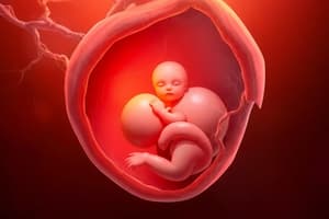
- The placenta is described as a discoidal organ, which indicates its disc-like shape. It is referred to as temporary because it forms during gestation and is expelled after birth. Functionally, it is vital as it serves as a critical vascular feto-maternal organ, connecting the developing fetus to the uterine wall of the mother. This connection enables the transfer of nutrients, gases, and wastes, playing a crucial role in fetal development.
- Its primary responsibilities include the regulation of metabolism and facilitating the exchange of essential chemical substances between the mother and the developing fetus or embryo. This includes nutrients required for growth and development and the removal of metabolic waste products.
Embryo vs. Fetus
- The terms "embryo" and "fetus" are often used interchangeably in casual conversation; however, they denote different stages of development. Technically, a fetus is defined as an embryo that has entered the fetal stage, which begins after the ninth week of development following fertilization. This distinction is important in the context of developmental biology and medicine, as various growth and developmental processes occur during these stages.
Need for Placenta
- Bird embryos typically develop inside eggs, where they access nutrients stored in the egg yolk. This yolk provides all the necessary sustenance for embryonic development while allowing the embryo to grow in a protective environment.
- In contrast, human embryos develop inside the womb, where they do not rely on egg yolk for nutrition. Instead, they obtain all essential nutrients directly from their mother's bloodstream through the placenta. This organ effectively takes on the dual role of nutrient supplier and waste disposal system, crucial for the embryo's growth and health.
- As human embryos lack significant yolk reserves, the placenta emerges as a critical requirement. It enables human embryos to not only extract necessary nutrients from the mother but also facilitates the elimination of waste materials, thus maintaining a nurturing and viable environment for fetal development.
Placenta Development Timeline
- The development of the placenta commences early in the pregnancy process. At the morula stage, which occurs shortly after fertilization, divisions of the fertilized egg result in a cluster of cells known as blastomeres. These cells will differentiate into two distinct groups: the inner cells, which will develop into the embryo proper, and the outer cells, which are designated to form structures vital for nutrient absorption and placental development.
- As the morula enters the uterine cavity, it undergoes several transformations. The zona pellucida, a protective layer surrounding the early embryo, ruptures, leading to the formation of a cavity within the morula. This stage is crucial as the inner cell mass, or embryoblast, will evolve into the embryo itself.
- On the other hand, the outer cell mass, known as the trophoblast, is responsible for developing the placental structures. The trophoblast is the major contributor to the fetal placenta, playing an active role in implantation and nutrient transfer.
- In summary, trophoblasts form the primary structural component of the fetal placenta during the early stages of development.
Implantation
- For the embryo to develop successfully, both the inner cell mass and the outer cell mass must engage in the implantation process within the endometrium, the inner lining of the uterus. Implantation is a critical step in establishing a connection between the mother's body and the developing fetus.
- Initially, the embryo establishes a loose attachment to the uterine wall using specific adhesion molecules known as selectins. This binding process aids in the initial adhesion necessary for further development and integration.
- The next phase involves a series of changes in the uterine cells, referred to as the decidual reaction. This reaction results in the formation of specialized decidual cells that alter the uterine environment in support of implantation and later fetal development.
- The outermost luminal layer of the endometrium, known as the decidua, undergoes significant modifications to facilitate implantation. This adaptation is essential for creating a conducive environment for the growing embryo.
- Once the embryo establishes itself, it binds to the uterine wall more firmly through the action of extracellular matrix proteins such as fibronectin and integrins, which help secure the embryo within the endometrium.
- Further compounding this process, trophoblast cells proliferate and amalgamate, losing their individual cell membranes to form a multinucleated structure known as the syncytium. This transformation allows the cells to function collectively.
- As part of its development, the trophoblast differentiates into two types of cells: cytotrophoblasts, which retain distinct cellular boundaries, and syncytiotrophoblasts, which lack defined cell borders and possess invasive capabilities, allowing them to erode into the uterine wall.
- Up to the end of the first week, this intricate developmental sequence occurs, establishing the foundation for the placenta and the embryo’s further growth.
Mid-Second Week Development
- By the mid-second week of development, the trophoblast is comprised of two distinct components: the syncytiotrophoblast and the cytotrophoblast, which play complementary roles in placental formation.
- The syncytiotrophoblast next begins forming a lacunar network—this structure involves the creation of small interconnected spaces that emerge as the syncytiotrophoblast invades deeper into the uterine tissue.
- Through this invasion, the syncytiotrophoblast erodes blood vessels in the maternal uterine wall, facilitating the establishment of maternal blood flow into the spaces created by the lacunar network.
- As arterial blood flows into these lacunae, it ultimately returns via venules, establishing a primitive form of uteroplacental circulation, which is essential for nutrient transfer between mother and embryo.
- The embryo, at this stage referred to as the embryo proper, consists of an amniotic cavity and a yolk sac cavity, both of which are encased within the chorionic cavity, also known as the extra embryonic coelom. This elaborate arrangement supports the ongoing development of the embryo.
- The chorion, another critical structure in this context, comprises extra embryonic somatopleuric mesoderm along with cytotrophoblast cells. Some sources also consider the syncytiotrophoblast as an integral part of the chorion, emphasizing its importance.
- Nutrient molecules diffuse into the chorionic cavity, where they are subsequently absorbed by the yolk sac and amniotic cavity, ultimately nourishing the developing bilaminar embryonic disc.
- Simultaneously, metabolically active cells in the embryo secrete waste materials into both the yolk sac and amniotic cavity. These wastes are then released into the chorionic cavity and transferred to the maternal blood for elimination.
- During this early period, the embryo relies entirely on simple diffusion to meet its nutrient needs, as there are no fully formed blood vessels available at this stage.
Decidua
- By the end of the second week of gestation, complete implantation of the embryo into the decidua is achieved. The decidua represents a modified form of the endometrium adapted to support pregnancy.
- There are three distinct types of decidua:
- Decidua capsularis: This portion of the decidua forms a capsule-like structure that covers the developing conceptus.
- Decidua basalis: This section lies underneath the conceptus and forms a basal plate that supports the placenta.
- Decidua parietalis: This portion remains unaffected and comprises the rest of the decidua that is not directly involved with the conceptus.
- Among these types, only the decidua basalis contributes to the formation of the maternal portion of the placenta. This unique contribution is essential for the establishment and function of the placenta as the pregnancy advances.
Further Placenta Development
- As the second week progresses, primary villi begin to emerge. These structures are finger-like projections that extend from the trophoblast and penetrate into the syncytiotrophoblast, essential for nutrient exchange and anchoring the placenta.
- Primary villi contain a core of extra embryonic somatopleuric mesoderm along with the projections of cytotrophoblast. This configuration is pivotal for the formation and elaboration of the placental structure.
- During the third week of gestation, extensions of the extra embryonic somatopleuric mesoderm invade the interior of the primary villus, forming a mesenchymal core, thereby converting the primary villus into a secondary villus.
- Subsequently, this mesenchyme develops into blood vessels within the villi, transforming them into tertiary villi. This progression is crucial for accommodating the increasing metabolic demands of the developing embryo.
- The blood vessels within these tertiary villi feature arteriolar ends (carrying oxygen-deficient blood) and venous ends (carrying oxygen-rich blood), facilitating efficient gas exchange and nutrient transfer.
Chorionic Villi
- Initially, chorionic villi encapsulate the embryo from all angles, which ensures optimal contact with the maternal blood supply. Over time, however, some villi along the luminal surface begin to degenerate due to diminishing blood supply.
- As development continues, the majority of chorionic villi are predominantly located on the side of the trophoblast that faces the endometrium, where they can engage directly with maternal blood and enhance nutrient transfer.
- The chorion/trophoblast exhibits two distinct anatomical regions:
- Chorionic frondosum: Characterized as a rough, leaf-like structure with numerous villi, it plays a significant role in the placenta's function.
- Chorionic laeve: This area presents a smoother surface with few to no chorionic villi, reflecting its diminished role in nutrient exchange.
- Only the chorionic frondosum is involved in contributing to the fetal component of the placenta, underscoring its importance in fetal development.
- The maternal endometrial spiral arteries open into the lacunae, allowing for material exchange between the mother and fetus. This interaction is critical for sustaining the developing fetus throughout gestation.
- After the exchange occurs in the lacunar spaces, blood is returned to the maternal circulatory system via endometrial veins, completing the cycle of maternal-fetal circulation.
Chorionic and Umbilical Vessels
- The development of chorionic vessels occurs within the extra embryonic somatopleuric mesoderm, which connects with the vascular network of the tertiary villi, thus facilitating the passage of nutrients and waste.
- By the end of the third week of pregnancy, the primordial heart and blood vessels have started to form within the embryo as well as in the wall of the yolk sac, which is derived from the extra embryonic splanchnopleuric mesoderm.
- The functioning primordial heart begins its pumping activity, marking the initiation of embryonic circulation.
- The establishment of a circulatory system becomes imperative to meet the increasing nutritional and metabolic needs of the growing embryo, as diffusion alone is no longer sufficient.
- At this stage, the blood vessels present in both the embryo and the primordial placenta are not yet connected, representing a necessary stage of vascular development.
- The connection between these vessels is established in the connecting stalk, paving the way for the formation of what will eventually become the primordial umbilical cord.
Umbilical Cord Formation
- As development progresses, a notable extension of the yolk sac into the connecting stalk forms a structure known as the allantois, which plays a significant role in waste management and respiratory exchange in embryos.
- Moreover, embryonic folding incorporates a segment of the yolk sac into the embryo, connected via a structure termed the vitelline duct (also known as the omphaloenteric duct), which is important for nutrient transfer.
- As the embryo undergoes further development, the yolk sac gradually diminishes in size, reflecting the increasing reliance on maternal nutrient supply.
- The collective structures formed from the vitelline duct, allantois, connecting stalk, and accompanying blood vessels come together to create the primitive umbilical cord, linking the mother and developing fetus.
Amniotic Cavity
- The growth of the amniotic cavity occurs, eventually surrounding the umbilical cord from all sides, providing a protective environment for the developing fetus.
- As the pregnancy progresses, the chorionic cavity becomes obliterated, leading to the amnion fusing with the chorion, creating a structure known as the amniochorion, which is integral for pregnancy maintenance.
- Both the embryo, along with its extra embryonic membranes and the primitive placenta, becomes securely implanted into the decidua, ensuring a stable attachment during the course of pregnancy.
- However, as the embryo grows, the blood supply to the decidua capsularis becomes compromised. This leads to degeneration and eventual loss of the decidua capsularis around the sixth month of development.
- Throughout development, the embryo initially grows while nestled within the maternal uterus; however, as it develops further, it continues to adapt, ultimately growing within the expanding uterine cavity.
Studying That Suits You
Use AI to generate personalized quizzes and flashcards to suit your learning preferences.