Podcast
Questions and Answers
What type of fibers stimulate dilation of the pupil in dim light?
What type of fibers stimulate dilation of the pupil in dim light?
- Parasympathetic fibers
- Motor fibers
- Sensory fibers
- Sympathetic fibers (correct)
Which type of muscle is involved in the constriction of the pupil?
Which type of muscle is involved in the constriction of the pupil?
- Cardiac muscle
- Smooth muscle (correct)
- Skeletal muscle
- Epithelial tissue
What is the function of the ciliary body in the eye?
What is the function of the ciliary body in the eye?
- To regulate blood pressure
- To transmit nerve signals
- To control the amount of light entering the eye
- To focus on near objects (correct)
What happens to the lens when the ciliary muscle contracts?
What happens to the lens when the ciliary muscle contracts?
Which artery supplies the orbit and eye?
Which artery supplies the orbit and eye?
What is the function of the central artery of the retina?
What is the function of the central artery of the retina?
Which of the following statements about the oculomotor nerve (CN III) is true?
Which of the following statements about the oculomotor nerve (CN III) is true?
Which branch of the ophthalmic artery supplies the eyelids?
Which branch of the ophthalmic artery supplies the eyelids?
Where do the postganglionic parasympathetic fibers from the ciliary ganglion course to?
Where do the postganglionic parasympathetic fibers from the ciliary ganglion course to?
What is the function of the postganglionic parasympathetic fibers that innervate the ciliary muscle?
What is the function of the postganglionic parasympathetic fibers that innervate the ciliary muscle?
What is the function of the dorsal nasal arteries?
What is the function of the dorsal nasal arteries?
Where do sympathetic preganglionic fibers ascend to synapse?
Where do sympathetic preganglionic fibers ascend to synapse?
What is the origin of sympathetic innervation to the eyeball?
What is the origin of sympathetic innervation to the eyeball?
Which muscle is innervated by postganglionic sympathetic fibers?
Which muscle is innervated by postganglionic sympathetic fibers?
What is the function of the postganglionic sympathetic fibers that innervate the dilator pupillae?
What is the function of the postganglionic sympathetic fibers that innervate the dilator pupillae?
How many dural layers cover the optic nerve (CN II)?
How many dural layers cover the optic nerve (CN II)?
What is the primary function of the bony orbit?
What is the primary function of the bony orbit?
Which of the following structures is NOT primarily located in the orbit?
Which of the following structures is NOT primarily located in the orbit?
What is the direction of the orbital openings?
What is the direction of the orbital openings?
Which of the following is a clinical correlation related to head anatomy?
Which of the following is a clinical correlation related to head anatomy?
What is the location of the apices of the orbits?
What is the location of the apices of the orbits?
What is the primary function of the lacrimal apparatus?
What is the primary function of the lacrimal apparatus?
Which cranial nerve is responsible for mediating the special sense of sight?
Which cranial nerve is responsible for mediating the special sense of sight?
What is the primary function of the postganglionic sympathetic fibers from the superior cervical ganglion?
What is the primary function of the postganglionic sympathetic fibers from the superior cervical ganglion?
Which nerve is responsible for conveying general sensory information from the orbit and eye?
Which nerve is responsible for conveying general sensory information from the orbit and eye?
What is the primary function of the lacrimal branch of CN V1?
What is the primary function of the lacrimal branch of CN V1?
Which muscle is responsible for eyelid movement and is innervated by postganglionic sympathetic fibers?
Which muscle is responsible for eyelid movement and is innervated by postganglionic sympathetic fibers?
What is the primary function of the H pattern test?
What is the primary function of the H pattern test?
Which nerve is responsible for innervating the extraocular skeletal muscles?
Which nerve is responsible for innervating the extraocular skeletal muscles?
What is the result of interruption of the sympathetic pathway to the smooth muscle of the eyelid?
What is the result of interruption of the sympathetic pathway to the smooth muscle of the eyelid?
What is the function of the trabecular meshwork?
What is the function of the trabecular meshwork?
What type of cells are responsible for low-light conditions and night vision?
What type of cells are responsible for low-light conditions and night vision?
What is the function of the ora serrata?
What is the function of the ora serrata?
What is the purpose of the fovea centralis?
What is the purpose of the fovea centralis?
What is the function of the ciliary body?
What is the function of the ciliary body?
What is the purpose of the optic disc?
What is the purpose of the optic disc?
What is the function of the iris?
What is the function of the iris?
What is the characteristic of the photoreceptor layer in the fovea centralis?
What is the characteristic of the photoreceptor layer in the fovea centralis?
Flashcards are hidden until you start studying
Study Notes
Orbits
- The orbits are pyramidal, bony cavities in the facial skeleton
- Their bases (orbital openings) are directed anterolaterally, while their apices are located posteromedially
- They contain and protect the eyeballs and their muscles, nerves, and vessels, along with most of the lacrimal apparatus
Clinical Testing of the Extraocular Muscles (EOM)
- The examiner uses an H pattern to assess how each eye tracks movement of an object
- The examiner can bring the finger directly to the midline to test convergence (medial rectus muscles of both eyes)
- If an eye movement disorder is detected, a clinical specialist may be consulted for further evaluation
Nerves in the Orbit
- Cranial nerves III, IV, and VI innervate the extra-ocular skeletal muscles
- CN II mediates the special sense of sight
- CN V1 (ophthalmic nerve) conveys general sensory information from the orbit and eye
- The oculomotor nerve (CN III) provides parasympathetic fibers, which exhibit the following features:
- Parasympathetic fibers arise centrally from the nucleus of Edinger-Westphal (preganglionic fibers) and course along CN III and its inferior division to synapse in the ciliary ganglion on postganglionic parasympathetic neurons
- Postganglionic parasympathetic fibers then course via short ciliary nerves to the eyeball
- These postganglionic fibers innervate the sphincter muscle of the pupil (sphincter pupillae) and the ciliary muscle for accommodation
- Sympathetic innervation to the eyeball is arranged as follows:
- Sympathetic innervation arises from the upper thoracic intermediolateral cell column of the spinal cord (T1-T2) and sends preganglionic fibers into the sympathetic trunk
- These fibers ascend to synapse in the superior cervical ganglion (SCG)
- Postganglionic sympathetic fibers course along the internal carotid artery, enter the orbit on the ophthalmic artery and ophthalmic nerve, and pass through the ciliary ganglion or along the long and short ciliary nerves to the eyeball
- These postganglionic fibers innervate the dilator muscle of the pupil (dilator pupillae) and the superior tarsal muscle of the upper eyelid
Anterior Segment of the Eye
- The anterior segment of the eye consists of:
- The anterior chamber between the cornea and the iris
- The posterior chamber between the iris and the lens
- Filled with aqueous humor, which is produced by the ciliary body and circulates from the posterior chamber, through the pupil, and into the anterior chamber, where it is absorbed by the trabecular meshwork into the scleral venous sinus (canal of Schlemm), at the angle of the cornea and iris
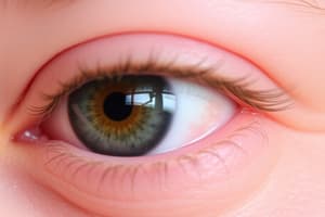
Retina
- The retina consists of:
- Nonvisual retina, which lines the internal surface of the ciliary body and iris
- Neural retina, composed of:
- An outer retinal pigmented epithelium lying adjacent to the vascular choroid
- A photosensitive region consisting of photoreceptive cells:
- Rods (night vision) are more sensitive to light and the receptors for low-light conditions (gray tones)
- Cones (daylight vision) are less sensitive to low light but very sensitive to red, green, and blue regions of the visual spectrum
- Interspersed layers of conducting and association neurons and supporting cells lie more internally in the retina, closer to the vitreous body
- The axons of ganglion cells ultimately convey the photosensory information to the optic disc, where the axons course in the optic nerve and are relayed centrally
- The optic disc is the "blind spot" because no cones or rods are present in this region of the retina
- The fovea centralis is the central focusing area and most sensitive portion of the retina, consisting only of cones, specialized for color vision and acute discrimination
Accommodation of the Lens
- The ciliary body contains smooth muscle arranged in a circular fashion like a sphincter
- When relaxed, it pulls a set of zonular fibers attached to the elastic lens taut and flattens the lens for viewing objects at some distance from the eye
- When focusing on near objects, the sphincter-like ciliary muscle (parasympathetically innervated by CN III) contracts and constricts closer to the lens, relaxing the zonular fibers and allowing the elastic lens to round up for accommodation (near vision)
Blood Supply to the Orbit and Eye
- Ophthalmic arteries arise from the internal carotid artery just as it exits the cavernous sinus, and supply the orbit and eye by the following branches:
- Central artery of the retina: travels in the optic nerve; occlusion leads to blindness
- Short and long posterior ciliary arteries: pierce the sclera and supply the ciliary body, iris, and choroid
- Lacrimal arteries: supply the gland, conjunctiva, and eyelids
- Ethmoidal arteries: supply the ethmoid and frontal sinuses, nasal cavity, and external anterior nose
- Medial palpebrae arteries: supply the eyelids
- Muscular arteries: supply skeletal muscles of the orbit and smooth muscles of the eyeball
- Dorsal nasal arteries: supply the lateral nose and lacrimal sac
- Supra-orbital artery: passes through supra-orbital notch and supplies the forehead and scalp
- Supratrochlear artery: supplies the forehead and scalp
Studying That Suits You
Use AI to generate personalized quizzes and flashcards to suit your learning preferences.