Podcast
Questions and Answers
A 55-year-old male presents with insidious onset shoulder pain, exhibiting a capsular pattern restriction and reports significant night pain disrupting sleep. Examination reveals intact rotator cuff strength but limited external rotation. Given these findings, which intervention is MOST appropriate at this stage?
A 55-year-old male presents with insidious onset shoulder pain, exhibiting a capsular pattern restriction and reports significant night pain disrupting sleep. Examination reveals intact rotator cuff strength but limited external rotation. Given these findings, which intervention is MOST appropriate at this stage?
- Aggressive end-range stretching exercises combined with eccentric strengthening of the rotator cuff.
- Administration of local anesthetic and commencement of translational manipulation under anesthesia to restore ROM.
- Pendulum exercises with gentle GH joint distraction and Grade I/II joint mobilizations to address pain and maintain joint integrity. (correct)
- Initiation of high-velocity, low-amplitude (HVLA) thrust mobilizations to address capsular restrictions.
A patient diagnosed with adhesive capsulitis is in the 'frozen' stage. Which clinical presentation is MOST indicative of this stage, guiding the physical therapist's intervention strategy?
A patient diagnosed with adhesive capsulitis is in the 'frozen' stage. Which clinical presentation is MOST indicative of this stage, guiding the physical therapist's intervention strategy?
- Significant, intense pain even at rest, with motion limited in all directions and inability to restore motion with intra-articular injection.
- Mild discomfort with painful end ranges of motion, primarily affecting external rotation and abduction.
- Minimal pain with significant capsular restrictions, and gradual improvement in motion observed over time.
- Large decrease in pain, experienced predominantly with movement, coupled with limited glenohumeral motion and marked scapular substitutions. (correct)
A patient is diagnosed with glenohumeral joint hypomobility secondary to immobilization following a distal radius fracture. Which of the following findings would be LEAST likely to be directly attributable to the GH joint hypomobility itself?
A patient is diagnosed with glenohumeral joint hypomobility secondary to immobilization following a distal radius fracture. Which of the following findings would be LEAST likely to be directly attributable to the GH joint hypomobility itself?
- Decreased arm swing during gait.
- Night pain and disturbed sleep during acute flares.
- Impaired grip strength in the ipsilateral hand. (correct)
- Protracted and anteriorly tilted scapula on the affected side.
A physical therapist is developing a non-operative management plan for a patient in the acute phase of glenohumeral joint hypomobility. Considering the APTA's Clinical Practice Guideline for Adhesive Capsulitis (2013), what combination of interventions would be MOST appropriate?
A physical therapist is developing a non-operative management plan for a patient in the acute phase of glenohumeral joint hypomobility. Considering the APTA's Clinical Practice Guideline for Adhesive Capsulitis (2013), what combination of interventions would be MOST appropriate?
A patient in Stage 4 ('Thawing' stage) of idiopathic frozen shoulder reports minimal pain but continues to exhibit significant capsular restrictions. Which of the following exercise progressions would be MOST appropriate to restore functional range of motion, while mitigating the risk of exacerbating symptoms?
A patient in Stage 4 ('Thawing' stage) of idiopathic frozen shoulder reports minimal pain but continues to exhibit significant capsular restrictions. Which of the following exercise progressions would be MOST appropriate to restore functional range of motion, while mitigating the risk of exacerbating symptoms?
A patient post-rTSA presents at 8 weeks with satisfactory deltoid activation and AAROM tolerance, but exhibits compensatory scapulothoracic movements during attempted glenohumeral abduction within the scapular plane. Which intervention would be MOST appropriate?
A patient post-rTSA presents at 8 weeks with satisfactory deltoid activation and AAROM tolerance, but exhibits compensatory scapulothoracic movements during attempted glenohumeral abduction within the scapular plane. Which intervention would be MOST appropriate?
A patient undergoing rehabilitation following a TSA with RTC repair is currently in the maximum protection phase. Considering the specific constraints dictated by the tendon repair, which of the following interventions is LEAST appropriate at this stage?
A patient undergoing rehabilitation following a TSA with RTC repair is currently in the maximum protection phase. Considering the specific constraints dictated by the tendon repair, which of the following interventions is LEAST appropriate at this stage?
A patient 6 weeks post-rTSA is demonstrating consistent pain (VAS 4/10) with active assisted elevation beyond 75 degrees, accompanied by audible crepitus upon palpation of the scapulothoracic articulation. Radiographic imaging reveals no hardware complications. What is MOST likely contributing to the patient's symptomatic presentation?
A patient 6 weeks post-rTSA is demonstrating consistent pain (VAS 4/10) with active assisted elevation beyond 75 degrees, accompanied by audible crepitus upon palpation of the scapulothoracic articulation. Radiographic imaging reveals no hardware complications. What is MOST likely contributing to the patient's symptomatic presentation?
A patient presents 10 weeks following a rTSA. Examination reveals 110 degrees of active elevation, 30 degrees of active external rotation, and 60 degrees of active internal rotation at the side. The patient's primary complaint is decreased functional reach overhead and difficulty with activities such as reaching into a high cupboard. Based on this presentation, what modification to the patient's exercise program would be MOST justifiable?
A patient presents 10 weeks following a rTSA. Examination reveals 110 degrees of active elevation, 30 degrees of active external rotation, and 60 degrees of active internal rotation at the side. The patient's primary complaint is decreased functional reach overhead and difficulty with activities such as reaching into a high cupboard. Based on this presentation, what modification to the patient's exercise program would be MOST justifiable?
During a follow-up examination 14 weeks post-TSA (without RTC repair), a patient reports experiencing a sudden onset of sharp pain and instability in the operated shoulder while attempting to lift a 10-pound object. Examination reveals palpable crepitus upon gentle palpation of the long head of the biceps tendon within the bicipital groove. What is the MOST appropriate immediate course of action?
During a follow-up examination 14 weeks post-TSA (without RTC repair), a patient reports experiencing a sudden onset of sharp pain and instability in the operated shoulder while attempting to lift a 10-pound object. Examination reveals palpable crepitus upon gentle palpation of the long head of the biceps tendon within the bicipital groove. What is the MOST appropriate immediate course of action?
A 28-year-old competitive swimmer presents with multidirectional instability of the glenohumeral joint, primarily anterior and inferior. Pre-operative assessment reveals significant capsular redundancy. Considering the established surgical procedures for GH joint stabilization, which of the following approaches would be MOST judicious?
A 28-year-old competitive swimmer presents with multidirectional instability of the glenohumeral joint, primarily anterior and inferior. Pre-operative assessment reveals significant capsular redundancy. Considering the established surgical procedures for GH joint stabilization, which of the following approaches would be MOST judicious?
In the context of open Bankart repair for anterior glenohumeral instability, meticulous surgical technique often involves addressing the subscapularis muscle. Which of the following statements BEST encapsulates the contemporary surgical considerations regarding the subscapularis during this procedure?
In the context of open Bankart repair for anterior glenohumeral instability, meticulous surgical technique often involves addressing the subscapularis muscle. Which of the following statements BEST encapsulates the contemporary surgical considerations regarding the subscapularis during this procedure?
A patient, 14 weeks post-operative following a total shoulder arthroplasty (TSA), demonstrates pain-free PROM with flexion to 135 degrees, abduction to 125 degrees, ER to 65 degrees, and IR to 75 degrees in the plane of the scapula. Active elevation against gravity is 110 degrees with appropriate mechanics. MMT reveals 4+/5 strength in RTC and deltoid. According to the criteria, what is the MOST appropriate progression for this patient?
A patient, 14 weeks post-operative following a total shoulder arthroplasty (TSA), demonstrates pain-free PROM with flexion to 135 degrees, abduction to 125 degrees, ER to 65 degrees, and IR to 75 degrees in the plane of the scapula. Active elevation against gravity is 110 degrees with appropriate mechanics. MMT reveals 4+/5 strength in RTC and deltoid. According to the criteria, what is the MOST appropriate progression for this patient?
A patient undergoing electrothermally assisted capsulorrhaphy for glenohumeral instability reports persistent pain and stiffness six months postoperatively. MRI reveals regions of capsular fibrosis and restricted range of motion. Which cellular mechanism is MOST implicated in the observed capsular changes following this procedure?
A patient undergoing electrothermally assisted capsulorrhaphy for glenohumeral instability reports persistent pain and stiffness six months postoperatively. MRI reveals regions of capsular fibrosis and restricted range of motion. Which cellular mechanism is MOST implicated in the observed capsular changes following this procedure?
A patient, 10 weeks post-op from rTSA, presents with the following limitations: passive flexion to 90 degrees, abduction to 70 degrees, external rotation to 30 degrees, and internal rotation to 40 degrees. Active elevation is limited to 60 degrees against gravity with noticeable scapular hiking. MMT shows 3/5 strength for the deltoid and RTC. Which of the following interventions would be MOST appropriate at this stage?
A patient, 10 weeks post-op from rTSA, presents with the following limitations: passive flexion to 90 degrees, abduction to 70 degrees, external rotation to 30 degrees, and internal rotation to 40 degrees. Active elevation is limited to 60 degrees against gravity with noticeable scapular hiking. MMT shows 3/5 strength for the deltoid and RTC. Which of the following interventions would be MOST appropriate at this stage?
A high-level overhead athlete is considering capsulolabral reconstruction following recurrent anterior shoulder instability events. He is particularly concerned about the potential impact of the surgery on his proprioceptive acuity and subsequent athletic performance. Which of the following statements BEST reflects the current understanding of the relationship between capsulolabral repair and proprioception in the glenohumeral joint?
A high-level overhead athlete is considering capsulolabral reconstruction following recurrent anterior shoulder instability events. He is particularly concerned about the potential impact of the surgery on his proprioceptive acuity and subsequent athletic performance. Which of the following statements BEST reflects the current understanding of the relationship between capsulolabral repair and proprioception in the glenohumeral joint?
A Physical Therapist is designing a home exercise program for a patient following a TSA. The patient is currently in the 'Minimum Protection/Return to Function Phase'. Which of the following exercises would be MOST appropriate to include in the program to address both scapular and glenohumeral joint stabilization?
A Physical Therapist is designing a home exercise program for a patient following a TSA. The patient is currently in the 'Minimum Protection/Return to Function Phase'. Which of the following exercises would be MOST appropriate to include in the program to address both scapular and glenohumeral joint stabilization?
During an arthroscopic Bankart repair, a surgeon encounters a significant glenoid bone defect along the anterior-inferior rim, exceeding 25% of the glenoid width. Concurrently, the patient exhibits Hill-Sachs lesion involving approximately 30% of the humeral head. What is the MOST appropriate surgical strategy to address the combined glenoid bone loss and humeral head defect in this patient?
During an arthroscopic Bankart repair, a surgeon encounters a significant glenoid bone defect along the anterior-inferior rim, exceeding 25% of the glenoid width. Concurrently, the patient exhibits Hill-Sachs lesion involving approximately 30% of the humeral head. What is the MOST appropriate surgical strategy to address the combined glenoid bone loss and humeral head defect in this patient?
A patient status post-reverse total shoulder arthroplasty (rTSA) demonstrates limited active elevation. Considering the altered biomechanics of the rTSA, which muscle's strengthening should be prioritized to MOST effectively improve active elevation?
A patient status post-reverse total shoulder arthroplasty (rTSA) demonstrates limited active elevation. Considering the altered biomechanics of the rTSA, which muscle's strengthening should be prioritized to MOST effectively improve active elevation?
When comparing Total Shoulder Arthroplasty (TSA) and Reverse Total Shoulder Arthroplasty (rTSA), which of the following statements BEST characterizes a key biomechanical difference affecting rehabilitation strategies?
When comparing Total Shoulder Arthroplasty (TSA) and Reverse Total Shoulder Arthroplasty (rTSA), which of the following statements BEST characterizes a key biomechanical difference affecting rehabilitation strategies?
Flashcards
Stage 1: Pre-adhesive Stage
Stage 1: Pre-adhesive Stage
Characterized by mild synovitis, gradual pain increase, and intact RTC strength. Duration is less than 3 months.
Stage 2: Freezing Stage
Stage 2: Freezing Stage
Marked by thickened synovitis, intense pain at rest, and limited motion in all directions. Lasts 3-9 months.
Stage 3: Frozen Stage
Stage 3: Frozen Stage
Features mature fibrosis with reduced pain, but limited GH motion. Duration is 9-15 months.
Stage 4: Thawing Stage
Stage 4: Thawing Stage
Signup and view all the flashcards
Non-operative Management Acute Phase
Non-operative Management Acute Phase
Signup and view all the flashcards
TSA Recovery Timeline
TSA Recovery Timeline
Signup and view all the flashcards
rTSA Lifting Restriction
rTSA Lifting Restriction
Signup and view all the flashcards
Shoulder ROM Progression
Shoulder ROM Progression
Signup and view all the flashcards
Submaximal Isometrics
Submaximal Isometrics
Signup and view all the flashcards
Dynamic Stability Exercises
Dynamic Stability Exercises
Signup and view all the flashcards
Criteria for Minimal Protection Phase
Criteria for Minimal Protection Phase
Signup and view all the flashcards
Minimum Protection Phase Duration
Minimum Protection Phase Duration
Signup and view all the flashcards
Goals of Minimum Protection Phase
Goals of Minimum Protection Phase
Signup and view all the flashcards
Active Elevation Requirement
Active Elevation Requirement
Signup and view all the flashcards
MMT Requirement for Strength
MMT Requirement for Strength
Signup and view all the flashcards
Capsulorrhaphy (Capsular Shift)
Capsulorrhaphy (Capsular Shift)
Signup and view all the flashcards
Electrothermally Assisted Capsulorrhaphy
Electrothermally Assisted Capsulorrhaphy
Signup and view all the flashcards
Bankart Repair
Bankart Repair
Signup and view all the flashcards
Postoperative Management of Capsulorrhaphy
Postoperative Management of Capsulorrhaphy
Signup and view all the flashcards
Capsulo-labral Reconstruction
Capsulo-labral Reconstruction
Signup and view all the flashcards
Study Notes
Therapeutic Exercise II: The Shoulder and Shoulder Girdle (Part 1 of 2)
- This course covers the anatomy and function of the shoulder girdle, common shoulder pathologies, and effective therapeutic exercise program design to manage musculoskeletal lesions.
Road Map
- Students should be able to describe the anatomy and function of the shoulder girdle.
- Students should understand common shoulder pathologies (e.g., hypomobility, hypermobility, soft tissue overuse conditions) that benefit from therapeutic exercise.
- Students should be able to effectively teach and progress/regress a therapeutic exercise program to manage musculoskeletal, soft tissue, and joint lesions, based on tissue healing stages.
- Students should be able to list common surgical procedures and precautions for shoulder pathologies, including hypomobility and instability.
- Students should understand the goals and appropriate interventions for post-operative shoulder and shoulder girdle dysfunction in each phase of rehabilitation.
- Students should demonstrate exercise progressions to improve range of motion (ROM), muscle performance, and functional use of the shoulder and shoulder girdle.
Outline
- Structure and Function of the Shoulder Girdle
- Hypomobility: Non-operative management and TSA
- Hypermobility: Instability of the GH joint
- Painful Biomechanical Shoulder Syndromes
Structure and Function of the Shoulder Girdle
- Joints of the shoulder girdle complex
- Shoulder girdle function
- Force couples of the shoulder complex
- Balance, mobility, and stability are crucial for the shoulder complex.
- Specific aspects of glenohumeral joint stability
Shoulder Girdle Complex
- Synovial Joints
- Glenohumeral (GH) joint
- Acromioclavicular (AC) joint
- Sternoclavicular (SC) joint
- Functional Articulations
- Scapulothoracic articulation (ST)
Anatomy Review
- The glenohumeral joint, acromioclavicular joint, and sternoclavicular joint comprise the shoulder girdle complex.
- The key anatomical parts are the clavicle, scapula, humerus, sternum and related structures.
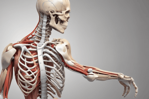
Glenohumeral Joint
- Incongruous ball and socket joint
- Triaxial
- Lax joint capsule
- Supported by:
- rotator cuff (RTC)
- glenohumeral ligaments
- coracohumeral ligaments
- Glenoid fossa: shallow, deepened by fibrocartilaginous lip (labrum)
- Humeral head is larger than glenoid fossa
- Function: deepens fossa for joint congruency and creates a negative intraarticular vacuum effect. Serves as attachment site for capsule.
Glenohumeral Joint: Stability
- Provided by static and dynamic restraints.
- Static restraints: bony anatomy, ligaments, glenoid labrum, adhesive/cohesive forces
- Dynamic restraints: RTC tendons blend with ligaments and glenoid labrum, providing stability through contraction.
- Coordinated response of the RTC muscles (supraspinatus, infraspinatus, teres minor, subscapularis)
Long Head of the Biceps & Long Head of the Triceps
- Important in GH joint stability.
- Long head of biceps: stabilizes against humeral elevation and contributes to anterior stability.
- Long head of triceps: provides support for inferior shoulder.
Acromioclavicular Joint
- Plane, triaxial joint
- May or may not have a disc
- Reinforced by superior and inferior AC ligaments
- Stability: supported by the coracoclavicular ligament.
- No direct dynamic support
Acromioclavicular Joint: Arthrokinematics
- Acromion: concave facet
- Clavicle: convex facet
- Scapular motions affecting the AC joint: upward/downward rotation, winging of the vertebral border, tipping of the inferior angle.
Sternoclavicular Joint
- Incongruent, triaxial, saddle-shaped joint with a disc.
- Stability provided by: anterior and posterior SC ligaments, interclavicular ligaments, costoclavicular ligaments.
- Movement of the clavicle results from scapular motions (e.g., elevation, depression, protraction, retraction).
Clavicular Elevation and Rotation
- Posterior rotation of the clavicle occurs as an accessory motion when the humerus is elevated above 90 degrees, and the scapula is upward rotating.
- Coracoclavicular ligament becomes taut to provide passive stability.
Scapulothoracic Articulation
- Motions of the scapula: elevation, depression, adduction, abduction, upward/downward rotation, protraction, and retraction.
Subacromial Space/Suprahumeral Space
- Coracoacromial arch: formed by acromion and coracoacromial ligament.
- Contents: subacromial/subdeltoid bursa, supraspinatus tendon, and part of the muscle, long head of biceps tendon.
- Compromise of this space = impingement syndrome.
Shoulder Girdle Function
- Review Force Couples: deltoid-rotator cuff, trapezius/serratus anterior, and anterior/posterior rotator cuff.
- Understanding why each force couple is important, and what happens if there is an imbalance.
Deltoid-RTC Relationship
- Deltoid leads to upward translation of the humerus (impingement).
- Downward force/translation/stabilization produced by infraspinatus, teres minor and subscapularis.
- Supraspinatus: significant stabilizing, compressive and slight upward translation effects on humerus with UE elevation.
Trapezius/Serratus Anterior
- Work together to create upward rotation of the scapula.
- Assists in elevation of the UE.
- Lack of function results in increased impingement.
Anterior/Posterior RTC
- Helps keep the humeral head properly positioned in the glenoid.
- Includes subscapularis, infraspinatus, and teres minor.
- Opposes anterior-posterior glenohumeral joint translation.
Shoulder Girdle Function: Failure
- Interruption of deltoid-RTC coordination
- Soft tissues overuse or compression
- Lead to shoulder dysfunction
Scapulohumeral Rhythm
- 3 phases that assist with 180 degrees of elevation.
- General 2:1 ratio (GH joint vs. scapula).
- Discrepancies in ratio 1:1 vs 2:1
Scapulohumeral Rhythm (Phases)
- Phase 1 (0-30° of abduction/ 0-60° flexion): primarily GH motion, scapula finding stable position.
- Phase 2 (30-90°): improved joint congruency as the length-tension relationship of muscles is maintained, nearing 1:1 ratio.
- Phase 3 (last 90°): dominated by glenohumeral motion; GH joint must ER to clear the subacromial arch.
Review Questions
- How does forward head and shoulder position affect the scapula and glenohumeral joint position?
- What are the various motions of the scapula?
- What are the major downward and upward rotators of the scapula?
- What is the importance of the long head of biceps and triceps?
- Why is ER of the humerus with shoulder elevation important?
Shoulder Pathology and Management
- The section outlines common shoulder disorders and surgical approaches.
- It covers non-operative and surgical management of each condition.
- Specific pathologies discussed include hypomobility and instability and painful shoulder syndromes.
- The interventions and procedures for each condition are detailed.
Shoulder Pathologies - General Interventions
- Hypomobility: Restore mobility/range of motion
- Hypermobility: Restore stability (strength)
- Painful Disorders: Restore proper biomechanics.
GH Joint Pathologies: Management
- Initial treatment: conservative approaches to avoid surgery.
- Treatment involves pharmacology, physical therapy, and therapeutic exercise.
- Surgical intervention may be considered if symptoms worsen.
Joint Hypomobility: Non-operative Management
- Hypomobility can occur in any of the shoulder complex joints: glenohumeral (GH) joint, acromioclavicular (AC) joint, sternoclavicular (SC) joints.
- Possible causes of restricted GH joint mobility include rheumatoid arthritis, prolonged immobilization, traumatic arthritis, osteoarthritis, and idiopathic frozen shoulder.
GH Joint Hypomobility: Interventions
- Acute Phase (Protection): Control pain, edema, muscle guarding, maintain soft tissue and joint integrity and mobility (e.g., pendulum exercises, PROM, passive joint distractions, Grade I/II joint mobilizations, gentle muscle setting).
- Subacute Phase (Controlled Motion): Control pain, edema, joint effusion, functional activities, AAROM to AROM progression, gradually increasing joint and soft tissue mobility, passive joint mobilization, self-mobilization, manual stretching, self-stretching.
- Chronic Phase (Return to Function): progressively incrementing flexibility and strength, functional activities (sport-specific training, work-specific training).
GH Arthroplasty: Post Operative Management
- Maximum Protection Phase: Day 1-6 weeks, Hospitalization overview, Control pain and inflammation, Maintain adjacent joint mobility, Restore and maintain shoulder mobility, Minimize muscle inhibition, guarding and atrophy.
- Precautions: PROM/AAROM only, rTSA: ER to neutral, TSA: ER <30 degrees, Avoid extension/horizontal abduction beyond neutral, No combined extension/adduction/IR, avoid lifting w/ the arm, and No driving for 4-6 weeks.
- Gentle Post-op AAROM Exercise (Gear Shift Exercise)
GH Arthroplasty: Post Operative Management
- Criteria for Progression to Moderate Protection/Controlled Motion Phase: Pain-free PROM/AAROM, Flexion to 130-140 degrees, Abduction to 120 degrees, ER to 60, IR to 70 degrees in the scapular plane, Active elevation, and MMT of 4/5 for RTC and deltoids.
- Minimum Protection/Return to Function Phase: 12-16 weeks, continue improving or maintaining shoulder mobility, improve neuromuscular control and muscle performance, and return to most functional activities.
- rTSA Special Considerations, No WB through the arm, Pain free submaximal isometrics, Dynamic low resistance exercises, and Late in phase progresses to scapular and shoulder muscle strengthening. Isometric stability exercises.
Shoulder Instability (Hypermobility):
- Atraumatic hypermobility: Generalized connective tissue laxity, repetitive microtrauma.
- Traumatic hypermobility: high force events compromise stabilizing structures, often resulting in dislocation (unidirectional or multidirectional).
- Causes: Physiologic laxity of connective tissue, repetitive microtrauma due to non-uniform loading of the joint, and the humeral head continuing to dislocate, leading to degeneration of tissue eventually.
- Anterior Dislocation: Excessive humeral head translation due to the forces against the arm in abduction and external rotation.
- Posterior Dislocation: Repetitive or forceful forces against a forward-flexed, adducted, internally rotated humerus.
- Inferior Dislocation: Primarily caused by RTC weakness, most commonly hemiplegia.
- Multidirectional Instability: GH joint stability compromised in more than one direction.
Recurrent Dislocations
- Significant ligamentous and capsular laxity: any motion that reproduces original instability.
- Voluntary dislocations: worse prognosis, minimal discomfort, and can dislocate without apprehension.
Closed Reduction of Anterior/Posterior Dislocations: Management
- Acute Phase (protection): Protect healing tissue, promote tissue health.
- Subacute Phase (controlled motion): Provide protection, increase shoulder mobility, increase stability/strength of RTC and scapular muscles.
- Chronic Phase (return to function): Restore functional control, return to activity.
- Important guidelines for posterior dislocations: Avoid humeral flexion with adduction and IR, and consider contraindications.
Muscle Activation Evidence (Exercises):
- Shoulder external/internal rotation/abduction, and scapular movements.
- Specific exercises for serratus anterior, lower trapezius, rhomboids, and levator scapulae, are highlighted.
Painful Shoulder Syndromes: (Nonsurgical approach)
- Impingement: Mechanical compression/irritation of the RTC and subacromial bursa. Painful arc with overhead reaching, positive impingement tests.
- Intrinsic Factors: vascular changes, tissue tension overload, and collagen disorientation/degeneration.
- Extrinsic Factors: Mechanical wearing of the RTC against the anterior/inferior 1/3 of the acromion caused by issues in the anatomy or biomechanical factors.
- Acromion shape: Type III = extrinsic impingement, Type I/II = mostly intrinsic.
- Faulty posture: Slouching, forward head, thoracic kyphosis, alters scapular/humeral muscle relationship and mechanics, leading to impingement/pain.
- Tendonitis/Bursitis: pain/inflammation of tendons/bursa of the rotator cuff, bicipital groove, subdeltoid/subacromial bursa.
Review (Important Questions)
- What are typical patient complaints with GH impingement?
- What are appropriate exercises for subdeltoid, proximal bicipital, or supraspinatus tendonitis in the acute phase of healing?
- What are patient complaints with shoulder impingement?
- What are specific exercises that would be appropriate for acute phase or subacute phase healing for these conditions?
Studying That Suits You
Use AI to generate personalized quizzes and flashcards to suit your learning preferences.