Podcast
Questions and Answers
In the absence of light, the influx of calcium ions ($Ca^{2+}$) into the rod cells leads to the inhibition of neurotransmitter release.
In the absence of light, the influx of calcium ions ($Ca^{2+}$) into the rod cells leads to the inhibition of neurotransmitter release.
False (B)
The pigmented epithelium plays a crucial role in vision by:
The pigmented epithelium plays a crucial role in vision by:
- Refracting incoming light to focus it onto the retina.
- Enhancing light absorption and preventing light scatter within the eye. (correct)
- Converting light energy into neural signals.
- Providing structural support to the photoreceptor cells.
Explain how light energy is converted into a brain signal during phototransduction, detailing major steps.
Explain how light energy is converted into a brain signal during phototransduction, detailing major steps.
Light reduces cGMP levels, closing Na+ channels, hyperpolarizing the rod, reducing $Ca^{2+}$ influx, and halting neurotransmitter release.
In the scotopic system, the pigment ______ in rods is responsible for absorbing light, and its deactivation under excessive light requires a ______ minute reset period.
In the scotopic system, the pigment ______ in rods is responsible for absorbing light, and its deactivation under excessive light requires a ______ minute reset period.
Match the visual system to the correct description:
Match the visual system to the correct description:
How does the circuitry differ in the photopic system compared to the scotopic system, and what is the functional consequence of this difference?
How does the circuitry differ in the photopic system compared to the scotopic system, and what is the functional consequence of this difference?
The fovea, located within the macula, is predominantly populated by rods, making it specialized for peripheral vision in low-light conditions.
The fovea, located within the macula, is predominantly populated by rods, making it specialized for peripheral vision in low-light conditions.
Describe the distribution of rods and cones in the retina, and how does this distribution contribute to the characteristics of central versus peripheral vision?
Describe the distribution of rods and cones in the retina, and how does this distribution contribute to the characteristics of central versus peripheral vision?
Unlike cones, rods are specialized to only absorb light in the ______.
Unlike cones, rods are specialized to only absorb light in the ______.
Which of the following statements best describes the function of Opsin?
Which of the following statements best describes the function of Opsin?
Which of the following best describes the functional consequence of the optic disc lacking photoreceptors?
Which of the following best describes the functional consequence of the optic disc lacking photoreceptors?
The inner nuclear layer of the retina is primarily composed of ganglion cells, which are responsible for transmitting action potentials to the brain.
The inner nuclear layer of the retina is primarily composed of ganglion cells, which are responsible for transmitting action potentials to the brain.
Explain how the ratio of photoreceptors to bipolar cells to ganglion cells differs in bright light versus dim light, and why this difference is functionally important.
Explain how the ratio of photoreceptors to bipolar cells to ganglion cells differs in bright light versus dim light, and why this difference is functionally important.
The central area of the retina responsible for sharp, detailed vision, densely packed with cones, is known as the ______.
The central area of the retina responsible for sharp, detailed vision, densely packed with cones, is known as the ______.
Match the retinal layer with its primary cellular component:
Match the retinal layer with its primary cellular component:
Which of the following best describes the path of light as it enters the eye and interacts with different layers of the retina?
Which of the following best describes the path of light as it enters the eye and interacts with different layers of the retina?
The fovea, located within the optic disc, has the highest concentration of cones and is responsible for the sharpest vision.
The fovea, located within the optic disc, has the highest concentration of cones and is responsible for the sharpest vision.
Describe the function of horizontal and amacrine cells in the retina, and explain how they contribute to visual processing.
Describe the function of horizontal and amacrine cells in the retina, and explain how they contribute to visual processing.
Axons from retinal ______ cells form the optic nerve, which carries visual information to the brain, exiting the eye at the optic disc.
Axons from retinal ______ cells form the optic nerve, which carries visual information to the brain, exiting the eye at the optic disc.
What is the functional significance of the pigmented epithelium's location as the deepest layer of the retina?
What is the functional significance of the pigmented epithelium's location as the deepest layer of the retina?
Which of the following statements accurately describes the relationship between the ciliary body and the lens during accommodation for near vision?
Which of the following statements accurately describes the relationship between the ciliary body and the lens during accommodation for near vision?
A point of light in the right nasal hemifield is processed by which of the following pathways?
A point of light in the right nasal hemifield is processed by which of the following pathways?
The primary function of the cornea is to adjust focal length for focusing on objects at varying distances, similar to the lens.
The primary function of the cornea is to adjust focal length for focusing on objects at varying distances, similar to the lens.
Explain how the interplay between the parasympathetic and sympathetic nervous systems affects pupil size, and what muscles each system controls.
Explain how the interplay between the parasympathetic and sympathetic nervous systems affects pupil size, and what muscles each system controls.
Information from the left visual hemifield is exclusively processed by the left hemisphere of the brain.
Information from the left visual hemifield is exclusively processed by the left hemisphere of the brain.
Light rays from objects closer than 9 meters are considered __________, requiring the lens and iris to adjust for clear vision.
Light rays from objects closer than 9 meters are considered __________, requiring the lens and iris to adjust for clear vision.
What is the primary function of the optic chiasm in visual processing?
What is the primary function of the optic chiasm in visual processing?
The densest concentration of cones is found in the ______.
The densest concentration of cones is found in the ______.
Match each component of the eye with its primary function.
Match each component of the eye with its primary function.
What is the key difference between photopic and scotopic vision?
What is the key difference between photopic and scotopic vision?
Match each retinal location with its corresponding density of photoreceptors:
Match each retinal location with its corresponding density of photoreceptors:
The ophthalmic artery and vein are located within the sclera, ensuring direct blood supply to the outer layers of the eye.
The ophthalmic artery and vein are located within the sclera, ensuring direct blood supply to the outer layers of the eye.
Which layers of the LGN (lateral geniculate nucleus) receive input from the temporal retina?
Which layers of the LGN (lateral geniculate nucleus) receive input from the temporal retina?
The optic tract carries visual information from the retina directly to the cortex.
The optic tract carries visual information from the retina directly to the cortex.
Explain how the ciliary body facilitates both near and distant vision through its actions on the lens and zonular fibers.
Explain how the ciliary body facilitates both near and distant vision through its actions on the lens and zonular fibers.
The anterior chamber of the eye is located between the __________ and the lens, and it is filled with fluid.
The anterior chamber of the eye is located between the __________ and the lens, and it is filled with fluid.
What type of ganglion cell primarily activates the alpha layer within layer 4C of the visual cortex, and what sensory information does it process?
What type of ganglion cell primarily activates the alpha layer within layer 4C of the visual cortex, and what sensory information does it process?
The nasal part of the visual field is ______, meaning it is seen by both eyes.
The nasal part of the visual field is ______, meaning it is seen by both eyes.
Which layer of the eye contains the photoreceptor cells responsible for converting light into neural signals?
Which layer of the eye contains the photoreceptor cells responsible for converting light into neural signals?
If there was damage to someone's left optic tract, what visual field deficits would you expect?
If there was damage to someone's left optic tract, what visual field deficits would you expect?
Flashcards
Vitreous Humor
Vitreous Humor
Gel-like substance filling the space behind the lens in the posterior chamber of eye
Macula
Macula
The central area of the retina for detailed vision.
Cones
Cones
Photoreceptor cells that detect color in the Photopic System of retina
Fovea
Fovea
Signup and view all the flashcards
Optic Disc (Blind Spot)
Optic Disc (Blind Spot)
Signup and view all the flashcards
Retinal Ganglion Cells
Retinal Ganglion Cells
Signup and view all the flashcards
Ganglion Cell Layer
Ganglion Cell Layer
Signup and view all the flashcards
Inner Nuclear Layer
Inner Nuclear Layer
Signup and view all the flashcards
Outer Nuclear Layer
Outer Nuclear Layer
Signup and view all the flashcards
Horizontal Cells
Horizontal Cells
Signup and view all the flashcards
Pigmented Epithelium
Pigmented Epithelium
Signup and view all the flashcards
Rod Activity in Darkness
Rod Activity in Darkness
Signup and view all the flashcards
Rod Activity in Light
Rod Activity in Light
Signup and view all the flashcards
Phototransduction
Phototransduction
Signup and view all the flashcards
Scotopic System
Scotopic System
Signup and view all the flashcards
Rhodopsin
Rhodopsin
Signup and view all the flashcards
Photopic System
Photopic System
Signup and view all the flashcards
Opsin
Opsin
Signup and view all the flashcards
Photopic Vision
Photopic Vision
Signup and view all the flashcards
Scotopic Vision
Scotopic Vision
Signup and view all the flashcards
Refracting Light
Refracting Light
Signup and view all the flashcards
Diverging Light
Diverging Light
Signup and view all the flashcards
Accommodation (Eye)
Accommodation (Eye)
Signup and view all the flashcards
Iris Function
Iris Function
Signup and view all the flashcards
Sclera
Sclera
Signup and view all the flashcards
Optic Nerve
Optic Nerve
Signup and view all the flashcards
Choroid
Choroid
Signup and view all the flashcards
Ciliary Body
Ciliary Body
Signup and view all the flashcards
Rod Distribution
Rod Distribution
Signup and view all the flashcards
Cone Distribution
Cone Distribution
Signup and view all the flashcards
Central Vision
Central Vision
Signup and view all the flashcards
Hemifield
Hemifield
Signup and view all the flashcards
Nasal Hemifield
Nasal Hemifield
Signup and view all the flashcards
Temporal Hemifield
Temporal Hemifield
Signup and view all the flashcards
Thalamus
Thalamus
Signup and view all the flashcards
Optic Tract
Optic Tract
Signup and view all the flashcards
Optic Chiasm
Optic Chiasm
Signup and view all the flashcards
Study Notes
Distribution of Rods
- As you get closer to the Macula, rods become denser.
Distribution of Cones
- Cones are densest in the macula.
- Cones are most dense in the middle, called the fovea.
Visual Words
- Central vision relates to the middle of the world.
- Hemifield is half of visual world when looking at the nose.
- The left side correlates with the left hemifield.
- A point of light in the left temporal hemifield is focused onto the left nasal retina.
- A point of light in the left nasal hemifield is focused on the right temporal retina.
- The right side correlates with the right hemifield.
- A point of light in the right temporal hemifield is focused on the right nasal retina.
- A point of light in the right nasal hemifield is focused on the left temporal retina.
- The nasal part of the retina can be seen with both eyes and is binocular.
- The temporal part of the retina is closer to the ears and is monocular.
- The left visual world is processed by the right brain.
- The right visual world is processed by the left brain.
Retina to Brain (Thalamus) Pathway
- The optic nerve is exiting the retina.
- The optic tract goes into the thalamus.
- The optic chiasm allows information to cross over.
Decussation
- The left temporal hemifield focuses on the left nasal retina.
- Decussation must occur for the left temporal hemifield to go through the optic chiasm and down into the right LGN.
- Ipisilateral refers to the same side.
- The right temporal hemifield focuses on the right nasal retina.
- Decussation must occur for the right temporal hemifield to go through the optic chiasm and down into the left LGN (Thalamus nucleus).
- The optic chiasm is only activated through decussation, meaning crossing.
- The right nasal hemifield is focused on the left temporal retina.
- Contralateral refers to the opposite side.
- The left nasal hemifield is focused on the right temporal retina.
LGN
- Two regions exist in the LGN.
- Two layers exist in the 1st LGN region and 4 layers in the 2nd region, totaling 6.
- Nasal retina goes to layers 1, 4, and 6.
- Temporal retina goes to layers 2, 3, and 5.
- Ganglion cells are in the LGN.
- M type ganglion cells activate layers 1 and 2 and relate to movement.
- P type ganglion cells activate layers 3, 4, 5, and 6 and relate to color.
Cortex
- The cortex contains 6 layers.
- Layer 4 receives information from the thalamus (visual layer).
- Layer 4 consists of 3 layers called A, B, and C.
- Layer C contains alpha and beta.
- Alpha is activated by information processed by M type ganglion cells (movement).
- Beta is activated by information processed by P type ganglion cells (color).
Visual System
- Most parts of the eye are optical and focus light onto the retina.
- The retina converts it into brain signals.
- Retinal cells release neurotransmitters in response to light, generating action potentials.
- Photopic vision occurs in well-lit conditions, such as during sunlight.
- Scotopic vision occurs in low light conditions, detecting small amounts of light from external sources.
Optical properties
- The cornea is the main optical element and has a fixed focal length that matches the eye's length.
- The lens takes in light and focuses it with an adjustable focal length.
- Focal length is the distance from the lens to where the light is focused.
- Refracting light involves bending light.
- The cornea and lens focus light rays onto the retina.
- Diverging light refers to light rays from objects closer than 9 meters (30ft) and creates a pupillary reflex.
- The lens and iris help see near clearly.
- Accommodation involves adjusting the lens curvature to focus on close objects.
- The iris is a muscle with a hole (pupil) that controls the amount of light entering the retina, and it helps see near and far at the same time.
- Two parts of the iris function to control the size of the pupil.
- The parasympathetic part includes the circular muscle that contracts the pupil and makes it smaller.
- The sympathetic part includes the radial muscle that dilates the pupil and makes it larger.
- The parasympathetic and sympathetic parts work together to control the pupil.
Gross Anatomy of Eye
- The sclera is the tough, white, outer layer of the eye.
- The optic nerve carries visual information from the retina to the brain, surrounded by the sclera.
- The ophthalmic artery and vein are blood vessels running through the center of the optic nerve and supply blood to the eye.
- The choroid is a vascular layer underneath the sclera.
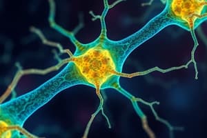
- The retina is the innermost layer containing photoreceptor cells (rods + cones) that convert light into neural signals.
- The ciliary body is a circular muscle that controls lens shape.
- Contraction of the ciliary body reduces tension on the zonular fibers allowing the lens to curve for near vision.
- Relaxation of the ciliary body pulls on zonular fibers flattening the lens for distant vision.
- Zonular fibers connect the lens to the ciliary body and exert tension on the lens.
- The anterior chamber is a fluid-filled space between the cornea and lens, containing aqueous humor.
- The posterior chamber (vitreous chamber) is a large space behind the lens, containing vitreous humor (gellike).
- The posterior chamber is primarily made up of sclera, choroid, and the retina
Photo Transduction
- In the absence of light, a rod increases cGMP, causing Na+ channels to open, leading to Na+ influx, depolarization, opening of voltage-gated Ca2+ channels, Ca2+ influx, and neurotransmitter release.
- In the presence of light, a rod decreases cGMP, preventing Na+ channels from opening and resulting in no neurotransmitter release.
- Photo transduction is converting light energy into a brain signal to bipolar and Ganglion Cells.
- Photoreceptors are the only cells to respond to light energy and transduce it.
- In no light conditions, rods produce a messenger called Cyclic Guanosine Monophosphate (cGMP).
- cGMP levels rise and open sodium channels, allowing sodium to enter rods.
- Sodium influx depolarizes the rod, which opens voltage-gated calcium channels, causing calcium influx in rods and release of neurotransmitters.
- Absence of light is converted into "no signal", rods are specialized to absorb light in the dark.
- In light conditions the rod builds up, blocking of cGMP and causing levels to go down that does not open channels
- This leads to no sodium influx, no deplore, no CA2+ channel, opening and no calcium influx
- Rod is a type of photo receptor.
Retina System Duplex
- The scotopic system allows to see in the dark.
- Rods are the photoreceptor in scotopic system.
- Rhodopsin is a pigment in the rod that allows it to absorb light.
- Rhodopsin is 1000x more sensitive to light than opsin.
- Rhodopsin is purple color, shines light ,white color and takes 4-5 minutes to reset to purple
- Conversion circuitry occurs, There may not be many photons of light, so only same rods are activated
- Mesoptic vision: ability to occur between scotopic/photopic conditions
Mesoptic vs Photopic condition
- Mesopic: Rods inactive, cones active.
- Mesopic: Rods never turn off.
- Mesopic: Cones need a lot more light to be active to recognize something, color but not vibrant
Photopic System
- Is the system allowing to see in light.
- cones are the photoreceptors used in the photopic system.
- Opsin is the photopigment used.
- 3 types of opsin absorbs different ranges: Blue, Green, Red
- There are enough photons of light to activate all cones.
- Circuitry here is 1:1 meaning: 1 cone >> 1 bipolar cell >> 1 ganglion cell.
Macula and Fovea
- Macula: regions where photoreceptors are dense.
- Fovea: central vision where cells converge and are exclusively are cones.
- Everything else is peripheral retina by vision and primarily rods.
Cellular Organization of Retina.
- Macula: the central area of the retina responsible for sharp, detailed vision and densely packed with cones.
- Cones: photoreceptor cells that detect color.
- Fovea: located within the macula and the central point that has the highest concentration of cones resulting in clearest sight.
- Optic Disc (Blind Spot): the area where the optic nerve exits the eye.
- Optic Disc: Lacks photoreceptors resulting in a blind spot in each eye.
- The brain compensates for the blind spot. Axons from retinal ganglion cells form the optic nerve that supply visual information to the brain, in relation to optic
- The blood supple to retina enter/exits via optic disc.
Retina Layers
- Surface to deep consists of: sclera >> choroid >> retina.
3 Primary Layers
- Ganglion Cell Layer is made of ganglion cells, that are a collection of cell bodies.
- Ganglion Cell Layer is the only cells that have action potentials and leave through optic disc enter optic nerve which (most superficial).
- Inner Nuclear Layer is primarily, made of bipolar cells.
- Outer Nuclear Layer is primarily deepest made and consists of photoreceptors.
- Photoreceptors have a photo pigment that absorbs light and outer segment part of the outer nuclear layer not separate.
- Light passes through other layers before being absorbed.
- Ratio to brighten light is 1:1:1
- Outer plexiform layer consists, with 1 photoreceptor, communicating with a 1 bipolar cell, that communicates with 1 ganglion.
Intermediate Layers
- Outer Plexiform Layer contains Horizontal cells.
- Horizontal cells take signal from 1 photo receptor to distribute it to multiple Bipolar cells that (between outer --> inner nuclear layer)
- Inner Plexiform Layer : Amacrine cells take signal from 1 bipolar cell and activate multiple ganglion cells (between ganglion to inner nuclear)
- In low light signalling, it to speak that it has to signal more cells, so output is greater, allowing us to see in low resolution lighting
Pigmented Epithelium
- Any light not absorbed by is absorbed in cells below the retina.
Studying That Suits You
Use AI to generate personalized quizzes and flashcards to suit your learning preferences.