Podcast
Questions and Answers
What is the proportion of adult body weight accounted for by the liver?
What is the proportion of adult body weight accounted for by the liver?
- 1.5%
- 10%
- 5%
- 2.5% (correct)
Which vein drains into the portal vein?
Which vein drains into the portal vein?
- Hepatic vein
- Inferior pancreaticoduodenal vein
- Splenic vein
- Superior mesenteric vein (correct)
What is the function of bile in the digestive process?
What is the function of bile in the digestive process?
- Regulation of blood sugar
- Production of insulin
- Emulsification of fat (correct)
- Emulsification of carbohydrates
What is the clinical consideration for hypertrophy of the head of the pancreas?
What is the clinical consideration for hypertrophy of the head of the pancreas?
Which ligament separates the diaphragmatic surface of the liver from the pleurae, lungs, pericardium, and heart?
Which ligament separates the diaphragmatic surface of the liver from the pleurae, lungs, pericardium, and heart?
In which area of the liver does the IVC traverse a deep groove?
In which area of the liver does the IVC traverse a deep groove?
What is the function of the gallbladder in relation to bile?
What is the function of the gallbladder in relation to bile?
Which structure forms the continuous groove formed anteriorly by the fossa for the gallbladder and posteriorly by the groove for the vena cava?
Which structure forms the continuous groove formed anteriorly by the fossa for the gallbladder and posteriorly by the groove for the vena cava?
What is the clinical consideration for degeneration of the islets of Langerhans?
What is the clinical consideration for degeneration of the islets of Langerhans?
What is the location of the liver in the abdomen?
What is the location of the liver in the abdomen?
Flashcards are hidden until you start studying
Study Notes
Retroperitoneal Organs
- Initially, some organs were intraperitoneal, suspended by a mesentery, including:
- Duodenum
- Pancreas
- Rectum
- Ascending and descending colon
Peritoneal Reflections or Folds
- Unlike other serous membranes, the peritoneum reflects inward to form a double layer of peritoneum
- Some folds contain:
- Vessels
- Nerves
- Supplying the viscera
- Maintaining proper positioning of the viscera
Mesentery
- A double layer of peritoneum that occurs due to invagination of the peritoneum by an organ
- Provides a means for neurovascular communication between the organ and the body wall
- Connects an intraperitoneal organ to the body wall (usually the posterior abdominal wall)
Omentum
- A double layer of peritoneum that attaches the stomach to neighboring organs
- Types:
- Lesser omentum (attached to lesser curvature of the stomach, derived from ventral mesentery)
- Greater omentum (attached to greater curvature of the stomach, derived from dorsal mesentery)
Greater Omentum
- Attaches to the greater curvature of the stomach and the first part of the duodenum
- Folds back up and attaches to the anterior surface of the transverse colon
- Extends inferiorly over the coils of the jejunum and ileum
- Contains:
- Accumulation of fat
- Two arteries (right and left gastro-omental vessels)
- Accompanying veins
- Role in immunity (abdominal policeman)
Lesser Omentum
- Extends from the lesser curvature of the stomach and the first part of the duodenum to the inferior surface of the liver
- Divided into:
- Medial hepatogastric ligament
- Lateral hepatoduodenal ligament
- Enclosed in this free edge are:
- Hepatic artery proper
- Bile duct
- Portal vein
- Additionally, the right and left gastric vessels are between the layers of the lesser omentum near the lesser curvature of the stomach
Peritoneal Ligaments
- A double fold of peritoneum that connects viscera together or viscera to the abdominal wall
- May form part of an omentum
- Named after the structures being connected, such as:
- Splenorenal ligament
- Gastrophrenic ligament
- Gastrosplenic ligament
- Gastrorenal ligament
- Gastrocolic ligament
- Falciform ligament
- Hepatogastric ligament
- Hepatoduodenal ligament
Peritoneal Folds and Fossa
- A peritoneal fold is a reflection of peritoneum that is raised from the body wall by underlying blood vessels, ducts, and ligaments
- Formed by obliterated fetal vessels
- Some peritoneal folds contain:
- Blood vessels
- Bleed if cut
- A peritoneal recess or fossa is a pouch of peritoneum formed by a peritoneal fold
Subdivisions of Peritoneal Cavity
- Greater sac:
- Further divided into two compartments by the mesentery of the transverse colon
- Supracolic compartment:
- Lies above the transverse mesocolon
- Contains the stomach, liver, and spleen
- Infracolic compartment:
- Lies below the transverse mesocolon
- Contains the small intestine, ascending and descending colon
- Lesser sac:
- Lies posterior to the stomach, liver, and lesser omentum
- Connected to the greater sac via the epiploic/omental foramen
Innervation of Peritoneum
- Parietal peritoneum:
- Similar to the anterior abdominal wall
- Sensitive to pain
- Visceral peritoneum:
- Similar to the abdominal viscera
- Insensitive to pain
Clinical Relevance
- Sampling of peritoneal fluid:
- Culdocentesis
- Paracentesis
- Disorders of the peritoneal cavity:
- Ascites
- Peritonitis
Abdominal Viscera
- The majority of the alimentary system
- Includes:
- Terminal part of the esophagus
- Stomach
- Intestines
- Spleen
- Pancreas
- Liver
- Gallbladder
- Kidneys
- Suprarenal glands
Division of Abdominal Viscera
- Foregut:
- Lower part of the esophagus
- Stomach
- First part of the duodenum
- Liver
- Pancreas
- Midgut:
- Remainder of the duodenum
- Jejunum
- Ileum
- Caecum
- Ascending and descending colon
- Hindgut:
- Remainder of the transverse colon
- Descending and sigmoid colon
- Rectum
- Anal canal
Abdominal Part of Esophagus
- Esophageal hiatus in the right crus of the diaphragm
- Relations:
- Anteriorly: left lobe of the liver
- Posteriorly: aorta and left crus of the diaphragm
Neurovasculature of Esophagus
- Esophageal branch of the left gastric artery
- Left inferior phrenic artery
- Veins:
- Submucosal veins of the esophagus
- Left gastric veins
- Portal venous system
- Azygous vein
- Systemic venous system
- Lymph nodes:
- Left gastric nodes
- Celiac nodes
- ANS:
- Sympathetic: greater splanchnic nerve
- Parasympathetic: vagus### Lymphatic Follicles and Intestinal Tract
- Lymphatic follicles are present in the midgut and are involved in the immune response.
- The intestinal tract is divided into two parts: midgut and hindgut.
- The midgut is derived from the endoderm and extends from the proximal two-thirds of the transverse colon to the distal one-third of the transverse colon.
- The hindgut is also derived from the endoderm and extends from the distal one-third of the transverse colon to the pectinate line.
Large Intestine
- The large intestine is approximately 1.5m long and extends from the ileocecal junction to the anus.
- It has three bands of longitudinal muscle fibers called taeniae coli, each about 5mm wide.
- The taeniae coli start at the base of the appendix and extend from the cecum to the rectum.
- Along the sides of the taeniae, there are tags of peritoneum filled with fat, called epiploic appendages.
- The sacculations, called haustra, are characteristic features of the large intestine and distinguish it from the rest of the intestinal tract.
Development of the Large Intestine
- The development of the large intestine is influenced by the endoderm of the midgut and hindgut.
- The midgut gives rise to the cecum, appendix, and ascending colon, while the hindgut gives rise to the descending colon, sigmoid colon, rectum, and anal canal.
Distinguishing Features of the Large Intestine
- Externally, the large intestine has taeniae coli, appendices epiploicae, and haustra.
- Internally, it has a larger caliber, absence of villi, widely dispersed mucosal folds, and more lymphatic tissue.
Cecum and Appendix
- The cecum is a cul-de-sac below the opening of the ileum into the colon and lies in the right iliac fossa.
- The appendix is a vermiform structure that hangs off the cecum.
- The appendix can be located in various positions, including subceacal, retroceacal, preileal, pelvic, and postileal.
Spleen
- The spleen is an ovoid, usually purplish, pulpy mass about the size and shape of one's fist.
- It is located in the superolateral part of the left upper quadrant of the abdomen, where it is protected by the inferior thoracic cage.
- The spleen is 12cm long, 8cm wide, and 3cm thick, and weighs around 200gm.
- It is not palpable below the left costal margin and is related to the 9th, 10th, and 11th ribs on the side.
Functions of the Spleen
- The spleen is a lymphatic organ that participates in the body's defense system as a site of lymphocyte proliferation and immune surveillance and response.
- It is also involved in the removal of expended red blood cells and broken-down platelets, and in the recycling of iron and globin.
- The spleen serves as a blood reservoir, storing red blood cells and platelets, and can provide a sort of "self-transfusion" in response to hemorrhage.
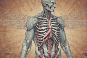
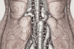
Liver
- The liver is the largest gland in the body and is located in the right upper quadrant of the abdomen.
- It weighs approximately 1500g and accounts for around 2.5% of adult body weight.
- The liver is involved in many metabolic activities, including the storage of glycogen and the secretion of bile.
- Bile is produced continuously by the liver and is stored in the gallbladder between meals.
- When food arrives in the duodenum, the gallbladder sends concentrated bile through the biliary ducts to the duodenum.
Location and Surfaces of the Liver
- The liver lies mainly in the right upper quadrant of the abdomen, where it is protected by the thoracic cage and the diaphragm.
- The normal liver lies deep to ribs 7-11 on the right side and crosses the midline toward the left nipple.
- The liver has a convex diaphragmatic surface and a relatively flat or even concave visceral surface.
- The diaphragmatic surface is smooth and dome-shaped, and is related to the concavity of the inferior surface of the diaphragm.
Clinical Considerations
- Hypertrophy of the head of the pancreas may cause portal or bile duct obstruction.
- Cancer of the head of the pancreas is usually a fatal pathology.
- Degeneration of the islets of Langerhans leads to diabetes mellitus.
- Pancreatitis is a serious inflammatory condition of the exocrine pancreas.
Studying That Suits You
Use AI to generate personalized quizzes and flashcards to suit your learning preferences.