Podcast
Questions and Answers
What term is used to refer to the functional system of tissues that surrounds and attaches teeth to the jawbone?
What term is used to refer to the functional system of tissues that surrounds and attaches teeth to the jawbone?
- Periodontium (correct)
- Gingival sulcus
- Cementoenamel junction
- Alveolar process
Which part of the periodontium is responsible for covering the cervical portions of teeth?
Which part of the periodontium is responsible for covering the cervical portions of teeth?
- Gingival sulcus
- Interdental gingiva
- Free gingiva (correct)
- Attached gingiva
What structure includes the demarcations like the free gingival groove and mucogingival junction?
What structure includes the demarcations like the free gingival groove and mucogingival junction?
- Gingival margin
- Alveolar mucosa
- Attached gingiva
- Gingiva (correct)
What does the attached gingiva primarily protect?
What does the attached gingiva primarily protect?
What anatomical area of the gingiva is characterized by its scalloped curvature?
What anatomical area of the gingiva is characterized by its scalloped curvature?
Which of the following is NOT one of the four anatomical areas of the gingiva?
Which of the following is NOT one of the four anatomical areas of the gingiva?
What is the role of the junctional epithelium in the structure of the gingiva?
What is the role of the junctional epithelium in the structure of the gingiva?
How is the position of the gingival margin influenced as one ages?
How is the position of the gingival margin influenced as one ages?
What is the primary composition of the periodontal ligament?
What is the primary composition of the periodontal ligament?
Which statement correctly describes a function of the periodontal ligament?
Which statement correctly describes a function of the periodontal ligament?
How does cementum compare to dentin and enamel?
How does cementum compare to dentin and enamel?
What percentage of cementum is organic material?
What percentage of cementum is organic material?
What characteristic of cementum makes orthodontic treatment possible?
What characteristic of cementum makes orthodontic treatment possible?
Which of the following statements is true about the periodontal ligament’s blood vessels?
Which of the following statements is true about the periodontal ligament’s blood vessels?
What can cementum do in response to pressure?
What can cementum do in response to pressure?
Which type of cementum primarily undergoes continuous resorption and repair throughout the life of a tooth?
Which type of cementum primarily undergoes continuous resorption and repair throughout the life of a tooth?
What is the primary function of the attached gingiva?
What is the primary function of the attached gingiva?
Which structure fills the space between two adjacent teeth?
Which structure fills the space between two adjacent teeth?
What is the depth of a clinically healthy gingival sulcus?
What is the depth of a clinically healthy gingival sulcus?
What is the name given to the depression connecting the facial and lingual papillae?
What is the name given to the depression connecting the facial and lingual papillae?
What does the gingival crevicular fluid do in a healthy sulcus?
What does the gingival crevicular fluid do in a healthy sulcus?
Which of the following statements about the col is true?
Which of the following statements about the col is true?
What characterizes the gingival sulcus?
What characterizes the gingival sulcus?
Which term refers to the most coronal portion of the junctional epithelium?
Which term refers to the most coronal portion of the junctional epithelium?
What is the main feature of the free gingival margin?
What is the main feature of the free gingival margin?
What characteristic mainly distinguishes attached gingiva from free gingiva?
What characteristic mainly distinguishes attached gingiva from free gingiva?
In which region is the width of attached gingiva typically the narrowest?
In which region is the width of attached gingiva typically the narrowest?
Which statement regarding the color of attached gingiva when healthy is correct?
Which statement regarding the color of attached gingiva when healthy is correct?
What was previously believed regarding keratinized gingiva and periodontal health?
What was previously believed regarding keratinized gingiva and periodontal health?
What is one characteristic of healthy attached gingiva texture?
What is one characteristic of healthy attached gingiva texture?
What is the role of junctional epithelium in relation to free gingiva?
What is the role of junctional epithelium in relation to free gingiva?
Which factor can cause physiologic pigmentation of the attached gingiva?
Which factor can cause physiologic pigmentation of the attached gingiva?
What is the primary function of cementum in relation to periodontal ligament fibers?
What is the primary function of cementum in relation to periodontal ligament fibers?
What occurs to alveolar bone when teeth are extracted?
What occurs to alveolar bone when teeth are extracted?
What is the composition percentage of inorganic material in alveolar bone?
What is the composition percentage of inorganic material in alveolar bone?
What is the role of the alveolar bone proper?
What is the role of the alveolar bone proper?
Which type of bone appears on a radiograph and is visible?
Which type of bone appears on a radiograph and is visible?
What is the cortical bone's function in relation to the alveolar bone?
What is the cortical bone's function in relation to the alveolar bone?
What effect does attrition have on the tooth, and how is it compensated for?
What effect does attrition have on the tooth, and how is it compensated for?
Which of the following is true about Sharpey’s fibers?
Which of the following is true about Sharpey’s fibers?
Flashcards
Periodontium
Periodontium
The functional system of tissues surrounding and attaching teeth to the jawbone.
Supporting tissues of teeth
Supporting tissues of teeth
Another name for the periodontium, referring to the structures that support the teeth.
Attachment apparatus
Attachment apparatus
A term for the periodontium, highlighting its role in attaching teeth to the jawbone.
Gingiva
Gingiva
Signup and view all the flashcards
What is the relationship between the gingival margin and the CEJ?
What is the relationship between the gingival margin and the CEJ?
Signup and view all the flashcards
What are the four anatomical areas of the gingiva?
What are the four anatomical areas of the gingiva?
Signup and view all the flashcards
Function of the gingiva
Function of the gingiva
Signup and view all the flashcards
Boundaries of the gingiva
Boundaries of the gingiva
Signup and view all the flashcards
Attached gingiva function
Attached gingiva function
Signup and view all the flashcards
What is interdental gingiva?
What is interdental gingiva?
Signup and view all the flashcards
Interdental col
Interdental col
Signup and view all the flashcards
Function of the interdental col
Function of the interdental col
Signup and view all the flashcards
Gingival sulcus
Gingival sulcus
Signup and view all the flashcards
Gingival sulcus depth
Gingival sulcus depth
Signup and view all the flashcards
Junctional epithelium, epithelial attachment, base of sulcus
Junctional epithelium, epithelial attachment, base of sulcus
Signup and view all the flashcards
Gingival crevicular fluid
Gingival crevicular fluid
Signup and view all the flashcards
Free Gingiva
Free Gingiva
Signup and view all the flashcards
Free Gingival Margin
Free Gingival Margin
Signup and view all the flashcards
Attached Gingiva
Attached Gingiva
Signup and view all the flashcards
Width of the Attached Gingiva
Width of the Attached Gingiva
Signup and view all the flashcards
Keratinized Gingiva
Keratinized Gingiva
Signup and view all the flashcards
Color of the Attached Gingiva
Color of the Attached Gingiva
Signup and view all the flashcards
Texture of the Attached Gingiva
Texture of the Attached Gingiva
Signup and view all the flashcards
Healthy Gingiva
Healthy Gingiva
Signup and view all the flashcards
Cementum's Role
Cementum's Role
Signup and view all the flashcards
Sharpey's Fibers
Sharpey's Fibers
Signup and view all the flashcards
Alveolar Bone
Alveolar Bone
Signup and view all the flashcards
What happens to alveolar bone when teeth are removed?
What happens to alveolar bone when teeth are removed?
Signup and view all the flashcards
Alveolar Bone Components
Alveolar Bone Components
Signup and view all the flashcards
Alveolar Bone Proper
Alveolar Bone Proper
Signup and view all the flashcards
Cortical Bone
Cortical Bone
Signup and view all the flashcards
What can be seen on a radiograph?
What can be seen on a radiograph?
Signup and view all the flashcards
Gingival Fluid
Gingival Fluid
Signup and view all the flashcards
Periodontal Ligament (PDL)
Periodontal Ligament (PDL)
Signup and view all the flashcards
What does PDL do?
What does PDL do?
Signup and view all the flashcards
More PDL Functions
More PDL Functions
Signup and view all the flashcards
Cementum
Cementum
Signup and view all the flashcards
Key Feature of Cementum
Key Feature of Cementum
Signup and view all the flashcards
Continuous Changes in Cementum
Continuous Changes in Cementum
Signup and view all the flashcards
Cementum's Permeability
Cementum's Permeability
Signup and view all the flashcards
Study Notes
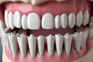
Periodontium: The Tooth Supporting Structures
- The periodontium is a functional system of tissues surrounding teeth and attaching them to the jawbone.
- It's also called the supporting tissues of teeth and the attachment apparatus.
- The periodontium is a key anatomical structure for oral health.
Healthy Periodontium
- A healthy periodontium features a gingival sulcus that is 1-3mm deep.
- The gingival margin approximates the curvatures of the cementoenamel junction (CEJ).
- The position of the gingival margin can vary based on age.
Structures of the Periodontium and Their Functions
- Gingiva: Seals around the tooth's neck, covers jaw alveolar processes, and holds tissue against the tooth during mastication.
- Periodontal Ligament: Suspends and maintains the tooth in its socket; anchors periodontal ligament fibers to secure the tooth. Protects the root dentin.
- Cementum: A thin, hard mineralized tissue covering the tooth root, softer than dentin or enamel. It's 55% organic (collagen matrix proteins) and 45% inorganic (calcium, hydroxyapatite, trace elements). Resists resorption better than bone.
- Alveolar Bone: Supports and surrounds the roots of teeth. It's a mineralized connective tissue composed of 60% inorganic, 25% organic, and 15% water.
The Gingiva
- The gingiva is part of the mucosa that surrounds the cervical portions of teeth and covers alveolar processes.
- It has different anatomical areas like free gingiva, gingival sulcus, interdental gingiva, and attached gingiva.
- The gingiva protects underlying structures from the oral environment.
- The gingiva's boundaries consist of the gingival margin and alveolar mucosa.
- It's demarcated by the free gingival groove and mucogingival junction.
Free Gingiva
- This is the unattached portion of the gingiva that surrounds the tooth at the cementoenamel junction (CEJ).
- It's also known as unattached or marginal gingiva.
- Free gingiva forms a soft tissue wall of the gingival sulcus.
- The free gingiva follows the scalloped, curved contours of the underlying CEJ.
Attached Gingiva
- This gingiva is continuous with free gingiva.
- It is tightly attached to the underlying cementum on the cervical thrid of the root and periosteum of alveolar bone.
- Incisors and molars have the widest attached gingiva. The thinnest is found in premolar regions.
Width of the Attached Gingiva
- The width of attached gingiva is widest in the incisor and molar regions and narrowest in premolar regions.
Color of the Attached Gingiva
- Healthy attached gingiva is pale or light coral pink.
- Pigmentation is a physiological trait that often happens from increased melanin production.
Texture of the Attached Gingiva
- Healthy attached and interdental gingiva may exhibit stippling, but it's not always present, and absent in many periodontally healthy adults.
Function of the Attached Gingiva
- This part of gingiva withstands the forces during chewing, speaking, and toothbrushing.
- It prevents free gingiva from pulling apically from the tooth when tension is placed on the alveolar mucosa.
Interdental Gingiva
- This part of the gingiva fills the embrasures between adjacent teeth, apical to the contact area.
- It has facial and lingual papillae.
- A depression appears apically to the contact area of adjacent teeth.
- This area prevents food from packing between teeth while chewing.
Gingival Sulcus
- This is a V-shaped shallow space between the free gingiva and the tooth surface, encircling the tooth neck.
- It's up to 3 mm deep in clinically healthy individuals, measured by a periodontal probe.
- The base of the sulcus interacts with the most coronal portion of the junctional epithelium.
Gingival Crevicular Fluid
- Also known as gingival sulcular fluid.
- It seeps from underlying connective tissue and is very minimal in healthy individuals.
- Levels increase with gingival stimulation, and even more when inflamed. It can be measured using a filter strip.
Periodontal Ligament (PDL)
- Network of soft connective tissue that attaches the root of a tooth to the bony walls of its socket.
- The PDL is primarily composed of dense fibrous connective tissue whose fibers connect root cementum and alveolar bone.
- It connects the tooth to the alveolar process and supports the tooth within the socket.
- PDL absorbs mechanical loads on the tooth.
- It maintains the tooth and contains nerve endings relaying sensory information, blood vessels providing oxygen and nutrients, as well as structures that aid periodontal regeneration.
Cementum
- A thin layer of hard, mineralized tissue that covers the tooth root surface
- It overlies the dentin.
- It's softer than dentin or enamel and consists of 55% organic material (collagen matrix proteins) and 45% inorganic material (calcium, hydroxyapatite, trace elements).
- Two types exist: cellular and acellular.
Alveolar Bone
- The jawbone that surrounds and supports the teeth' roots. It's a mineralized connective tissue.
- The width depends on the region of the mouth, widest at incisors and molars, and least thick at premolars.
- Presence of teeth relates to this bone's existence.
- It resorbs when teeth are extracted. It's absent if teeth do not erupt.
Layers of the Alveolar Process
- The alveolar process consists of alveolar bone proper, cortical bone, and cancellous bone.
- Alveolar bone proper is a thin layer lining the socket that contains the alveolus, foramina, and Sharpey's fibers.
- Cortical bone (cortical plate) is a dense outer layer.
- Cancellous bone (spongy bone) fills the inner portion of the alveolar process.
Studying That Suits You
Use AI to generate personalized quizzes and flashcards to suit your learning preferences.