Podcast
Questions and Answers
What is the periodontal pocket?
What is the periodontal pocket?
- A result of gingival margin movement towards the root of the tooth
- A clinically healthy gingival sulcus
- A deepened gingival sulcus due to apical displacement of the gingival attachment (correct)
- A shallow area between the teeth and gums
What are the learning outcomes related to the periodontal pocket?
What are the learning outcomes related to the periodontal pocket?
- Understanding the role of dental floss in preventing periodontal pockets
- Explaining the importance of fluoride in treating periodontal pockets
- Describing the association of periodontal pocket histopathology with the clinic (correct)
- Listing the surgical procedures for treating periodontal pockets
How do the types of periodontitis differ?
How do the types of periodontitis differ?
- In their etiology
- In their response to therapy
- In their natural history and progression (correct)
- In their response to fluoride treatment
What causes deepening of the gingival sulcus?
What causes deepening of the gingival sulcus?
What is the histopathology of the periodontal pocket associated with?
What is the histopathology of the periodontal pocket associated with?
What distinguishes attachment loss clinically?
What distinguishes attachment loss clinically?
How can one define periodontal disease activity?
How can one define periodontal disease activity?
What is the importance of periodontal pocket in periodontal diseases?
What is the importance of periodontal pocket in periodontal diseases?
What is the pathogenesis of the periodontal pocket?
What is the pathogenesis of the periodontal pocket?
What are the clinical features of an edematous pocket wall?
What are the clinical features of an edematous pocket wall?
What does the presence of pus in a periodontal pocket indicate?
What does the presence of pus in a periodontal pocket indicate?
What is the principal component of the debris in periodontal pockets?
What is the principal component of the debris in periodontal pockets?
What is observed in the root surface wall of periodontal pockets with light and electron microscopy?
What is observed in the root surface wall of periodontal pockets with light and electron microscopy?
What is the result of exposure to oral fluid and bacterial plaque on the cementum of root surfaces?
What is the result of exposure to oral fluid and bacterial plaque on the cementum of root surfaces?
What characterizes active root caries lesions?
What characterizes active root caries lesions?
What zone of a periodontal pocket is covered by attached plaque?
What zone of a periodontal pocket is covered by attached plaque?
What characterizes periods of quiescence in periodontal disease activity?
What characterizes periods of quiescence in periodontal disease activity?
What does site specificity refer to in periodontal disease?
What does site specificity refer to in periodontal disease?
What is the effect of diseased root fragments when placed in tissue culture?
What is the effect of diseased root fragments when placed in tissue culture?
Which of the following is true about the relationship between attachment loss and pocket depth?
Which of the following is true about the relationship between attachment loss and pocket depth?
What is the characteristic type of bone loss in suprabony pockets?
What is the characteristic type of bone loss in suprabony pockets?
How does periodontal abscess formation occur when trauma to the tooth or perforation of the lateral wall of the root in endodontic therapy happens?
How does periodontal abscess formation occur when trauma to the tooth or perforation of the lateral wall of the root in endodontic therapy happens?
What is a periodontal abscess classified as when it occurs in the soft-tissue wall of a deep periodontal pocket?
What is a periodontal abscess classified as when it occurs in the soft-tissue wall of a deep periodontal pocket?
What type of microorganisms have been reported to primarily colonize the periodontal abscess?
What type of microorganisms have been reported to primarily colonize the periodontal abscess?
What causes pathologic changes in the pulp associated with periodontal pockets?
What causes pathologic changes in the pulp associated with periodontal pockets?
In which type of pockets does the base lie coronal to the crest of the alveolar bone?
In which type of pockets does the base lie coronal to the crest of the alveolar bone?
What is meant by 'lateral periodontal cyst' as mentioned in the text?
What is meant by 'lateral periodontal cyst' as mentioned in the text?
What is found normally between the apical end of junctional epithelium and alveolar bone?
What is found normally between the apical end of junctional epithelium and alveolar bone?
What is generally but not always correlated with pocket depth?
What is generally but not always correlated with pocket depth?
Which type of pocket occurs when the bottom of the pocket is coronal to the underlying alveolar bone?
Which type of pocket occurs when the bottom of the pocket is coronal to the underlying alveolar bone?
What clinical signs suggest the presence of periodontal pockets?
What clinical signs suggest the presence of periodontal pockets?
What has been described in human chronic periodontitis as invading the apical and lateral areas of the pocket wall?
What has been described in human chronic periodontitis as invading the apical and lateral areas of the pocket wall?
What microorganisms are mostly associated with healthy gingiva?
What microorganisms are mostly associated with healthy gingiva?
What type of lesion are periodontal pockets?
What type of lesion are periodontal pockets?
What do the clinical signs of periodontal pockets include?
What do the clinical signs of periodontal pockets include?
What is true about suprabony (supracrestal or supraalveolar) pockets?
What is true about suprabony (supracrestal or supraalveolar) pockets?
What is the initial lesion in the development of periodontitis?
What is the initial lesion in the development of periodontitis?
What is associated with healthy gingiva?
What is associated with healthy gingiva?
What has been found in intercellular spaces of the epithelium in human chronic periodontitis?
What has been found in intercellular spaces of the epithelium in human chronic periodontitis?
Define the periodontal pocket.
Define the periodontal pocket.
What is the importance of periodontal pocket in periodontal diseases?
What is the importance of periodontal pocket in periodontal diseases?
List the types of periodontal pockets.
List the types of periodontal pockets.
How is periodontal pocket measured and how is it clinically distinguished from gingival pockets?
How is periodontal pocket measured and how is it clinically distinguished from gingival pockets?
List the histopathology of the periodontal pocket and its association with the clinic.
List the histopathology of the periodontal pocket and its association with the clinic.
Define attachment loss and how it is clinically distinguished.
Define attachment loss and how it is clinically distinguished.
Define periodontal disease activity and how it is clinically distinguished.
Define periodontal disease activity and how it is clinically distinguished.
What is the pathogenesis of the periodontal pocket?
What is the pathogenesis of the periodontal pocket?
What is meant by site specificity in periodontal disease?
What is meant by site specificity in periodontal disease?
How do the types of periodontitis differ?
How do the types of periodontitis differ?
What are the two types of periodontal pockets?
What are the two types of periodontal pockets?
What are the clinical signs that suggest the presence of periodontal pockets?
What are the clinical signs that suggest the presence of periodontal pockets?
What types of microorganisms are associated with diseased gingiva?
What types of microorganisms are associated with diseased gingiva?
What has been described in human chronic periodontitis as invading the apical and lateral areas of the pocket wall?
What has been described in human chronic periodontitis as invading the apical and lateral areas of the pocket wall?
What are the areas noted in the soft-tissue wall of the periodontal pocket?
What are the areas noted in the soft-tissue wall of the periodontal pocket?
What are the clinical features that suggest the presence of periodontal pockets?
What are the clinical features that suggest the presence of periodontal pockets?
What is the initial lesion in the development of periodontitis?
What is the initial lesion in the development of periodontitis?
What are the mechanisms of tissue destruction triggered by bacterial plaque?
What are the mechanisms of tissue destruction triggered by bacterial plaque?
What has scanning electron microscopy permitted the description of in the soft-tissue wall of the periodontal pocket?
What has scanning electron microscopy permitted the description of in the soft-tissue wall of the periodontal pocket?
What is the significance of periodontal pockets as healing lesions?
What is the significance of periodontal pockets as healing lesions?
What are the pathologic changes that may occur in the pulp due to the spread of infection from periodontal pockets?
What are the pathologic changes that may occur in the pulp due to the spread of infection from periodontal pockets?
What is the mean length of the distance between the apical extent of calculus and the alveolar crest in human periodontal pockets?
What is the mean length of the distance between the apical extent of calculus and the alveolar crest in human periodontal pockets?
In what type of pockets does the base lie between the tooth and the bone, with bone loss being mostly vertical?
In what type of pockets does the base lie between the tooth and the bone, with bone loss being mostly vertical?
What is the classification of a periodontal abscess that occurs in the soft-tissue wall of a deep periodontal pocket?
What is the classification of a periodontal abscess that occurs in the soft-tissue wall of a deep periodontal pocket?
What are the microorganisms that have been reported to primarily colonize the periodontal abscess?
What are the microorganisms that have been reported to primarily colonize the periodontal abscess?
What is the characteristic type of bone loss in suprabony pockets?
What is the characteristic type of bone loss in suprabony pockets?
What is the term used to describe the localized destruction of periodontal tissues along a lateral root surface, most often in the mandibular canine–premolar area?
What is the term used to describe the localized destruction of periodontal tissues along a lateral root surface, most often in the mandibular canine–premolar area?
What is the result of incomplete removal of calculus during treatment of a periodontal pocket?
What is the result of incomplete removal of calculus during treatment of a periodontal pocket?
What is the term used to describe the severity of attachment loss in pocket formation, which is generally but not always correlated with the depth of the pocket?
What is the term used to describe the severity of attachment loss in pocket formation, which is generally but not always correlated with the depth of the pocket?
What may cause painful symptoms or affect the response of the pulp to restorative procedures in the context of periodontal pockets?
What may cause painful symptoms or affect the response of the pulp to restorative procedures in the context of periodontal pockets?
What are the clinical features of a fibrotic pocket wall?
What are the clinical features of a fibrotic pocket wall?
What is the principal component of the debris in periodontal pockets?
What is the principal component of the debris in periodontal pockets?
What are the zones found in the bottom of a periodontal pocket?
What are the zones found in the bottom of a periodontal pocket?
What is the effect of diseased root fragments when placed in tissue culture?
What is the effect of diseased root fragments when placed in tissue culture?
What is the characteristic type of bone loss in suprabony pockets?
What is the characteristic type of bone loss in suprabony pockets?
What causes pathologic changes in the pulp associated with periodontal pockets?
What causes pathologic changes in the pulp associated with periodontal pockets?
What characterizes active root caries lesions?
What characterizes active root caries lesions?
What does site specificity refer to in periodontal disease?
What does site specificity refer to in periodontal disease?
What is the condition of the pocket wall when inflammatory fluid and cellular exudate predominate?
What is the condition of the pocket wall when inflammatory fluid and cellular exudate predominate?
What are the components of purulent exudate in the periodontal pocket?
What are the components of purulent exudate in the periodontal pocket?
The periodontal pocket is defined as a pathologically deepened gingival ______.
The periodontal pocket is defined as a pathologically deepened gingival ______.
Deepening of the gingival sulcus may occur as a result of coronal movement of the gingival margin, apical displacement of the gingival attachment, or a combination of the two ______.
Deepening of the gingival sulcus may occur as a result of coronal movement of the gingival margin, apical displacement of the gingival attachment, or a combination of the two ______.
The periodontal pocket is one of the most important clinical features of ______ disease.
The periodontal pocket is one of the most important clinical features of ______ disease.
The different types of periodontitis differ with regard to their etiology, natural history, progression, and response to ______.
The different types of periodontitis differ with regard to their etiology, natural history, progression, and response to ______.
The deepening of the gingival sulcus may occur as a result of coronal movement of the gingival margin, apical displacement of the gingival attachment, or a combination of the two ______.
The deepening of the gingival sulcus may occur as a result of coronal movement of the gingival margin, apical displacement of the gingival attachment, or a combination of the two ______.
The periodontal pocket is one of the most important clinical features of ______ disease.
The periodontal pocket is one of the most important clinical features of ______ disease.
The different types of periodontitis differ with regard to their etiology, natural history, progression, and response to ______.
The different types of periodontitis differ with regard to their etiology, natural history, progression, and response to ______.
The periodontal pocket, which is defined as a pathologically deepened gingival sulcus, is one of the most important clinical features of ______ disease.
The periodontal pocket, which is defined as a pathologically deepened gingival sulcus, is one of the most important clinical features of ______ disease.
The periodontal pocket, which is defined as a pathologically deepened gingival sulcus, is one of the most important clinical features of periodontal ______.
The periodontal pocket, which is defined as a pathologically deepened gingival sulcus, is one of the most important clinical features of periodontal ______.
The periodontal pocket is defined as a pathologically deepened gingival ______.
The periodontal pocket is defined as a pathologically deepened gingival ______.
Sites of periodontal destruction are often found next to sites with little or no destruction. Therefore, the severity of periodontitis increases with the development of new disease sites and with the increased breakdown of existing sites. Pulp Changes Associated With Periodontal Pockets The spread of infection from periodontal pockets may cause pathologic changes in the ______.
Sites of periodontal destruction are often found next to sites with little or no destruction. Therefore, the severity of periodontitis increases with the development of new disease sites and with the increased breakdown of existing sites. Pulp Changes Associated With Periodontal Pockets The spread of infection from periodontal pockets may cause pathologic changes in the ______.
Relationship of Attachment Loss and Bone Loss to Pocket Depth The severity of the attachment loss in pocket formation is generally but not always correlated with the depth of the pocket. This is because the degree of attachment loss depends on the location of the base of the pocket on the root surface, whereas pocket depth is the distance between the base of the pocket and the crest of the gingival margin. Pockets of the same depth may be associated with different degrees of attachment loss, and pockets of different depths may be associated with the same amount of ______ loss.
Relationship of Attachment Loss and Bone Loss to Pocket Depth The severity of the attachment loss in pocket formation is generally but not always correlated with the depth of the pocket. This is because the degree of attachment loss depends on the location of the base of the pocket on the root surface, whereas pocket depth is the distance between the base of the pocket and the crest of the gingival margin. Pockets of the same depth may be associated with different degrees of attachment loss, and pockets of different depths may be associated with the same amount of ______ loss.
Area Between Base of Pocket and Alveolar Bone Normally, the distance between the apical end of the junctional epithelium and the alveolar bone is relatively constant. The distance between the apical extent of calculus and the alveolar crest in human periodontal pockets is most constant, having a mean length of 1.97 mm (±33.16%).The distance from attached plaque to bone is never less than 0.5 mm and never more than 2.7 mm. These findings suggest that the bone-resorbing activity induced by the bacteria is exerted within these distances. However, the finding of isolated bacteria or clumps of bacteria in the connective tissue and on the bone surface may modify these considerations. Relationship of ______ to Bone In infrabony pockets, the base of the pocket is apical to the crest of the alveolar bone, and the pocket wall lies between the tooth and the bone.
Area Between Base of Pocket and Alveolar Bone Normally, the distance between the apical end of the junctional epithelium and the alveolar bone is relatively constant. The distance between the apical extent of calculus and the alveolar crest in human periodontal pockets is most constant, having a mean length of 1.97 mm (±33.16%).The distance from attached plaque to bone is never less than 0.5 mm and never more than 2.7 mm. These findings suggest that the bone-resorbing activity induced by the bacteria is exerted within these distances. However, the finding of isolated bacteria or clumps of bacteria in the connective tissue and on the bone surface may modify these considerations. Relationship of ______ to Bone In infrabony pockets, the base of the pocket is apical to the crest of the alveolar bone, and the pocket wall lies between the tooth and the bone.
Periodontal Abscess A periodontal abscess is a localized purulent inflammation in the periodontal tissues. It is also known as a lateral abscess or a ______ abscess. Abscesses that are localized in the gingiva, that are caused by injury to the outer surface of the gingiva, and that do not involve the supporting structures are called gingival abscesses. Gingival abscesses may occur in the presence or absence of a periodontal ______.
Periodontal Abscess A periodontal abscess is a localized purulent inflammation in the periodontal tissues. It is also known as a lateral abscess or a ______ abscess. Abscesses that are localized in the gingiva, that are caused by injury to the outer surface of the gingiva, and that do not involve the supporting structures are called gingival abscesses. Gingival abscesses may occur in the presence or absence of a periodontal ______.
What has been described in human chronic periodontitis as invading the apical and lateral areas of the pocket ______?
What has been described in human chronic periodontitis as invading the apical and lateral areas of the pocket ______?
What is observed in the root surface wall of periodontal pockets with light and electron ______?
What is observed in the root surface wall of periodontal pockets with light and electron ______?
What is found normally between the apical end of junctional epithelium and ______ bone?
What is found normally between the apical end of junctional epithelium and ______ bone?
What are the mechanisms of tissue destruction triggered by bacterial ______?
What are the mechanisms of tissue destruction triggered by bacterial ______?
What are the clinical features of a fibrotic pocket ______?
What are the clinical features of a fibrotic pocket ______?
What characterizes active root caries ______?
What characterizes active root caries ______?
In periodontal pockets, if the inflammatory fluid and cellular exudate predominate, the pocket wall is bluish-red, soft, spongy, and friable, with a smooth, shiny surface; at the clinical level, this is generally referred to as an ______ pocket wall.
In periodontal pockets, if the inflammatory fluid and cellular exudate predominate, the pocket wall is bluish-red, soft, spongy, and friable, with a smooth, shiny surface; at the clinical level, this is generally referred to as an ______ pocket wall.
If there is a relative predominance of newly formed connective tissue cells and fibers, the pocket wall is more firm and pink and clinically referred to as a ______ pocket wall.
If there is a relative predominance of newly formed connective tissue cells and fibers, the pocket wall is more firm and pink and clinically referred to as a ______ pocket wall.
Purulent exudate, if present in the patient, consists of living, degenerated, and necrotic leukocytes; living and dead bacteria; serum; and a scant amount of ______.
Purulent exudate, if present in the patient, consists of living, degenerated, and necrotic leukocytes; living and dead bacteria; serum; and a scant amount of ______.
The root surface wall of periodontal pockets often undergoes changes that are significant because they may perpetuate the periodontal infection, cause pain, and complicate periodontal treatment. Pathologic granules have been observed with light and electron microscopy, and they may represent areas of collagen degeneration or areas in which collagen fibrils have not been fully ______ initially.
The root surface wall of periodontal pockets often undergoes changes that are significant because they may perpetuate the periodontal infection, cause pain, and complicate periodontal treatment. Pathologic granules have been observed with light and electron microscopy, and they may represent areas of collagen degeneration or areas in which collagen fibrils have not been fully ______ initially.
When root fragments from teeth with periodontal disease are placed in tissue culture, they induce irreversible morphologic changes in the cells of the culture. Such changes are not produced by normal roots. Diseased root fragments also prevent the in vitro attachment of human gingival fibroblasts, whereas normal root surfaces allow the cells to attach ______.
When root fragments from teeth with periodontal disease are placed in tissue culture, they induce irreversible morphologic changes in the cells of the culture. Such changes are not produced by normal roots. Diseased root fragments also prevent the in vitro attachment of human gingival fibroblasts, whereas normal root surfaces allow the cells to attach ______.
Areas of increased ______ are probably a result of an exchange of minerals and organic components at the cementum saliva interface after exposure to the oral cavity.
Areas of increased ______ are probably a result of an exchange of minerals and organic components at the cementum saliva interface after exposure to the oral cavity.
Exposure to oral fluid and bacterial plaque results in proteolysis of the embedded remnants of Sharpey fibers; the cementum may be softened, and it may undergo fragmentation and ______.
Exposure to oral fluid and bacterial plaque results in proteolysis of the embedded remnants of Sharpey fibers; the cementum may be softened, and it may undergo fragmentation and ______.
Unlike enamel caries, root surface caries tend to progress around rather than into the tooth. Active root caries lesions appear as well-defined yellowish or light-brown areas; they are frequently covered by plaque, and they have a softened or ______ consistency on probing.
Unlike enamel caries, root surface caries tend to progress around rather than into the tooth. Active root caries lesions appear as well-defined yellowish or light-brown areas; they are frequently covered by plaque, and they have a softened or ______ consistency on probing.
Periodontal pockets go through periods of exacerbation and quiescence as a result of episodic bursts of activity followed by periods of ______.
Periodontal pockets go through periods of exacerbation and quiescence as a result of episodic bursts of activity followed by periods of ______.
Periods of quiescence are characterized by a reduced inflammatory response and little or no loss of bone and connective tissue ______.
Periods of quiescence are characterized by a reduced inflammatory response and little or no loss of bone and connective tissue ______.
Pockets can be classified as follows: Gingival pocket is formed by gingival enlargement without destruction of the underlying ______ tissues. The sulcus is deepened because of the increased bulk of the gingiva. Periodontal pocket produces destruction of the supporting ______ tissues, thereby leading to the loosening and exfoliation of the teeth. The remainder of this chapter refers to this type of pocket. Two types of ______ pockets exist, as follows: Suprabony (supracrestal or supraalveolar) occurs when the bottom of the pocket is coronal to the underlying alveolar bone. Intrabony (infrabony, subcrestal, or intraalveolar) occurs when the bottom of the pocket is apical to the level of the adjacent alveolar bone. With this second type, the lateral pocket wall lies between the tooth surface and the alveolar bone. Pockets can involve one, two, or more tooth surfaces, and they can be of different depths and types on different surfaces of the same tooth and on approximal surfaces of the same interdental space. Pockets can also be spiral (i.e., originating on one tooth surface and twisting around the tooth to involve one or more additional surfaces)These types of pockets are most common in furcation areas. Clinical Features Clinical signs that suggest the presence of ______ pockets include a bluish-red thickened marginal gingiva; a bluish-red vertical zone from the gingival margin to the alveolar mucosa; gingival bleeding and suppuration; tooth mobility; diastema formation; and symptoms such as localized pain or pain “deep in the bone.” The only reliable method of locating ______ pockets and determining their extent is careful probing of the gingival margin along each tooth surface. Pathogenesis The initial lesion in the development of periodontitis is the inflammation of the gingiva in response to a bacterial challenge. Changes involved in the transition from the normal gingival sulcus to the pathologic ______ pocket are associated with different proportions of bacterial cells in dental plaque. Healthy gingiva is associated with few microorganisms, mostly coccoid cells and straight rods. Diseased gingiva is associated with increased numbers of spirochetes and motile rods. However, the microbiota of diseased sites cannot be used as a predictor of future attachment or bone loss, because their presence alone is not sufficient for disease to start or progress. Bacterial Invasion Bacterial invasion of the apical and lateral areas of the pocket wall has been described in human chronic periodontitis. Filaments, rods, and coccoid organisms with predominant gram-negative cell walls have been found in intercellular spaces of the epithelium. Hillmann and colleagues have reported the presence of Porphyromonas gingivalis and Prevotella intermedia in the gingiva of aggressive periodontitis cases. Actinobacillus actinomycetemcomitans has also been found in the tissues.Bacteria may invade the intercellular space under exfoliating epithelial cells, but they are also found between deeper epithelial cells as well as accumulating on the basement lamina. Some bacteria traverse the basement lamina and invade the subepithelial connective tissue. Mechanisms of Tissue Destruction The inflammatory response triggered by bacterial plaque unleashes a complex cascade of events aimed at destroying and removing bacteria, necrotic cells, and deleterious agents. However, this process is nonspecific; in an attempt to restore health, the host’s cells (e.g., neutrophils, macrophages, fibroblasts, epithelial cells) produce proteinases, cytokines, and prostaglandins that can damage or destroy the tissues. Microtopography of the Gingival Wall Scanning electron microscopy has permitted the description of several areas in the soft-tissue (gingival) wall of the ______ pocket in which different types of activity take place. These areas are irregularly oval or elongated and adjacent to one another, and they measure about 50 to 200 μm. These findings suggest that the pocket wall is constantly changing as a result of the interaction between the host and the bacteria. The following areas have been noted: 1. Areas of relative quiescence, showing a relatively flat surface with minor depressions and mounds and occasional shedding of cells. 2. Areas of bacterial accumulation, which appear as depressions on the epithelial surface, with abundant debris and bacterial clumps penetrating into the enlarged intercellular spaces. These bacteria are mainly cocci, rods, and filaments, with a few spirochetes. 3. Areas of emergence of leukocytes, in which leukocytes appear in the pocket wall through holes located in the intercellular spaces. 4. Areas of leukocyte–bacteria interaction, in which numerous leukocytes are present and covered with bacteria in an apparent process of phagocytosis. Bacterial plaque associated with the epithelium is seen either as an organized matrix covered by a fibrinlike material in contact with the surface of cells or as bacteria penetrating into the intercellular spaces 5. Areas of intense epithelial desquamation, which consist of semiattached and folded epithelial squames, which are sometimes partially covered with bacteria 6. Areas of ulceration, with exposed connective tissue 7. Areas of hemorrhage, with numerous erythrocytes. The transition from one area to another could result from bacteria accumulating in previously quiescent areas and triggering the emergence of leukocytes and the leukocyte–bacteria interaction. Periodontal Pockets as Healing Lesions Periodontal pockets are chronic inflammatory lesions and thus are constantly undergoing repair. Complete healing does not occur because of the persistence of the bacterial attack, which continues to stimulate an inflammatory response, thereby causing degeneration of the new tissue elements formed during the continuous effort at repair.
Pockets can be classified as follows: Gingival pocket is formed by gingival enlargement without destruction of the underlying ______ tissues. The sulcus is deepened because of the increased bulk of the gingiva. Periodontal pocket produces destruction of the supporting ______ tissues, thereby leading to the loosening and exfoliation of the teeth. The remainder of this chapter refers to this type of pocket. Two types of ______ pockets exist, as follows: Suprabony (supracrestal or supraalveolar) occurs when the bottom of the pocket is coronal to the underlying alveolar bone. Intrabony (infrabony, subcrestal, or intraalveolar) occurs when the bottom of the pocket is apical to the level of the adjacent alveolar bone. With this second type, the lateral pocket wall lies between the tooth surface and the alveolar bone. Pockets can involve one, two, or more tooth surfaces, and they can be of different depths and types on different surfaces of the same tooth and on approximal surfaces of the same interdental space. Pockets can also be spiral (i.e., originating on one tooth surface and twisting around the tooth to involve one or more additional surfaces)These types of pockets are most common in furcation areas. Clinical Features Clinical signs that suggest the presence of ______ pockets include a bluish-red thickened marginal gingiva; a bluish-red vertical zone from the gingival margin to the alveolar mucosa; gingival bleeding and suppuration; tooth mobility; diastema formation; and symptoms such as localized pain or pain “deep in the bone.” The only reliable method of locating ______ pockets and determining their extent is careful probing of the gingival margin along each tooth surface. Pathogenesis The initial lesion in the development of periodontitis is the inflammation of the gingiva in response to a bacterial challenge. Changes involved in the transition from the normal gingival sulcus to the pathologic ______ pocket are associated with different proportions of bacterial cells in dental plaque. Healthy gingiva is associated with few microorganisms, mostly coccoid cells and straight rods. Diseased gingiva is associated with increased numbers of spirochetes and motile rods. However, the microbiota of diseased sites cannot be used as a predictor of future attachment or bone loss, because their presence alone is not sufficient for disease to start or progress. Bacterial Invasion Bacterial invasion of the apical and lateral areas of the pocket wall has been described in human chronic periodontitis. Filaments, rods, and coccoid organisms with predominant gram-negative cell walls have been found in intercellular spaces of the epithelium. Hillmann and colleagues have reported the presence of Porphyromonas gingivalis and Prevotella intermedia in the gingiva of aggressive periodontitis cases. Actinobacillus actinomycetemcomitans has also been found in the tissues.Bacteria may invade the intercellular space under exfoliating epithelial cells, but they are also found between deeper epithelial cells as well as accumulating on the basement lamina. Some bacteria traverse the basement lamina and invade the subepithelial connective tissue. Mechanisms of Tissue Destruction The inflammatory response triggered by bacterial plaque unleashes a complex cascade of events aimed at destroying and removing bacteria, necrotic cells, and deleterious agents. However, this process is nonspecific; in an attempt to restore health, the host’s cells (e.g., neutrophils, macrophages, fibroblasts, epithelial cells) produce proteinases, cytokines, and prostaglandins that can damage or destroy the tissues. Microtopography of the Gingival Wall Scanning electron microscopy has permitted the description of several areas in the soft-tissue (gingival) wall of the ______ pocket in which different types of activity take place. These areas are irregularly oval or elongated and adjacent to one another, and they measure about 50 to 200 μm. These findings suggest that the pocket wall is constantly changing as a result of the interaction between the host and the bacteria. The following areas have been noted: 1. Areas of relative quiescence, showing a relatively flat surface with minor depressions and mounds and occasional shedding of cells. 2. Areas of bacterial accumulation, which appear as depressions on the epithelial surface, with abundant debris and bacterial clumps penetrating into the enlarged intercellular spaces. These bacteria are mainly cocci, rods, and filaments, with a few spirochetes. 3. Areas of emergence of leukocytes, in which leukocytes appear in the pocket wall through holes located in the intercellular spaces. 4. Areas of leukocyte–bacteria interaction, in which numerous leukocytes are present and covered with bacteria in an apparent process of phagocytosis. Bacterial plaque associated with the epithelium is seen either as an organized matrix covered by a fibrinlike material in contact with the surface of cells or as bacteria penetrating into the intercellular spaces 5. Areas of intense epithelial desquamation, which consist of semiattached and folded epithelial squames, which are sometimes partially covered with bacteria 6. Areas of ulceration, with exposed connective tissue 7. Areas of hemorrhage, with numerous erythrocytes. The transition from one area to another could result from bacteria accumulating in previously quiescent areas and triggering the emergence of leukocytes and the leukocyte–bacteria interaction. Periodontal Pockets as Healing Lesions Periodontal pockets are chronic inflammatory lesions and thus are constantly undergoing repair. Complete healing does not occur because of the persistence of the bacterial attack, which continues to stimulate an inflammatory response, thereby causing degeneration of the new tissue elements formed during the continuous effort at repair.
Pockets can be classified as follows: Gingival pocket is formed by gingival enlargement without destruction of the underlying periodontal tissues. The sulcus is deepened because of the increased bulk of the gingiva. ______ pocket produces destruction of the supporting periodontal tissues, thereby leading to the loosening and exfoliation of the teeth. The remainder of this chapter refers to this type of pocket.
Pockets can be classified as follows: Gingival pocket is formed by gingival enlargement without destruction of the underlying periodontal tissues. The sulcus is deepened because of the increased bulk of the gingiva. ______ pocket produces destruction of the supporting periodontal tissues, thereby leading to the loosening and exfoliation of the teeth. The remainder of this chapter refers to this type of pocket.
Two types of ______ pockets exist, as follows: Suprabony (supracrestal or supraalveolar) occurs when the bottom of the pocket is coronal to the underlying alveolar bone. Intrabony (infrabony, subcrestal, or intraalveolar) occurs when the bottom of the pocket is apical to the level of the adjacent alveolar bone. With this second type, the lateral pocket wall lies between the tooth surface and the alveolar bone.
Two types of ______ pockets exist, as follows: Suprabony (supracrestal or supraalveolar) occurs when the bottom of the pocket is coronal to the underlying alveolar bone. Intrabony (infrabony, subcrestal, or intraalveolar) occurs when the bottom of the pocket is apical to the level of the adjacent alveolar bone. With this second type, the lateral pocket wall lies between the tooth surface and the alveolar bone.
Clinical signs that suggest the presence of ______ pockets include a bluish-red thickened marginal gingiva; a bluish-red vertical zone from the gingival margin to the alveolar mucosa; gingival bleeding and suppuration; tooth mobility; diastema formation; and symptoms such as localized pain or pain “deep in the bone.”
Clinical signs that suggest the presence of ______ pockets include a bluish-red thickened marginal gingiva; a bluish-red vertical zone from the gingival margin to the alveolar mucosa; gingival bleeding and suppuration; tooth mobility; diastema formation; and symptoms such as localized pain or pain “deep in the bone.”
The initial lesion in the development of ______ is the inflammation of the gingiva in response to a bacterial challenge. Changes involved in the transition from the normal gingival sulcus to the pathologic ______ pocket are associated with different proportions of bacterial cells in dental plaque.
The initial lesion in the development of ______ is the inflammation of the gingiva in response to a bacterial challenge. Changes involved in the transition from the normal gingival sulcus to the pathologic ______ pocket are associated with different proportions of bacterial cells in dental plaque.
Bacterial invasion of the apical and lateral areas of the pocket wall has been described in human chronic ______. Filaments, rods, and coccoid organisms with predominant gram-negative cell walls have been found in intercellular spaces of the epithelium.
Bacterial invasion of the apical and lateral areas of the pocket wall has been described in human chronic ______. Filaments, rods, and coccoid organisms with predominant gram-negative cell walls have been found in intercellular spaces of the epithelium.
The inflammatory response triggered by bacterial plaque unleashes a complex cascade of events aimed at destroying and removing bacteria, necrotic cells, and deleterious agents. However, this process is nonspecific; in an attempt to restore health, the host’s cells (e.g., neutrophils, macrophages, fibroblasts, epithelial cells) produce proteinases, cytokines, and prostaglandins that can damage or destroy the tissues. Mechanisms of Tissue Destruction
The inflammatory response triggered by bacterial plaque unleashes a complex cascade of events aimed at destroying and removing bacteria, necrotic cells, and deleterious agents. However, this process is nonspecific; in an attempt to restore health, the host’s cells (e.g., neutrophils, macrophages, fibroblasts, epithelial cells) produce proteinases, cytokines, and prostaglandins that can damage or destroy the tissues. Mechanisms of Tissue Destruction
Scanning electron microscopy has permitted the description of several areas in the soft-tissue (gingival) wall of the ______ pocket in which different types of activity take place.
Scanning electron microscopy has permitted the description of several areas in the soft-tissue (gingival) wall of the ______ pocket in which different types of activity take place.
Periodontal pockets are chronic inflammatory lesions and thus are constantly undergoing ______. Complete healing does not occur because of the persistence of the bacterial attack, which continues to stimulate an inflammatory response, thereby causing degeneration of the new tissue elements formed during the continuous effort at repair.
Periodontal pockets are chronic inflammatory lesions and thus are constantly undergoing ______. Complete healing does not occur because of the persistence of the bacterial attack, which continues to stimulate an inflammatory response, thereby causing degeneration of the new tissue elements formed during the continuous effort at repair.
The periodontal pocket is defined as a pathologically deepened gingival ______.
The periodontal pocket is defined as a pathologically deepened gingival ______.
In which type of pockets does the base lie coronal to the crest of the alveolar bone?
In which type of pockets does the base lie coronal to the crest of the alveolar bone?
What characterizes active root caries ______?
What characterizes active root caries ______?
What are the mechanisms of tissue destruction triggered by bacterial ______?
What are the mechanisms of tissue destruction triggered by bacterial ______?
The periodontal pocket is one of the most important clinical features of ______ disease.
The periodontal pocket is one of the most important clinical features of ______ disease.
What is the characteristic type of bone loss in suprabony pockets?
What is the characteristic type of bone loss in suprabony pockets?
What characterizes periods of quiescence in periodontal disease activity?
What characterizes periods of quiescence in periodontal disease activity?
What are the clinical features of an edematous pocket wall?
What are the clinical features of an edematous pocket wall?
What is found normally between the apical end of junctional epithelium and ______ bone?
What is found normally between the apical end of junctional epithelium and ______ bone?
What causes deepening of the gingival sulcus?
What causes deepening of the gingival sulcus?
The condition of the soft-tissue wall of the periodontal pocket results from the interplay of the destructive and constructive tissue changes. Their balance determines clinical features such as color, consistency, and surface texture of the pocket wall. If the inflammatory fluid and cellular exudate predominate, the pocket wall is bluish-red, soft, spongy, and friable, with a smooth, shiny surface; at the clinical level, this is generally referred to as an edematous pocket wall. If there is a relative predominance of newly formed connective tissue cells and fibers, the pocket wall is more firm and pink and clinically referred to as a ______ pocket wall.
The condition of the soft-tissue wall of the periodontal pocket results from the interplay of the destructive and constructive tissue changes. Their balance determines clinical features such as color, consistency, and surface texture of the pocket wall. If the inflammatory fluid and cellular exudate predominate, the pocket wall is bluish-red, soft, spongy, and friable, with a smooth, shiny surface; at the clinical level, this is generally referred to as an edematous pocket wall. If there is a relative predominance of newly formed connective tissue cells and fibers, the pocket wall is more firm and pink and clinically referred to as a ______ pocket wall.
Purulent exudate, if present in the patient, consists of living, degenerated, and necrotic leukocytes; living and dead bacteria; serum; and a scant amount of ______.
Purulent exudate, if present in the patient, consists of living, degenerated, and necrotic leukocytes; living and dead bacteria; serum; and a scant amount of ______.
The periodontal pocket is defined as a pathologically deepened gingival ______.
The periodontal pocket is defined as a pathologically deepened gingival ______.
The root surface wall of periodontal pockets often undergoes changes that are significant because they may perpetuate the periodontal infection, cause pain, and complicate periodontal treatment. Pathologic granules have been observed with light and electron microscopy, and they may represent areas of collagen degeneration or areas in which collagen fibrils have not been fully ______ initially.
The root surface wall of periodontal pockets often undergoes changes that are significant because they may perpetuate the periodontal infection, cause pain, and complicate periodontal treatment. Pathologic granules have been observed with light and electron microscopy, and they may represent areas of collagen degeneration or areas in which collagen fibrils have not been fully ______ initially.
Scanning electron microscopy has permitted the description of several areas in the soft-tissue (gingival) wall of the ______ pocket in which different types of activity take place.
Scanning electron microscopy has permitted the description of several areas in the soft-tissue (gingival) wall of the ______ pocket in which different types of activity take place.
Periodontal pockets go through periods of exacerbation and quiescence as a result of episodic bursts of activity followed by periods of ______.
Periodontal pockets go through periods of exacerbation and quiescence as a result of episodic bursts of activity followed by periods of ______.
Pathologic granules have been observed with light and electron microscopy, and they may represent areas of collagen degeneration or areas in which collagen fibrils have not been fully ______ initially.
Pathologic granules have been observed with light and electron microscopy, and they may represent areas of collagen degeneration or areas in which collagen fibrils have not been fully ______ initially.
The periodontal pocket, which is defined as a pathologically deepened gingival sulcus, is one of the most important clinical features of periodontal ______ disease.
The periodontal pocket, which is defined as a pathologically deepened gingival sulcus, is one of the most important clinical features of periodontal ______ disease.
Pus is a common feature of periodontal disease, but it is only a secondary sign. The presence of pus or the ease with which it can be expressed from the pocket merely reflects the nature of the ______ changes in the pocket wall.
Pus is a common feature of periodontal disease, but it is only a secondary sign. The presence of pus or the ease with which it can be expressed from the pocket merely reflects the nature of the ______ changes in the pocket wall.
Sites of periodontal destruction are often found next to sites with little or no destruction. Therefore, the severity of periodontitis increases with the development of new disease sites and with the increased breakdown of existing sites. Pulp Changes Associated With Periodontal Pockets The spread of infection from periodontal pockets may cause pathologic changes in the ______.
Sites of periodontal destruction are often found next to sites with little or no destruction. Therefore, the severity of periodontitis increases with the development of new disease sites and with the increased breakdown of existing sites. Pulp Changes Associated With Periodontal Pockets The spread of infection from periodontal pockets may cause pathologic changes in the ______.
Sites of periodontal destruction are often found next to sites with little or no destruction. Therefore, the severity of periodontitis increases with the development of new disease sites and with the increased breakdown of existing sites. Pulp Changes Associated With Periodontal Pockets The spread of infection from periodontal pockets may cause pathologic changes in the pulp. Such changes may give rise to painful symptoms, or they may adversely affect the response of the pulp to restorative procedures. Involvement of the pulp in periodontal disease occurs through either the apical foramen or the lateral pulp canals after pocket infection reaches them. Atrophic and inflammatory pulpal changes occur in such cases (see Chapters 52 and 63). Relationship of Attachment Loss and Bone Loss to Pocket Depth The severity of the ______ loss in pocket formation is generally but not always correlated with the depth of the pocket. This is because the degree of ______ loss depends on the location of the base of the pocket on the root surface, whereas pocket depth is the distance between the base of the pocket and the crest of the gingival margin. Pockets of the same depth may be associated with different degrees of ______ loss, and pockets of different depths may be associated with the same amount of ______ loss. The severity of bone loss is generally but not always correlated with pocket depth. Extensive ______ and bone loss may be associated with shallow pockets if the ______ loss is accompanied by recession of the gingival margin, and slight bone loss can occur with deep pockets. Area Between Base of Pocket and Alveolar Bone Normally, the distance between the apical end of the junctional epithelium and the alveolar bone is relatively constant. The distance between the apical extent of calculus and the alveolar crest in human periodontal pockets is most constant, having a mean length of 1.97 mm (±33.16%).The distance from attached plaque to bone is never less than 0.5 mm and never more than 2.7 mm. These findings suggest that the bone-resorbing activity induced by the bacteria is exerted within these distances. However, the finding of isolated bacteria or clumps of bacteria in the connective tissue and on the bone surface may modify these considerations. However, the finding of isolated bacteria or clumps of bacteria in the connective tissue and on the bone surface may modify these considerations. Relationship of Pocket to Bone In infrabony pockets, the base of the pocket is apical to the crest of the alveolar bone, and the pocket wall lies between the tooth and the bone.
Sites of periodontal destruction are often found next to sites with little or no destruction. Therefore, the severity of periodontitis increases with the development of new disease sites and with the increased breakdown of existing sites. Pulp Changes Associated With Periodontal Pockets The spread of infection from periodontal pockets may cause pathologic changes in the pulp. Such changes may give rise to painful symptoms, or they may adversely affect the response of the pulp to restorative procedures. Involvement of the pulp in periodontal disease occurs through either the apical foramen or the lateral pulp canals after pocket infection reaches them. Atrophic and inflammatory pulpal changes occur in such cases (see Chapters 52 and 63). Relationship of Attachment Loss and Bone Loss to Pocket Depth The severity of the ______ loss in pocket formation is generally but not always correlated with the depth of the pocket. This is because the degree of ______ loss depends on the location of the base of the pocket on the root surface, whereas pocket depth is the distance between the base of the pocket and the crest of the gingival margin. Pockets of the same depth may be associated with different degrees of ______ loss, and pockets of different depths may be associated with the same amount of ______ loss. The severity of bone loss is generally but not always correlated with pocket depth. Extensive ______ and bone loss may be associated with shallow pockets if the ______ loss is accompanied by recession of the gingival margin, and slight bone loss can occur with deep pockets. Area Between Base of Pocket and Alveolar Bone Normally, the distance between the apical end of the junctional epithelium and the alveolar bone is relatively constant. The distance between the apical extent of calculus and the alveolar crest in human periodontal pockets is most constant, having a mean length of 1.97 mm (±33.16%).The distance from attached plaque to bone is never less than 0.5 mm and never more than 2.7 mm. These findings suggest that the bone-resorbing activity induced by the bacteria is exerted within these distances. However, the finding of isolated bacteria or clumps of bacteria in the connective tissue and on the bone surface may modify these considerations. However, the finding of isolated bacteria or clumps of bacteria in the connective tissue and on the bone surface may modify these considerations. Relationship of Pocket to Bone In infrabony pockets, the base of the pocket is apical to the crest of the alveolar bone, and the pocket wall lies between the tooth and the bone.
The severity of ______ loss is generally but not always correlated with pocket depth. Extensive attachment and ______ loss may be associated with shallow pockets if the attachment loss is accompanied by recession of the gingival margin, and slight ______ loss can occur with deep pockets.
The severity of ______ loss is generally but not always correlated with pocket depth. Extensive attachment and ______ loss may be associated with shallow pockets if the attachment loss is accompanied by recession of the gingival margin, and slight ______ loss can occur with deep pockets.
Infrabony ______, the base of the pocket is apical to the crest of the alveolar bone, and the pocket wall lies between the tooth and the bone.
Infrabony ______, the base of the pocket is apical to the crest of the alveolar bone, and the pocket wall lies between the tooth and the bone.
Atrophic and inflammatory ______al changes occur in such cases (see Chapters 52 and 63).
Atrophic and inflammatory ______al changes occur in such cases (see Chapters 52 and 63).
The transition from the normal gingival sulcus to the pathologic ______ pocket are associated with different proportions of bacterial cells in dental plaque.
The transition from the normal gingival sulcus to the pathologic ______ pocket are associated with different proportions of bacterial cells in dental plaque.
The ______ between the apical extent of calculus and the alveolar crest in human periodontal pockets is most constant, having a mean length of 1.97 mm (±33.16%).
The ______ between the apical extent of calculus and the alveolar crest in human periodontal pockets is most constant, having a mean length of 1.97 mm (±33.16%).
In ______ pockets, the base is coronal to the crest of the alveolar bone, and the pocket wall lies coronal to the bone.
In ______ pockets, the base is coronal to the crest of the alveolar bone, and the pocket wall lies coronal to the bone.
The distance from attached ______ to bone is never less than 0.5 mm and never more than 2.7 mm.
The distance from attached ______ to bone is never less than 0.5 mm and never more than 2.7 mm.
In suprabony pockets, the alveolar crest gradually attains a more apical position in relation to the tooth, but it retains its general ______ and architecture.
In suprabony pockets, the alveolar crest gradually attains a more apical position in relation to the tooth, but it retains its general ______ and architecture.
Abscesses that are localized in the gingiva, that are caused by injury to the outer surface of the gingiva, and that do not involve the supporting structures are called ______ abscesses.
Abscesses that are localized in the gingiva, that are caused by injury to the outer surface of the gingiva, and that do not involve the supporting structures are called ______ abscesses.
Pockets can be classified as follows: ______
Pockets can be classified as follows: ______
Flashcards are hidden until you start studying
Study Notes
Periodontal Pocket Overview
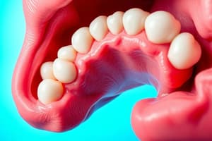
- A periodontal pocket is defined as a pathologically deepened gingival sulcus.
- The deepening may result from coronal movement of the gingival margin or apical displacement of the gingival attachment.
- Periodontal pockets are crucial clinical features of periodontal disease.
Types and Differences of Periodontitis
- Different types of periodontitis vary in etiology, natural history, progression, and response to treatment.
- Types of periodontal pockets include suprabony (where the base lies coronal to the alveolar bone) and infrabony (where the base is apical to the alveolar bone).
Causes of Pocket Deepening
- Deepening of the gingival sulcus can occur due to:
- Coronal movement of the gingival margin
- Apical displacement of the gingival attachment
- A combination of both
Histopathology and Clinical Features
- Histopathology of periodontal pockets shows inflammation, tissue destruction, and microbial invasion.
- Clinical features of an edematous pocket wall include a bluish-red, soft, spongy, and friable appearance with a smooth, shiny surface.
Attachment Loss and Clinical Distinctions
- Attachment loss is generally correlated with pocket depth, but the level of attachment loss is contingent on the base location on the root surface.
- Periodontal disease activity is clinically assessed through attachment loss, pocket depth, and inflammation.
Microbial Involvement
- Specific microorganisms have been linked to periodontal disease, including those that primarily colonize periodontal abscesses.
- Healthy gingiva is typically associated with different microbiota compared to diseased conditions.
Pathogenesis and Pulp Changes
- The pathogenesis of periodontal pockets involves bacterial infiltration leading to inflammation and tissue destruction.
- Infection from periodontal pockets can trigger pathological changes in the pulp tissue.
Clinical Examination of Periodontal Pockets
- Clinical signs of the presence of periodontal pockets include:
- Increased pocket depth
- Bleeding on probing
- Suppuration (presence of pus)
- The debris in periodontal pockets predominantly consists of bacterial byproducts, inflammatory cells, and necrotic tissue.
Unique Characteristics and Treatment Implications
- Suprabony pockets exhibit horizontal bone loss, whereas infrabony pockets show vertical bone loss.
- Incomplete removal of calculus during treatment can exacerbate pocket formation and tissue destruction.
- Active root caries lesions may be present in areas with periodontal disease activity.
Site Specificity in Periodontal Disease
- Site specificity refers to the pattern of periodontal destruction, often observed as localized disease activity adjacent to healthy areas.
Microscopic Observations
- Light and electron microscopy reveal changes in the root surface within periodontal pockets, including alterations in the attachment to the cementum.
- The presence of calculus correlates with the distance from the crest of the alveolar bone, typically averaging around 1.97 mm.
Outcomes of Incomplete Treatment
- Pathologic changes in the pulp and ongoing disease progression can result from inadequate pocket management.
- Clinical monitoring and assessment of pockets are critical for effective periodontal disease management and intervention.
Studying That Suits You
Use AI to generate personalized quizzes and flashcards to suit your learning preferences.