Podcast
Questions and Answers
Which of the following best describes the role of the periosteum in the context of the scalp's anatomy?
Which of the following best describes the role of the periosteum in the context of the scalp's anatomy?
- A fibrous membrane covering the surface of the skull. (correct)
- A type of tissue that includes skin and hair follicles.
- A layer with many blood vessels within subcutaneous tissue.
- A connective tissue that provides cushioning.
How many bones make up the cranium or cranial vault?
How many bones make up the cranium or cranial vault?
- 14 bones
- 6 bones
- 8 bones (correct)
- 28 bones
What is the primary function of the auditory ossicles located within the skull?
What is the primary function of the auditory ossicles located within the skull?
- To regulate balance and spatial orientation.
- To form the structure of the cranial vault.
- To enable hearing by transmitting vibrations. (correct)
- To protect the brain from physical trauma.
How many bones make up the face?
How many bones make up the face?
What is the primary function of the cone-shaped fossae of the orbits?
What is the primary function of the cone-shaped fossae of the orbits?
What is the role of the nasal septum?
What is the role of the nasal septum?
What percentage of the cranial vault is occupied by the brain, cerebrospinal fluid (CSF), and blood?
What percentage of the cranial vault is occupied by the brain, cerebrospinal fluid (CSF), and blood?
Why is the brain considered a perfusion sensitive organ?
Why is the brain considered a perfusion sensitive organ?
What functions are primarily coordinated by the cerebellum?
What functions are primarily coordinated by the cerebellum?
Which lobe of the brain is primarily responsible for processing visual information such as color and movement?
Which lobe of the brain is primarily responsible for processing visual information such as color and movement?
Which part of the diencephalon processes sensory input and influences mood and body movements?
Which part of the diencephalon processes sensory input and influences mood and body movements?
Which component of the brainstem is responsible for coordinating vital signs such as heart rate and blood pressure?
Which component of the brainstem is responsible for coordinating vital signs such as heart rate and blood pressure?
Which of the following components of the brainstem operates as a relay system, transmitting information for vision, hearing, movement, and posture?
Which of the following components of the brainstem operates as a relay system, transmitting information for vision, hearing, movement, and posture?
What is the function of the arachnoid mater?
What is the function of the arachnoid mater?
Which cranial nerve is responsible for the sense of smell?
Which cranial nerve is responsible for the sense of smell?
Which cranial nerve controls pupillary constriction and eye movement?
Which cranial nerve controls pupillary constriction and eye movement?
Which cranial nerve's primary function relates to hearing and balance perception?
Which cranial nerve's primary function relates to hearing and balance perception?
What is the most common mechanism of injury (MOI) associated with head trauma?
What is the most common mechanism of injury (MOI) associated with head trauma?
Why do lacerations to the scalp bleed profusely?
Why do lacerations to the scalp bleed profusely?
What is the primary risk associated with open head injuries compared to closed head injuries?
What is the primary risk associated with open head injuries compared to closed head injuries?
Which type of skull fracture typically results from an extension of a linear fracture to the base of the skull?
Which type of skull fracture typically results from an extension of a linear fracture to the base of the skull?
What physical sign is associated with a basilar skull fracture?
What physical sign is associated with a basilar skull fracture?
What is a key characteristic of secondary brain injury?
What is a key characteristic of secondary brain injury?
What is the Monro-Kellie Doctrine?
What is the Monro-Kellie Doctrine?
What physiological change can lead to cerebral herniation?
What physiological change can lead to cerebral herniation?
What is considered a late sign of rising intracranial pressure (ICP)?
What is considered a late sign of rising intracranial pressure (ICP)?
What are the components of Cushing's Triad?
What are the components of Cushing's Triad?
What is the difference between decorticate and decerebrate posturing?*
What is the difference between decorticate and decerebrate posturing?*
Which type of traumatic brain injury (TBI) involves specific, clearly defined brain injury isolated to a certain brain region?
Which type of traumatic brain injury (TBI) involves specific, clearly defined brain injury isolated to a certain brain region?
What type of intracranial hemorrhage occurs between the dura mater and the skull?
What type of intracranial hemorrhage occurs between the dura mater and the skull?
What specific sign/symptom is associated with an epidural hemorrhage?
What specific sign/symptom is associated with an epidural hemorrhage?
Which type of intracranial hemorrhage results from bleeding between the arachnoid mater and the dura mater?
Which type of intracranial hemorrhage results from bleeding between the arachnoid mater and the dura mater?
Which type of traumatic brain injury occurs when the brain is jarred around inside the skull and typically resolves spontaneously without structural damage?
Which type of traumatic brain injury occurs when the brain is jarred around inside the skull and typically resolves spontaneously without structural damage?
The mnemonic EENT stands for
The mnemonic EENT stands for
What is the main concern regarding injuries in or near the mouth and nose when assessing maxillofacial trauma?
What is the main concern regarding injuries in or near the mouth and nose when assessing maxillofacial trauma?
What term describes a horizontal fracture of the maxilla involving the hard palate and inferior maxilla?
What term describes a horizontal fracture of the maxilla involving the hard palate and inferior maxilla?
In the context of eye anatomy, what does the term 'globe' refer to?
In the context of eye anatomy, what does the term 'globe' refer to?
Which cranial nerve controls eye movement and pupil constriction?
Which cranial nerve controls eye movement and pupil constriction?
What action should be taken with a patient experiencing epistaxis (nosebleed)?
What action should be taken with a patient experiencing epistaxis (nosebleed)?
What is the correct initial action to take when managing a patient with chemical burns to the eye?
What is the correct initial action to take when managing a patient with chemical burns to the eye?
In the context of eye injuries, what does 'extruded/avulsed' refer to?
In the context of eye injuries, what does 'extruded/avulsed' refer to?
What is the recommended course of action for managing a dental avulsion?
What is the recommended course of action for managing a dental avulsion?
Flashcards
Periosteum
Periosteum
The fibrous membrane covering the surface of the skull.
Cranium/Cranial Vault
Cranium/Cranial Vault
The structure that encases and protects the brain.
Auditory Ossicles
Auditory Ossicles
Tiny bones that allow us to hear, located in the middle ear.
Foramen Magnum
Foramen Magnum
Signup and view all the flashcards
Thalamus Function
Thalamus Function
Signup and view all the flashcards
Brainstem Function
Brainstem Function
Signup and view all the flashcards
Reticular Activating System (RAS)
Reticular Activating System (RAS)
Signup and view all the flashcards
Dura Mater
Dura Mater
Signup and view all the flashcards
Arachnoid Mater
Arachnoid Mater
Signup and view all the flashcards
Pia Mater
Pia Mater
Signup and view all the flashcards
Olfactory Nerve
Olfactory Nerve
Signup and view all the flashcards
Optic Nerve
Optic Nerve
Signup and view all the flashcards
Oculomotor Nerve
Oculomotor Nerve
Signup and view all the flashcards
Trigeminal Nerve
Trigeminal Nerve
Signup and view all the flashcards
Vagus Nerve
Vagus Nerve
Signup and view all the flashcards
Head Trauma
Head Trauma
Signup and view all the flashcards
Closed Head Injury
Closed Head Injury
Signup and view all the flashcards
Open Skull Fracture
Open Skull Fracture
Signup and view all the flashcards
Linear Skull Fracture
Linear Skull Fracture
Signup and view all the flashcards
Depressed Skull Fracture
Depressed Skull Fracture
Signup and view all the flashcards
Basilar Skull Fracture
Basilar Skull Fracture
Signup and view all the flashcards
Traumatic Brain Injury (TBI)
Traumatic Brain Injury (TBI)
Signup and view all the flashcards
Primary Brain Injury
Primary Brain Injury
Signup and view all the flashcards
Secondary Brain Injury
Secondary Brain Injury
Signup and view all the flashcards
Cerebral Perfusion Pressure (CPP)
Cerebral Perfusion Pressure (CPP)
Signup and view all the flashcards
Cushing's Triad
Cushing's Triad
Signup and view all the flashcards
Focal Brain Injury
Focal Brain Injury
Signup and view all the flashcards
Diffuse Brain Injury
Diffuse Brain Injury
Signup and view all the flashcards
Epidural Haemorrhage
Epidural Haemorrhage
Signup and view all the flashcards
Subdural Haemorrhage
Subdural Haemorrhage
Signup and view all the flashcards
Subarachnoid Haemorrhage
Subarachnoid Haemorrhage
Signup and view all the flashcards
Cerebral Contusion
Cerebral Contusion
Signup and view all the flashcards
Cerebral Concussion
Cerebral Concussion
Signup and view all the flashcards
Diffuse Axonal Injury (DAI)
Diffuse Axonal Injury (DAI)
Signup and view all the flashcards
Facial/EENT Trauma
Facial/EENT Trauma
Signup and view all the flashcards
Orbits
Orbits
Signup and view all the flashcards
Eyeball
Eyeball
Signup and view all the flashcards
Cornea
Cornea
Signup and view all the flashcards
External Ear
External Ear
Signup and view all the flashcards
Le Fort I Fracture
Le Fort I Fracture
Signup and view all the flashcards
Study Notes
- Patient Care Theory is also known as PARA1004
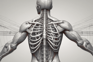
Anatomy of the Scalp
- The layers of the scalp include skin and hair, subcutaneous and connective tissue, periosteum, and lots of blood vessels
- The periosteum is a fibrous membrane that covers the surface of the skull.
Anatomy of the Skull
- The skull consists of 28 bones.
- Eight make up the cranium/cranial vault which encloses and protects the brain
- The auditory ossicles are composed of six tiny bones that facilitate hearing
- The face contains fourteen bones which do not form part of the cranial vault.
- Cranial Vault bones include 1 frontal, 2 parietal, 2 temporal, 1 occipital, 1 sphenoid, 1 ethmoid bone and 4 cranial sutures.
- The brain connects to the spinal cord and exits the cranial vault through the foramen magnum.
Anatomy of the Auditory Ossicles
- There are three auditory ossicles in each ear: Malleus, Incus, and Stapes
- These bones enable hearing via the transmission of vibration from the tympanic membrane.
Anatomy of the Facial Bones
- Key bones include: Nasal (2), Lacrimal (2), Palatine (2), Inferior nasal concha (2), Zygomatic (2), Maxilla (2), Mandible (1), and Vomer (1)
- There are 14 facial bones in total
- Cone-shaped fossae, known as orbits, enclose and protect the eyeballs
- The bridge of the nose is the nasal bone; most of the exterior nose is cartilage.
- The nasal septum separates the nose.
- The posterior condyle of the mandible joins with the temporal bone at the temporomandibular joint (TMJ).
Anatomy of the Brain
- The brain occupies 80% of the cranial vault, including cerebral spinal fluid (CSF) and blood.
- It is metabolically active and perfusion-sensitive.
- The brain consumes 25% of the body's glucose and 20% of the body's oxygen.
- The brain has no capacity to store oxygen or glucose, and is dependent on blood flow to supply these.
- In response to impaired oxygen/glucose supply, the brain attempts to compensate.
Cerebrum
- The largest part of the brain and largest portion of the cerebrum is the cerebral cortex.
- The cerebrum handles higher-level executive functions and regulates skeletal muscle movement and level of arousal (LOA).
- The cerebrum is divided into two hemispheres, each further divided into lobes.
Cerebellum and Lobe Function
- The Cerebellum is Latin for ‘little brain'
- It coordinates voluntary movements, posture, equilibrium, and coordination
- The frontal lobe is crucial for voluntary motor action, problem-solving, decision-making, impulse control, memory, learning and personality traits.
- The parietal lobe controls sensory and motor functions on the opposite side of the body as well as memory and emotion
- The occipital lobe processes visual information (color, movement, etc)
- The temporal lobe is responsible for speech, hearing, taste, smell, and long-term memory.
Diencephalon
- The diencephalon is located between the brainstem and cerebrum and contains the thalamus, subthalamus, hypothalamus, and epithalamus.
- The thalamus processes sensory input and influences mood and body movements
- The subthalamus controls motor functions
- The hypothalamus controls many of the body's functions (HR, digestion, temperature, emotion, hunger...) and the Epithalamus has an unknown function.
Brainstem
- The brainstem connects the brain and spinal cord.
- The Reticular Activating System (RAS) maintains consciousness and arousal.
- The brainstem controls vital signs.
- Damage to the brainstem can be fatal.
- The Medulla, Pons, and Midbrain make up the brainstem.
- The medulla coordinates vital signs, such as heart rate (HR), blood pressure (BP), blood vessel diameter, respiratory rate (RR), breathing pattern, vomiting, coughing, sneezing, and swallowing.
- The pons regulates sleep and breathing patterns.
- The midbrain contains basal ganglia, operates as a relay system, and transmits information for vision, hearing, movement, and posture.
Meninges
- Meninges are protective layers that surround the entire brain and spinal cord
- The meninges includes Pia Mater, Arachnoid Mater, and Dura Mater
- The dura mater is a tough outer layer that adheres to the inner wall of the skull.
- The arachnoid mater is a delicate, thin layer beneath which is a space with thin, delicate connective tissue and CSF (cerebrospinal fluid).
- The pia mater is a thin, translucent vascular layer that is adhered to the surface of the brain.
Cranial Nerves
- Olfactory (I) enables smell
- Optic (II) enables light perception and vsion
- Oculomotor (III) enables pupil constriction and eye movement
- Trochlear (IV) enables eye movement
- Trigeminal (V) enables motor chewing and sensory face, sinuses and teeth
- Abducens (VI) enables eye movements
- Facial (VII) enables facial movements
- Vestibulocochlear (VIII) enables hearing and balance perception
- Glossopharyngeal (IX) enables motor throat, swallowing, and gland seretion and sensory tongue and throat
- Vagus (X) enables heart, lungs, palate, pharynx, larynx, trachea, bronchi, GI tract, and ear performance
- Spinal Accessory (XI) enables shoulder and neck movements
- Hypoglossal (XII) enables tongue, throat and neck movements
Head Trauma
- Traumatic insults to the head may result in injury to the skin/soft tissues, skull or the brain.
- More than 50% of all traumatic deaths result from head injuries.
- The most common mechanism of injury (MOI) is motor vehicle collisions (MVCs).
- Over 2/3 of people in a MVC experience some type of head injury.
Scalp Injuries
- Lacerations to the scalp bleed profusely because of the amount of blood vessels in subcutaneous and connective tissues, and present a risk of hypovolemic shock, especially in children.
- Subgaleal hematoma creates a collection of blood between the skull and soft tissues of the scalp.
- Scalp injuries are usually caused by blunt trauma, and indicate the possibility of TBI (traumatic brain injury).
Types of Head injuries: Closed vs. Open
- Closed head injuries are the most common type and usually are caused by blunt trauma.
- In closed head injuries brain tissue is not exposed to the environment, but the fixed volume of the cranium increases the Intracranial Pressure (ICP)
- Open head injuries expose the dura mater and cranial contents to the environment and have a high mortality rate.
Skull Fractures
- Skull fractures are often associated with MVCs, falls, and assaults.
- There are four types of skull fractures: linear, depressed, basilar, and open.
- Linear fractures are non-displaced, occur in the temporal/parietal region 50% of the time, and may not show external signs. Eighty percents of Skull fractures are linear.
- Depressed fractures present with a soft, boggy area and bone fragments may be driven into the brain/meninges. Frontal/parietal regions with thinner bones are susceptible.
- Basilar fractures result from an extension of a linear fracture to the base of the skull and can cause CSF drainage from the ears/nose, periorbital ecchymosis (raccoon eyes), and Battle Sign (ecchymosis over mastoid process). These signs may develop rapidly or over 24 hours.
- Open fractures involve the contents of the cranial vault being exposed to the outside environment, very high mortality rate, and poses a high risk of bacterial meningitis.
Traumatic Brain Injuries
- These are defined as 'Traumatic insult to brain, producing physical, intellectual, emotional and social changes’.
- Common mechanisms include: Blunt force trauma, Deceleration injuries and Coup-contrecoup injury
- Two broad categories: Primary (direct) which injuries to brain/associated structures that result instantaneously from impact, and Secondary (Indirect) which are after-effects of a primary injury, that occur any time after the injury can include Cerebral edema, Intracranial hemorrhage, Increased ICP, Cerebral ischemia, Hypoxic brain injury, and Infection
Intracranial Pressure (ICP)
- Normal ICP: 0-15mmHg
- Increasing intracranial pressure may include: Oedema (swelling) or bleeding inside the cranial vault, which Squeezes brain against skull, and Decreases decreasing Cerebral Perfusion Pressure (CPP)
- CPP - pressure of blood flow through brain, CPP = MAP-ICP as ICP rises and becomes closer to, or surpasses MAP which decreases decreasing cerebral blood flow can decrease and lead to death due to no glucose or oxygen storage capacity
- CPP requires 60mmHg to perfuse the brain effictively, any drop results in cerebral ischemia
- CPP and ICP cannot be measured in the field, so focus is to reduce rises and Maintaining MAP
- Increasing ICP can cause herniation in which brain matter is forced through foramen magnum.
S&S: Rising ICP (Early)
- Vomiting (Often projectile and without warning/nausea)
- Headache
- Altered LOC
- Seizures
- Increased BP
- Decreasing HR
- Cheyne-Stokes Respirations (Irregular pattern)
S&S: Rising ICP (Late)
- Cushing’s Triad:
- Hypertension, Bradycardia, Irregular Respirations
- Unilaterally dilated and non-reactive or sluggish pupil
- Decorticate (Abnormal flexion)
- Decerebrate (Abnormal extension)
- Pulse Pressure: Difference between systolic and diastolic
Traumatic Brain Injuries Types: Focal vs Diffuse
- Focal Brain Injuries: cause a specific, clearly defined brain injury that is isolated to certain regions and includes: Intracranial Haemorrhage, Intracerebral Harmorrhage, and Cerebral Contusion
- Diffuse Brain Injuries: affect entire brain, Includes: Cerebral concussion and Diffuse Axonal Injury (DAI)
Intracranial Haemorrhage (Focal)
- Can cause increasing ICP
- Named by location of bleed:
- Epidural/Extradural haemorrhage, Subdural haemorrhage, Subarachnoid haemorrhage,Intracerebral haemorrhage
- Epidural/Extradural Haemorrhage is bleeding between the dura mater and the skull, usually resulting from a direct blow to the head or associated with linear skull fracture of temporal bone, and middle meningeal artery runs along the groove
- Specific S&S: Initial immediate LOC, followed by brief period of consciousness,Lucid interval, Subsequent deterioration into unconsciousness, Pupil on injures side becomes fixed and dilated
- Subdural Haemorrhage involves bleeding between the arachnoid mater and the dura mater, usually resulting from falls or injuries with strong deceleration forces, usually results from a vein rupturing and is more common with elderly patients and the alcohol dependand
- Specific S&S: fluctuating conciousness, focal neurological Signs
- Subarachnoid Haemorrhage is bleeding into the subarachnoid space where CSF is and can show traumatic or atraumatic mechanisms that may be ruptured
- Specific S&S: sudden, severe headache or meningeal irritation
- Intracerebral Haemorrhage is bleeding within the brain tissue itself
- Specific S&S may vary and can be associated with other injuries
Cerebral injuries: Contusion vs. Concussion vs DAI
- Cerebral Contusion is bruised and damaged brain tissue isolated to local area
- Specific S&S: headache and swelling indicate increased ICP
- Cerebral Concussion involves the brain jarring inside skull and usually resolves spontaneously with confusion/disorientation or amnesia
- Diffuse Axonal Injury(DAI) is similar to concussion but there is severe permanent neurological impairment from stretching/shearing/tearing of nerve fibers, typically resulting from severe coup and Contrecoup injuries
- Specific S&S: LOC (temporary or prolonged), Confusion, disorientation, amnesia (retro/antero), Cognitive impairment, mood swings, sensory/motor deficits
Assessment of Head injuries
- Head-injured patients are difficult to assess and are unreliable historians due to unconcioussness and amnesia and may be combative
- Consider the MOI and likelihood of head injury, maintan a high index of suspicion
- The assessment and management should be guided by injury severity, the level conciousness, haemodynamic stability, and airway stability; always be prepared for changes in condition
Management of Head injuries
- With exception of isolated penetrating head trauma, SMR is usually required
- Airway should be maintained and you must be prepared for vomitting and the likely need for suction
- Monitor for hypoxia and increased breathing rates. and if signs of Cerebral Herniation occur, ventilate <10 breaths per minute while maintaining ETCO2 of greater than 3-45mmHg
- If Cerebral Herniation is present maintain ETCO2 between 30-35mmHg to shunt oxygen and glucose
Circulatory Support
- If MAP and CPP drops a direct pressure control of haemorrhage is needed but be careful around open or depressed areas of the skull, because this pressure may cause more damage
90 - 90 - 9 Rule
- A single drop below 90% SpO2 increases mortality
- A single drop below 90mmHg systolic BP increass mortality
- A drop in GCS to <9 increases mortality
- Aim to prevent as possible by applying loose sterile dressing
Head Injuries: Summary
- Always consider potential for other life-threatening injuries, consider SMR (likely needed!), always be prepaired for rapid deterioration by monitoring airway, circulation, and breathing
Facial Trauma
- EENT stands for Ears, Eyes, Nose and Throat, and refers an number of structures in the head and neck other than the brain that may be traumatized
- Facial Trauma can be often overlooked because if more dominate injuries
Types of Anatomical Review
- Orbits are cone-shaped that contain eyeballs, blood vessels and thin, fractured bones
- Nose has multiple structures like a nasal bone, cartilages, and sinuses and is divided by membranes
Eye
- Eyes are also known "Gloves" is appoximatilly 3cm in diameter and controlled by III and II, can be easily damaged from movement
Ears
- Ears have multiple features, inner/outer, a membrane etc
Mouth
- The mouth includes teeth and the digestion and cranial nerves to support them
Thraot/Neck
- The Throat/neck are areas for a list a of items
Maillofocial Injuries
- Soft tissue injuries: May bleed alot but rarely life threatening/ May have swelling and injuries
General Symptoms are Ecchymosis, Swelling, pain, crepus,dental larracation and Fractures
Injuries to the: nose/ face and head
- Nosal -Fractures on the nose often complicated by ear splits
- Madubular
- Fractures that can cause pain /dantal avulsion/ or loss of mouth movements
###Maxillary Le Frote Fractures are broken bones with lots of bone damage
###Orbits
- Broken ethods may have orbit fractures as well - which causes eye damage
###Zygomatic Fracture:
-A broken Cheekbone is very painful and often has nerve damage to face and eye
###Assessments and General Care
- Always assume potential and closed head injuries - always Elevate head to drain, ICP can impact a patients outcome
###Nose assessment
- Control bleeds, leaning forward and prepare suction equipment
###Eye Assessment/ General
- They can range from foreign body to glove damage -Foreign object : it's protected orbit,eyelif and eyelashes and try flushing, never pull out
###Eye Laccaration And Heat burn -Control bleeds don't place direct force and heat burn will need chemical flush
###Ear assessment
- Soft tissues injuries are managed as other tissues. Do not attempt to remove objects and if there's blood always protect area
###Other injuries mouth and Airway
- Broken teeth are removed, if teeth can be reinserted go for it and reinserting has a strong rate
###Nock/Airway Penetrating Trauma require an inclusive dressing
-
Packing is needed, with caution around the airway -Stabilize and life saved interventions
-
Blunt Trauma, high likelihood of damage
-
Assessment includes crepis/ air sounds
###Summary:
Key Points
Always assume other life threatening is injuries Consider SMR as needed Prepaired fro Deterorations as monitoring is the key
Studying That Suits You
Use AI to generate personalized quizzes and flashcards to suit your learning preferences.