Podcast
Questions and Answers
What is the primary focus of mycology as a field of study?
What is the primary focus of mycology as a field of study?
- The study of bacteria
- The study of insects
- The study of fungi (correct)
- The study of viruses
Which characteristic is associated with molds?
Which characteristic is associated with molds?
- Unicellular growth
- Pasty growth
- Filamentous growth (correct)
- Mucoid growth
Which of the following best describes the cellular structure of fungi?
Which of the following best describes the cellular structure of fungi?
- Eukaryotic with a nucleus (correct)
- Prokaryotic without a nucleus
- Lacking cellular structures
- Similar to bacteria
How do fungi obtain their nutrients?
How do fungi obtain their nutrients?
What is the role of fungi in the environment?
What is the role of fungi in the environment?
Which of the following is an industrial use of fungi?
Which of the following is an industrial use of fungi?
Which of the following fungi is commonly used in the fermentation process for making bread and beer?
Which of the following fungi is commonly used in the fermentation process for making bread and beer?
What notable discovery is attributed to Alexander Fleming involving fungi?
What notable discovery is attributed to Alexander Fleming involving fungi?
What is a significant impact of fungi on plants??
What is a significant impact of fungi on plants??
What is the typical habitat of saprophytic fungi?
What is the typical habitat of saprophytic fungi?
What is the term for a fungal disease?
What is the term for a fungal disease?
Which factor has contributed to the increased incidence of fungal infections in recent times?
Which factor has contributed to the increased incidence of fungal infections in recent times?
What is a hypha?
What is a hypha?
What is the term for hyphae that are divided by cross walls?
What is the term for hyphae that are divided by cross walls?
What structure is formed by a mass of hyphae?
What structure is formed by a mass of hyphae?
What is the function of vegetative mycelium?
What is the function of vegetative mycelium?
Which of the following is a sexual spore formed in a sac-like cell?
Which of the following is a sexual spore formed in a sac-like cell?
What structure bears basidiospores?
What structure bears basidiospores?
Which type of spore is contained inside a sporangium?
Which type of spore is contained inside a sporangium?
What is a dematiaceous fungus?
What is a dematiaceous fungus?
What type of infections do dermatophytes cause?
What type of infections do dermatophytes cause?
What does it mean when a fungus is described as diphasic or dimorphic?
What does it mean when a fungus is described as diphasic or dimorphic?
What term describes the ability of a fungus to grow on the outside of a hair shaft?
What term describes the ability of a fungus to grow on the outside of a hair shaft?
Organisms that possess a true nucleus are called what?
Organisms that possess a true nucleus are called what?
What is a 'teleomorph'?
What is a 'teleomorph'?
What is the term for the vegetative body of a fungus?
What is the term for the vegetative body of a fungus?
Which term is used as a prefix to indicate a cutaneous mycosis?
Which term is used as a prefix to indicate a cutaneous mycosis?
What is the primary characteristic of yeasts?
What is the primary characteristic of yeasts?
What is geographic grouping in the classification of fungi based on?
What is geographic grouping in the classification of fungi based on?
Flashcards
Mycology
Mycology
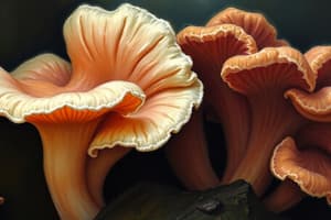
The study of fungi.
Molds
Molds
Filamentous type of fungal growth.
Yeasts
Yeasts
Pasty or mucoid form of fungal growth.
Eukaryotic
Eukaryotic
Signup and view all the flashcards
Heterotrophic
Heterotrophic
Signup and view all the flashcards
Saprophytes
Saprophytes
Signup and view all the flashcards
Parasites
Parasites
Signup and view all the flashcards
Ascospore
Ascospore
Signup and view all the flashcards
Basidiospore
Basidiospore
Signup and view all the flashcards
Zygospore
Zygospore
Signup and view all the flashcards
Conidia
Conidia
Signup and view all the flashcards
Arthroconidium
Arthroconidium
Signup and view all the flashcards
Blastoconidia / Blastospore
Blastoconidia / Blastospore
Signup and view all the flashcards
Chlamydospore
Chlamydospore
Signup and view all the flashcards
Sporangiospore
Sporangiospore
Signup and view all the flashcards
Thallospore
Thallospore
Signup and view all the flashcards
Ascus
Ascus
Signup and view all the flashcards
Chromoblastomycosis
Chromoblastomycosis
Signup and view all the flashcards
Coenocytic
Coenocytic
Signup and view all the flashcards
Conidiophore
Conidiophore
Signup and view all the flashcards
Dematiaceous
Dematiaceous
Signup and view all the flashcards
Dermatophyte
Dermatophyte
Signup and view all the flashcards
Diphasic
Diphasic
Signup and view all the flashcards
Ectothrixic
Ectothrixic
Signup and view all the flashcards
Endothrixic
Endothrixic
Signup and view all the flashcards
Eukaryotes
Eukaryotes
Signup and view all the flashcards
Fungemia
Fungemia
Signup and view all the flashcards
Germ Tube
Germ Tube
Signup and view all the flashcards
Mold
Mold
Signup and view all the flashcards
Mycosis
Mycosis
Signup and view all the flashcards
Study Notes
Overview of Mycology
- Mycology is the study of fungi, including molds and yeasts
- Molds exhibit filamentous growth, while yeasts have a pasty or mucoid form
- There are over 50,000 valid fungal species, with multiple names possibly due to variances in size, color, host, or geographical location
- Fungi are gram-positive and require oxygen
- Fungi are eukaryotic, containing a nucleus with a membrane, endoplasmic reticulum, and mitochondria
- Fungi are heterotrophic and need organic nutrients for energy, similar to animals and bacteria, whereas plants make their own food
- Fungi rely on enzymes to get energy from organic substrates
- Saprophytes live on dead organic matter
- Parasites live on other living organisms
- Fungi are important in carbon and element recycling
Role of Fungi in the Economy
- Mushrooms (Basidiomycetes) and truffles (Ascomycetes) are industrial uses of fungi
- Fungi serve as a natural food source for animals
- Yeast is a food supplement that provides vitamins
- Penicillium is used to ripen cheese, adding flavor to Roquefort, etc.
- Fungi are utilized to change texture and flavor in the manufacturing of food
- Fermentation is an industrial use of fungi
- Fruit juices go through fermentation to produce ethyl alcohol
- Saccharomyces cerevisiae is used as brewer's and baker's yeast in fermentation.
- Fats, proteins, acids, and industrial alcohol are fermented by fungi
Antibiotics and Plant Pathology
- In 1928, Alexander Fleming first discovered antibiotics, from observing how a fungus suppresses bacteria on a culture plate
- Fungi cause the majority of plant diseases
Medical Importance and Mycology
- 50-100 recognized species are human pathogens
- Many prefer to be free-living saprophytes, and only accidentally become pathogens
- Pathogenic fungi must tolerate the host site temperature and possess an enzymatic system to parasitize animal tissues
- Fungal infections have been on the rise recently
Medical Mycology - History
- From 1941 to 1973, reported deaths in the U.S. from scarlet fever, typhoid, whooping cough, diphtheria, dysentery, and malaria decreased from 10,165 to 107
- Deaths attributed to mycoses increased from 324 to 530 during the same period
Medical Mycology - Modern Man
- Increased mobility/travel to geographical areas facilitates exposure to endemic fungi
- Immunocompromised individuals are more susceptible to fungal infections
- AIDS patients, individuals taking tetracycline (acne) and birth control pills, those with excessive use of antibiotics, and people taking immunosuppressants are immunocompromised populations
- Environmental factors like air and water pollution, processed foods, and fad diets compromise the immune system, leading to fungal infections
- Incidence of fungal infections increase with ageing populations
Mycoses Immunology
- Antibody mediated immunity (B-cell humoral) stimulates antibody production in response to fungal infections
- Serological tests are used to identify fungal diseases
- Cellular mediated immunity (T-cell) is effective in resisting fungal infections
Fungal Structures: Hyphae and Mycelium
- Hyphae are fundamental, tube-like structural units of fungi
- Septate hyphae are divided by cross walls
- Aseptate hyphae lack cross walls
- Mycelium is a mass of hyphae that form the vegetative part of a fungus
- Aerial mycelium grows in the air
- Vegetative mycelium absorbs nutrients
- Fertile mycelium produces conidia or spores for reproduction
Fungal Spores
- Sporulation is the merging of nuclear material or genes to form spores
- Self-fertile fungi are homothallic; those with mating types are heterothallic
- Sexual spores result from the fusion of nuclei
Sexual Spores
- Ascospores form in a sac-like cell called an ascus, where eight spores are often formed (Ascomycetes)
- Basidiospores are formed through the union of two nuclei on a club-shaped structure called a basidium (Basidiomycetes)
- Zygospores are thick-walled and formed during sexual reproduction in Phycomycetes
Asexual Spores
- Asexual spores are the most common spore type
- Conidia are asexual fungal spores borne externally on a conidiophore, referred to as macro- and microconidia (Ascomycetes & Deuteromycetes)
- Macroconidia are multicellular; Microconidia are unicellular
- Arthroconidia (Arthrospores) are asexual spores formed by mycelium disarticulation
- Blastoconidia / Blastospores are asexual spores formed from a budding process along the mycelium or another blastospore (class Ascomycetes)
- Chlamydospores are thick-walled, resistant asexual spores formed by direct differentiation, concentrating protoplasm and nutrients in the mycelium
- Sporangiospores are asexual spores contained in a sporangium at the end of a sporangoiphore (Phycomycetes)
- Thallospores are asexual spores produced on a thallus (hypha), and can be found in Deuteromycetes
Miscellaneous Mycology Terms
- Ascus: Sac-like structure in Ascomycetes containing (usually eight) ascospores
- Asexual Reproduction: Spores formed directly from vegetative mycelium/specialized hyphae
- Chromoblastomycosis: Subcutaneous mycosis from traumatic inoculation of dematiaceous fungus
- Coenocytic: Aseptate hypha or cell with numerous nuclei
- Conidiophore: Specialized hypha branch where conidia develop
- Dematiaceous: Pigmented, dark color, usually gray to black
- Dermatophyte: Fungi causing superficial mycoses
- Diphasic (dimorphic): Ability of fungi to grow as yeast or filamentous stages
- Ectothrixic: Fungus grows on the outside of a hair shaft
- Endothrixic: Fungus grows and penetrates into the hair shaft
- Endogenous: Derived from an internal source
- Exogenous: Derived from an external source
- Eukaryotes: Organisms with a true nucleus (like fungi)
- Fungemia: Fungal blood infection
- Fungi Imperfecti: Fungi class with septate hyphae, known for asexual reproduction only
- Germ Tube: Small projections from yeast cells, indicating hyphal formation
- Hyaline: Colorless, clear
- Mold: General term for filamentous fungi
- Mycetoma: Clinical syndrome of localized, tumorous lesions in cutaneous/subcutaneous tissues; often on the foot
- Mycosis: A disease caused by a fungus
- Mycotoxins: Toxins of fungal origin
- Oospore: Zygospore - Sexual spore (Phycomycetes)
- Perfect Fungi: Fungi with sexual and asexual reproductive stages
- Phycomycetes: Fungi class with coenocytic mycelium/stiff sporangiophores
- Pseudohyphae: Chain of elongated budding cells that fail to detach
- Ringworm: Circular/ring-like skin lesions caused by dermatophytes
- Rhizoids: Root-like structures
- Saprobe (Saprophyte): Organism getting nourishment from dead organic matter
- Sexual Reproduction: Zygote/spore formation after fusion of two haploid nuclei
- Sporangiophore: Hypha or stalk bearing a sporangium
- Sporangium: Sac/cell containing asexually produced spores
- Spore: Reproductive body of a fungus; resistant body for adverse environment
- Sterigmata: Structure from a basidium, supporting basidiospores
- Sporotrichosis: Mycosis from Sporothrix schenckii inoculation, lymphocutaneous type
- Telemorph: Sexual form of a fungus
- Thallospore: Spore formed by parts of the thallus changing
- Thallus: Vegetative body of a fungus
- Tinea (Ringworm): Prefix with infected area to indicate cutaneous mycosis
- Yeast: Pasty/mucoid fungus, microscopically shows budding cells
- Zoophilic: Dermatophytes which are parasitic on animals/humans
Fungi Classification by Geography
- Classification is based on geographic distribution, noting that fungal diseases are localized to particular areas
- Histoplasmosis occurs in the Central Mississippi and Ohio River Valley
- Coccidioidomycosis occurs in the San Joaquin Valley
Fungi Classification by Epidemiology
- Epidemiologic grouping is based on how fungal diseases spread
- Few are contagious (e.g., ringworm of the scalp)
- Mycoses depends on host susceptibility
Fungi Classification by Taxonomy
- Scientific grouping according to morphologic and cultural characteristics
- KINGDOM: Plantae
- PHYLUM: Thallophyte - Entire plant is somatic; no roots, or leaves.
- SUBPHYLUM: Schizomycotina
- CLASS: Schizomycetes (bacteria)
- ORDER: Actinomycetales
- FAMILY: Actinomycetaceae
- GENUS: Actinomyces
- SPECIES: A. isarelii, A. bovis, A. naeslundii
- GENUS: Nocardia
- SPECIES: N. asteroides, N. brasiliensis, N. carviae
- FAMILY: Streptomycetaceae
- GENUS: Streptomyces
- SPECIES: S. madurae, S. pelletierii, S. paraquavensis, S. somaliensis
- ORDER: Actinomycetales
- CLASS: Schizomycetes (bacteria)
- SUBPHYLUM: Schizomycotina
- SUBPHYLUM: Myxomycotina
- CLASS: Myxomycetes (slime molds)
- SUBPHYLUM: Eumycotina (true fungi)
- CLASS: Phycomycetes (algae fungi)
- GENUS: Rhizopus sp., Mucor sp., Others
- CLASS: Ascomycetes
- GENUS: Penicillium sp., Aspergillus sp., Many others
- CLASS: Basidiomycetes
- GENUS: Amanita sp. (many poisonous toadstools), Agaricus sp. (edible mushrooms)
- CLASS: Deuteromycetes (fungi imperfecti)
- Yeast that reproduce by budding.
- GENUS & SPECIES:
- Cryptococcus neoformans, Candida albicans; Diphasic yeast (yeast-like when grown at 37 degrees)
- GENUS & SPECIES:
- Blastomyces dermatitidis, Blastomyces brasiliensis, Histoplasma capsulatum, Coccidioides immitis, Sporotrichum schenkii
- Filamentous fungi - no yeast stage
- GENUS & SPECIES:
- Geotrichum sp., Microsporum sp., Trichophyton sp., Epidermophyton sp.
- GENUS & SPECIES:
- CLASS: Phycomycetes (algae fungi)
- PHYLUM: Thallophyte - Entire plant is somatic; no roots, or leaves.
Topographic Grouping of Fungi
- Classification is based on the type of mycoses produced
Superficial Mycoses
- Confined to outermost skin/hair layers with no host cellular/inflammatory response
- Disease is recognized on a cosmetic level
Cutaneous Mycoses
- Fungi have an affinity for keratin in skin, nails, and hair
- Most cutaneous infections from dermatophytes
- They prefer non-living layers so host response is inflammation/eczema (named by body area)
- The three genera of cutaneous mycoses include Microsporum, Epidermophyton, and Trichophyton
- These cause infection of skin, hair and nails
Cutaneous Mycoses by Genera
- Microsporum affects hair and skin rarely, and causes nail infections
- Can remit spontaneously, and frequently occurs in children
- Trichophyton affects hair, skin and nails in both children and adults
- Causes chronic infections
- Epidermophyton affects skin and nails, with rare hair infections
- Commonly occurs in adults
Dermatophytes
- Dermatophytes produce septate, hyaline mycelium
- Each has a preferred host
- Microsporum affects children, rarely adults
- Trichophyton affects both children and adults
- Epidermophyton affects adults, rarely children
Subcutaneous Mycoses
- Subcutaneous mycoses - infections in deeper layers of skin and often muscle tissue
- Man is an accidental host
- Introduction usually from trauma
- Infection tentatively identified by tissue reaction
Systemic Mycoses
- Attack deep tissues and organ systems, create symptoms that resemble diseases
- The two categories of systemic disease:
- Those caused by truly pathogenic fungi with the ability to cause disease in the normal human host when the inoculum is of sufficient size.
- Histoplasma capsulatum, Blastomyces dermatitidis, Coccidioides immitis, Paracoccidioides brasiliensis*
- Opportunistic fungi, low virulence organisms, and infections will occur when patient's defenses are lowered.
- Aspergillus sp., Candida albicans, Cryptococcus neoformans*
Lab Methods: Specimen Collection
- Collection, handling, and processing of clinical mycology specimens
- Properly collect all specimens
- Physician or nursing staff collections
- Skin is cleaned with 70% alcohol, and surface saprophytes are removed
- Nails are cleaned and clipped, then minced before inoculation to media
- Collect hair from the scalp edge with a Wood's lamp (fluorescence)
- Obtain hair by plucking, brushing, or using sticky tape
- Collect body fluids via normal sterile procedures
Lab Methods: Specimen Preparation
- Hair & nails are sent in a dry envelope, in a proper container
- Send frozen, or on dry ice
- Package with biohazard regulations, use tube media (no plates)
- Use aluminum screw-capped mailing tubes with cardboard outer tubes
- Include inside labeling: patient ID, specimen source, suspected organism
- Include Outside labeling: "WARNING: POTENTIAL PATHOGEN"
Lab Methods: Processing Specimens and Body Fluids
- Skin, nails, & hair undergo direct exam via KOH preparation to recover fungus
- Appropriate body fluid processing:
- CSF is centrifuged; examine sediment microscopically and inoculate media
- Pleural fluid, sputum, and bronchial aspirations must be fresh; saprophytes would overgrow pathogens such as H. capsulatum, only refrigerate for 2 hours
- Gastric washings are processed like pleural fluids
- Genito-urinary specimens use the first morning specimen; centrifuge
- Blood/bone marrow is inoculated directly to BHI broth/slant, inoculate to other fungal media as well
- Wound abscess or drainage should be cultured anaerobically for suspected actinomycosis
- Tissue specimens are examined for pus, caseous material, or granules; mince aseptically and inoculate using sterile saline
Lab Methods: Direct Examination
- Direct examination required on any biological material sent to the lab for fungus culture
- Look for spores, hyphae, mycelial elements, budding yeast, and mycotic granules
- Wet mount prep (good for yeast examination) done in a natural medium minimizes structure loss
- KOH prep with potassium hydroxide is done on skin scraping, hail, nails, sputum, and vaginal specimens
- The KOH digests and clears tissue cells, mucus, etc. so fungal elements are visible
Lab Methods: Stains Used
- Lactophenol Cotton Blue (LPCB) quickly evaluates fungal structures and stains chitin
- Periodic Acid - Schiff stain (PAS) stains polysaccharide in cell walls of fungi, fungal stain pink-red with blue nuclei
- Gomori Methenamine Silver stain outlines fungi in black because silver nitrate precipitates on the cell wall
- The internal parts of hyphae are deep rose to black and the background is light green
- Gridley stains Hyphae and yeast dark blue or rose with a deep blue tissue background
- Mayer Mucicarmine stains capsules of Cryptococcus neoformans deep rose
- Fluorescent antibody stain is simple, specific and sensitive for detecting fungi and other organisms
- Papanicolaou stain is good for initial differentiation via dimorphic fungi, works well on sputum smears
- Gram stains are generally gram positive for fungi, variable for Actinomyces and Nocardia
- Modified Acid-Fast stain is used to differentiate acid-fast Nocardia from other aerobic Actinomyces
- Giemsa stains blood and bone marrow. Histoplasma capsulatum is an intracellular organism, which appears as small oval to pear-shaped yeast-like cells
Lab Methods: Yeast Stains and Fungal Culturing
- India Ink demonstrates the capsule of Cryptococcus neoformans in CSF specimens
- Tube media is generally used for fungal culturing to prevent spore release
- Tube media also has less dehydration and is easy to store
- Inoculate agar tubes in a straight line.
- Preliminary identification is based on differential growth patterns on various media
Culture Media
- Carefully select appropriate culture media
- Use more than one media type
- Sabouraud's dextrose agar (Sab-Dex) is a classic medium, is recommended for most studies
- Sabouraud's dextrose agar with chloramphenicol inhibits bacterial growth
- Mycosel agar inhibits bacterial growth and saprophytic fungi
- Cycloheximide inhibits saprophytes like Aspergillus and Scopulariopsis
- Cycloheximide also inhibits Cryptococcus neoformans
- It inhibits bacteria-like fungi (Actinomycetes)
Culture Recovery, and Special Agar
- Brain Heart Infusion slant is more enriched than Sab-Dex, and is used in H. capsulatum recovery
- Potato-dextrose agar (PDA) and Corn-meal agar induce spore formation, aiding identification
- Caffeic Acid Agar allows Cryptococcus neoformans to produce melanin
- Birdseed Agar isolates Cryptococcus neoformans from contaminated cultures
- KT Medium & Kelley Agar converts dimorphic fungus, Blastomycetes dermatitidis, from mycelial to yeast form
- Modified Converse Liquid Medium (Levine's) promotes spherule production by Coccidioides immitis
Growth Requirements
- Grow fungi at room temperature (25-30 C)
- Nocardia sp. and some dimorphic organisms grow best at 37°C
- Any fungus capable of growing at 37°C should be considered a potential pathogen
- True fungi are aerobic and there are a few anaerobes
- Some yeasts grow overnight, saprophytes are fast growers (several days), and cultures are held for 4 weeks
- Paracoccidioides brasiliensis may require 4-5 weeks, and Histoplasma capsulatum require 10 weeks
ID Techniques for Fungi
- Inoculation involves plating and slanting the specimen
- Plates are inoculated with a large "S" shape to allow rapid growing fungi to be removed
- Slants are inoculated with a straight line
- Incubation is aerobic, but can be anaerobic if Actinomycetes are suspected
- Expect dimorphic fungi, and set room temp roughly 37E
- Consider the type of media and growth rate
- Routine cultures are examined every other day for 4 weeks
- Systemic pathogens take 10 days to 2 week growth, but saprophytic fungi grow within 1 week
Colony Morphology and Mycelium
- Surface topography describes fungal colonies that are either free-growing, or grown in restricted manner
- Colonies can be cottony, granular, velvety, powdery, silky, smooth, creamy, or waxy in texture
- Pigmentation can be colorless or brightly colored on the fungus itself, its sporulating apparatus, on the agar, or on the bottom of the colony (reverse pigmentation)
- Pigment color is due to color the sporulating apparatus
- The top pigment and agar discoloration are crucial in determining fungi
- Vegetative mycelium provides nutrition and aerial mycelium is reproductive
Microscopic Evaluation and Hyphae
- Teased Preparation is a method of Microscopic evaluation
- Slide Culture Techniques are great because they allow for undisturbed microscopic morphology
- Transparent Tape Preparation is a method of Microscopic evaluation
- Hyphae structure has plural hyphae and single hyphae
- Hyphae is either septate or aseptate and is either dematiaceous and hyaline
Spore Structures and Tests
- Spores are used in reproduction and terms that address reproduction exist
- Biochemical studies can be performed to identify yeast and yeast-like organisms
- Grow and utilize carbohydrates under anaerobic-ly observing acid and gas production
- Inoculate under broth that is completely covered, as acid production will turn Bromcresol purple yellow
- Observe gas production in bubbles every 48 hours for 14 days
- Utilize carbohydrates as a carbon source in carbohydrate assimilation as seen when Bromcresol purple turns yellow
- Tubes can be negative if unchanged
- Utilize 3 tubes that differ in nitrogen sources in nitrogen assimilation, and bromthymol blue will become blue to yellow if the test is positive
- Use Christensen's urea agar to see if the urea hydrolyzed from yeast forms ammonia that turns media yellow to dark pink
- Conduct a germ tube test of Candida albicans or stellatoidea
- Inoculate with appropriate agar and observe every 24 hours to test for chlamydospores
Hypersensitivity and Saprophytes
- Hypersensitivity (seromycology; skin tests) and serological tests can determine T-cell and B-cell immunity
- Serological tests require drawing sera in pairs during the acute convalescent phases
- Test for complement fixation, agglutination, precipitin, immunofluorescence, immunodiffusion, and counterimmunoelectrophoresis
- The goals of testing antifungal susceptibility, and the serum concentration of antifungal agents are to examine any unusual fungal isolates
- Some progress in attempt to standardize susceptibility testing as a means in evaluating unusual fungal isolates, determining the cause(s) of patient relapse and treatment failure
- Determining serum concentration of antifungal agents is important when absorption factors in kidney or liver
- HPLC is highly accurate in drug analysis
- Saprophytes can be contaminants or opportunistic pathogens
- Most are inhibited by cycloheximide and are grouped by mycelia
- Mycelia have septate or aseptate hyaline versus dematiaceous properties
Hyaline Members
- Hyaline members can vary in growth rate. surface appearance, and color appearance
- Aspergillus mycelium is septate and hyaline with unbranched condiophores
- A. fumigatus from a pulmonary source grows well at 37C
- Paecilomyces sp.: is a rapid grower with Brush-like conidiophores and Long, tapered sterigmata
- Penicillium sp. is commonly and rapidly white that becomes bluish-green. Conidiophores characteristically form a brush-shaped structure and Sterigmata is flask shaped
- Scopulariopsis sp. are a moderately slow growing that becomes white turning brown with age. Branched or unbranched conidiophores; sterigmata are coarsely roughened
- Trichoderma sp. Shows a moderately rapid growth with Flask-shaped conidiophores and clustered conidia
Dematiaceous Members
- Fusarium sp. has a Rapid colonial growth and be come a brightly colored. Short conidiophores are often branched, have both macro- and micro-conidia, which are oval to sickle-shaped
- Has been reported in eye infections
- Alternaria sp. exhibits growth; colonies become very dark with age, can be overgrown
- conidiophores bear single chains of conidia
- Curvalaria sp. Show rapid growth as velvety colonies that vary in color from grayish-brown to black that form Spirally arranged brown conidia
- Cladosporium sp. has rapid growth and reverse black mycelium
- The dematiaceous mycelium is septate.
The Phycomycetes
- The Phycomycetes are asexual. and are always susceptible to cycloheximide.
- Some Phyomycetes are rapidly growing growers that have root-like rhizoids
- Spore (sporangia) bearing structures are called sporangiophores.
- Mucor fills a growth tube one day, lacks rhizoids, and has sporangiophores
- Rhizopus features rhizoids, and sporangiophores nodal are originally from the culture
- Syncephalastrum shows rapid growth as a white/dark colonies that grows high
- The high, septate mycelium is short, exhibits branched sporangiophores
Yeasts: Candida
- Candida albicans often comes with oral thrush, skin infections
- Testing, production of chlamydospore on agar can identify infection
- Torulopsis glabrata is always with the candida infection and always shows during carb fermentation
- Can be observed with elderly during diabetes
- Cryptococcus neoformans from wild pigeons that grown in melanin/warm temp
- Identified by India ink staining
- Geotrichum will show with respiration infections, and show on vagina
- Disease shows when immune is low
Bacteria-like Fungi
- Sulfar granulation can be observed by the Actinomycetes israeli
- To grow, require media, special staining and conditions
- They will always anaerobically grow
Biphasic Fungi and Mycoses: Sporothrix schenkii
- Sporothrix schenckii grows and can cause rose fever with gardeners are affected
Coccidioides immitis
- Coccidioides immitis Is The San Joaquin valley fever
- Exhibits as mycelila/tissue parasites
Histoplasma capsulatum
- Histoplasma capsulatum is Mississippi Valley fever (found in darling)
Deep Tissue Agents
- Blastomyces dermatitidis and North American blastomycosis / Gilcrest disease agent
- Paracoccidioides braziliensis agent of South American Blastomycosis
Types of Mycoses
- Types are based on body sites
Superficial Mycoses
- Affect skin/hair tissues, inflammatory and or destructive, but are the causation
- Malassezia furfur that cause/pityriasis
- Phaennellcomyes werneckii causes Exophiala
- Trichosporon
- Piedraia hortae
Treatment and Cutaneous Mycoses
- Cutaneous Mycoses include C. albicans & three genera are always from skin/hair tissue
-*Microsporum, Epidermophyton
- Septate and hairlike is common
Microscopic Types
- Skin and hair as Microsporum
- Tryhoohyton always shows in all children -Trychophyton infection
- Epidermohpyton appears in a beaver-wall infection
Subcutaneous Mycoses and Infections
- Causes from soil bacteria
- Chromoblastomycosis
- Chromoblastomycosis (fungal)
Chromoblastomysos Types
- A papule caused spreading/warty or tumor like Pedrosoi is common/wears shoes, wear hygiene/world, tropical
Etiological Agents
- Fungal agents
- Ameduromycotic agents and are clinical pictures with Amoxicilitic and death is a risk
- Is slow and progressive
- Body walled Lymph node are affected/body tries and immune
- systemic is common
Agents and Blastomycosis
- Dissemination of agents from yeast, affect the entire system
- Types of Agents -Dermatitides ,Paracoccidiodomycosis
Capsulation
- Capsulation and bronchio pneumonia shows and occurs with bird/ bat stool
- Coccidiodides is asymptomatic and airborne
- Histoplasmosis produces arthroconidia
- Brazilian Para and Brazilian blaster
Treatment
- Effectiveness caries
- Nystatin is work
- Pimarcin
- B and D flucytosin
- Mica oil
Studying That Suits You
Use AI to generate personalized quizzes and flashcards to suit your learning preferences.