Podcast
Questions and Answers
What is the most common cause of genu varum in infants?
What is the most common cause of genu varum in infants?
Developmental and physiologic causes.
What treatment is usually directed for children with genu varum caused by rickets?
What treatment is usually directed for children with genu varum caused by rickets?
Correcting the cause, usually vitamin D deficiency.
What is coxa vara?
What is coxa vara?
An angle of the femur less than 110°.
What surgical procedure is performed to correct cubitus varus?
What surgical procedure is performed to correct cubitus varus?
What are the main causes of osteogenesis imperfecta?
What are the main causes of osteogenesis imperfecta?
Which of the following is NOT a characteristic sign of infantile rickets?
Which of the following is NOT a characteristic sign of infantile rickets?
Vitamin D deficiency is a common cause of osteogenesis imperfecta.
Vitamin D deficiency is a common cause of osteogenesis imperfecta.
What is the primary cause of rickets?
What is the primary cause of rickets?
What are the two main types of bone cells?
What are the two main types of bone cells?
Which condition is primarily characterized by increased resorption of bone?
Which condition is primarily characterized by increased resorption of bone?
What is the treatment for severe deformities due to infantile rickets?
What is the treatment for severe deformities due to infantile rickets?
Flashcards are hidden until you start studying
Study Notes
Common Orthopaedic Deformities
- Deformities are deviations from the normal appearance of a body part.
- Orthopaedic deformities can be congenital or acquired.
- Many deformities may require surgical intervention.
Knee Deformities
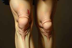
- Genu Varum (Bow Legs) in infants:
- Most commonly caused by development and is physiological.
- Usually corrects itself by age 4.
- No treatment is needed.
- Genu Varum (Bow Legs) in children aged 1.5-2 years:
- Most commonly caused by rickets due to vitamin D deficiency.
- Symptoms include systemic signs and lab findings.
- Treatment focuses on correcting the underlying cause, often with vitamin D supplements.
- Advanced cases may require osteoclasis or osteotomy.
- Genu Varum (Bow Legs) in adults:
- Causes include osteoarthritis, mal-united fractures, ligamentous injuries, and Paget's disease.
- Treatment involves surgical correction of the deformity.
- Genu Valgum (Knock Knees) up to age 4:
- Usually developmental or physiological.
- Typically corrects itself by ages 6-8.
- If progressive, supracondylar osteotomy of the femur is required.
Hip Deformities
- Normal neck-shaft angle of the proximal femur is 160° at birth, decreasing to 125° in adulthood.
- Coxa Vara: angle less than 110°.
- Coxa Valga: angle more than 130°.
- Both can lead to gait disability, spinal pain, and deformity.
- Treatment is by corrective subtrochanteric osteotomy.
Elbow Deformities
- Normal carrying angle is 10-15 degrees of valgus.
- Cubitus valgus: angle above 15°.
- Cubitus varus: angle below 10°.
- These deformities can be surgically corrected with a wedge osteotomy at the lower humerus.
Bone Softening Diseases
- Bone is connective tissue with cells and matrix.
- Bone Cells:
- Osteoblasts: form bone.
- Osteoclasts: resorb bone.
- Bone Matrix (Osteoid):
- Made of collagen and calcium salts.
- Causes of Bone Softening Diseases:
- Defective osteoid formation (e.g. osteogenesis imperfecta)
- Defective osteoid mineralization (e.g. rickets)
- Increased bone resorption by osteoclasts (e.g. hyperparathyroidism)
- Decreased bone formation by osteoblasts (e.g. osteoporosis)
Osteogenesis Imperfecta (OI)
- Generalized mesenchymal disorder characterized by:
- Defective osteoid production, resulting in fragile bone and multiple fractures.
- Defective collagen production, causing joint hyperlaxity and blue sclera.
- Types of OI:
- Congenital: stillborn or born with multiple fractures.
- Infantile: manifests early in infancy with multiple easy fractures.
- Tarda: manifests in late childhood.
- OI Treatment:
- No cure.
- Orthopedic management focuses on:
- Preventing fractures and deformities using supporting devices.
- Treating current fractures.
- Correcting deformities with surgery if needed.
Rickets
- Disease of growing bone due to inadequate calcification of bone matrix.
- Causes:
- Infantile: deficient vitamin D intake and lack of sunlight exposure.
- Coeliac: deficient absorption of fat and vitamin D due to gluten intolerance.
- Renal: disturbed calcium-phosphorus metabolism due to renal dysfunction.
- Clinical Picture:
- Bony deformities (bow legs, knock knees).
- Characteristic signs of infantile rickets:
- Boxy-bossy skull
- Rickety rosary
- Pigeon chest
- Harrison Sulcus
- Broad metaphysis
- Marfan sign
- 10 Important Clinical Features:
- Delayed closure of fontanelles
- Frontal bossing
- Dental hypoplasia
- Pectus carinatum
- Swelling in wrist and ankle joints
- Wide sutures
- Craniotabes
- Rachitic rosary
- Harrison's sulcus
- Bowing of legs
- X-ray:
- Bone rarefaction, broadened and cupped metaphyses with a brush border.
- Treatment:
- Infantile rickets: vitamin D, calcium, good diet, sunlight exposure.
- Renal rickets: active vitamin D (one alpha) and calcium.
- Severe deformities: surgical correction (osteotomy) may be needed.
Hyperparathyroidism
- Primary hyperparathyroidism: increased parathyroid hormone secretion due to hyperplasia or tumor.
- Causes increased bone resorption leading to:
- Hypercalcemia.
- Recurrent renal calculi.
- Cystic changes in bones, deformities, and pathological fractures.
- Treatment: surgical excision of parathyroid adenoma.
Osteoporosis
- Diminished bone mass due to decreased bone formation by osteoblasts with aging and menopause.
- Common Affected Sites:
- Vertebral column: causes vertebral collapse, back pain, kyphosis, and wedged or biconcave vertebrae on x-ray.
- Proximal femur.
- Proximal humerus.
- Distal radius (prone to fractures after minor trauma).
- Diagnosis: bone densitometry (DEXA).
- Treatment:
- Adequate diet (calcium, vitamin D, protein).
- Daily exercise and sunlight exposure.
- Medications:
- Calcium, vitamin D.
- Calcitonin (increases osteoblastic activity).
- Bisphosphonates (inhibit osteoclastic activity).
- Hormone replacement therapy (HRT) in some cases.
Studying That Suits You
Use AI to generate personalized quizzes and flashcards to suit your learning preferences.


