Podcast
Questions and Answers
Which type of oral mucosa is characterized by its adaptation to accommodate the sensation of taste?
Which type of oral mucosa is characterized by its adaptation to accommodate the sensation of taste?
- Lining mucosa
- Masticatory mucosa
- Attached gingiva
- Specialized mucosa (correct)
The gingiva and hard palate are primarily composed of which type of oral mucosa?
The gingiva and hard palate are primarily composed of which type of oral mucosa?
- Masticatory mucosa (correct)
- Specialized mucosa
- Alveolar mucosa
- Lining mucosa
Which of the following is a characteristic of lining mucosa, found on the cheeks and floor of the mouth?
Which of the following is a characteristic of lining mucosa, found on the cheeks and floor of the mouth?
- Keratinized surface
- Tightly bound to underlying structures
- Adapted for sensation of taste
- Non-keratinized epithelium (correct)
In the context of oral tissues, what is the primary difference between keratinized and nonkeratinized epithelium?
In the context of oral tissues, what is the primary difference between keratinized and nonkeratinized epithelium?
Which type of mucosa exhibits cells on its surface that tend to have nuclei, indicating a healthier and more normal cellular appearance?
Which type of mucosa exhibits cells on its surface that tend to have nuclei, indicating a healthier and more normal cellular appearance?
What tissues constitute the periodontium?
What tissues constitute the periodontium?
What are the two main divisions of the periodontium?
What are the two main divisions of the periodontium?
Which of the following is NOT a component of the gingival unit?
Which of the following is NOT a component of the gingival unit?
What is the clinical significance of stippling in the attached gingiva?
What is the clinical significance of stippling in the attached gingiva?
What is the normal depth of a healthy gingival sulcus, as measured with a periodontal probe?
What is the normal depth of a healthy gingival sulcus, as measured with a periodontal probe?
When does a gingival sulcus become a periodontal pocket?
When does a gingival sulcus become a periodontal pocket?
What is the primary function of the junctional epithelium?
What is the primary function of the junctional epithelium?
Which characteristic distinguishes the attached gingiva from the alveolar mucosa?
Which characteristic distinguishes the attached gingiva from the alveolar mucosa?
Which of these is a characteristic of a healthy gingival papilla?
Which of these is a characteristic of a healthy gingival papilla?
Which of the following best describes the function of the attachment unit?
Which of the following best describes the function of the attachment unit?
Which of the following would not be part of the attachment unit?
Which of the following would not be part of the attachment unit?
What is the primary role of cementum?
What is the primary role of cementum?
Acellular cementum is typically fond in which section of the tooth?
Acellular cementum is typically fond in which section of the tooth?
What is the significance of Sharpey's fibers within the periodontium?
What is the significance of Sharpey's fibers within the periodontium?
Which component of the attachment apparatus provides the direct bony support for the tooth socket?
Which component of the attachment apparatus provides the direct bony support for the tooth socket?
What is the term for the bony socket in which the tooth sits?
What is the term for the bony socket in which the tooth sits?
Which of the following best describes the structure of alveolar bone?
Which of the following best describes the structure of alveolar bone?
What happens to the alveolar bone after a tooth is extracted?
What happens to the alveolar bone after a tooth is extracted?
Which is the function of the periodontal ligament (PDL)?
Which is the function of the periodontal ligament (PDL)?
What type of tissue primarily constitutes the periodontal ligament?
What type of tissue primarily constitutes the periodontal ligament?
Which group of periodontal fibers extends from the cervical area of the tooth to the alveolar crest?
Which group of periodontal fibers extends from the cervical area of the tooth to the alveolar crest?
What is the function of the oblique group of periodontal fibers?
What is the function of the oblique group of periodontal fibers?
Which group of periodontal fibers is uniquely found between the roots of multirooted teeth?
Which group of periodontal fibers is uniquely found between the roots of multirooted teeth?
What is the main purpose of the arrangement of periodontal ligament fibers?
What is the main purpose of the arrangement of periodontal ligament fibers?
What influence does the periodontal ligament have on tooth movement within the alveolar socket?
What influence does the periodontal ligament have on tooth movement within the alveolar socket?
What can result from traumatic occlusal forces on teeth?
What can result from traumatic occlusal forces on teeth?
The mesial movement of molars to compensate for proximal abrasion is called
The mesial movement of molars to compensate for proximal abrasion is called
After periodontal surgery, a patient notices increased sensitivity to hot and cold. Which of the following structures is MOST likely responsible for this increased sensitivity?
After periodontal surgery, a patient notices increased sensitivity to hot and cold. Which of the following structures is MOST likely responsible for this increased sensitivity?
A dental hygienist is explaining the importance of keratinized gingiva to a patient. What is the MOST important reason for its presence?
A dental hygienist is explaining the importance of keratinized gingiva to a patient. What is the MOST important reason for its presence?
During a periodontal examination, the dentist notes that the gingival margin of tooth #8 (maxillary right central incisor) has receded, exposing part of the root surface. Which of the following is MOST likely missing in the exposed area?
During a periodontal examination, the dentist notes that the gingival margin of tooth #8 (maxillary right central incisor) has receded, exposing part of the root surface. Which of the following is MOST likely missing in the exposed area?
A patient complains of bleeding gums during brushing. Upon examination, the dental hygienist finds redness and swelling of the interdental papillae. Which periodontal structure is MOST directly affected in this condition?
A patient complains of bleeding gums during brushing. Upon examination, the dental hygienist finds redness and swelling of the interdental papillae. Which periodontal structure is MOST directly affected in this condition?
A patient has a history of grinding teeth. Which of the following periodontal structures helps the tooth withstand these forces?
A patient has a history of grinding teeth. Which of the following periodontal structures helps the tooth withstand these forces?
Which type of epithelium is uniquely associated with the lining mucosa?
Which type of epithelium is uniquely associated with the lining mucosa?
What is the primary characteristic of keratinized cells found in the oral mucosa?
What is the primary characteristic of keratinized cells found in the oral mucosa?
Which statement accurately describes the composition of the gingival unit?
Which statement accurately describes the composition of the gingival unit?
What is the typical depth of a healthy gingival sulcus, measured from the gingival margin to the base of the sulcus?
What is the typical depth of a healthy gingival sulcus, measured from the gingival margin to the base of the sulcus?
Which of the following is a key characteristic of the junctional epithelium?
Which of the following is a key characteristic of the junctional epithelium?
What distinguishes the attached gingiva from the alveolar mucosa?
What distinguishes the attached gingiva from the alveolar mucosa?
Which of the following describes the composition of the gingival papilla?
Which of the following describes the composition of the gingival papilla?
What is the main function of cementum within the attachment unit?
What is the main function of cementum within the attachment unit?
Where is acellular cementum predominantly located on the tooth?
Where is acellular cementum predominantly located on the tooth?
What do Sharpey's fibers represent within the attachment unit?
What do Sharpey's fibers represent within the attachment unit?
Which type of bone forms the immediate lining of the tooth socket?
Which type of bone forms the immediate lining of the tooth socket?
What typically happens to the alveolar bone following tooth extraction?
What typically happens to the alveolar bone following tooth extraction?
Which of the following accurately describes the function of the periodontal ligament (PDL)?
Which of the following accurately describes the function of the periodontal ligament (PDL)?
Which group of periodontal fibers runs from the cementum to the alveolar crest?
Which group of periodontal fibers runs from the cementum to the alveolar crest?
Which group of periodontal fibers is primarily responsible for resisting intrusive (vertical) forces on the tooth?
Which group of periodontal fibers is primarily responsible for resisting intrusive (vertical) forces on the tooth?
Flashcards
Oral Mucosa
Oral Mucosa
The lining of the oral cavity, also known as the oral mucous membrane.
Specialized Mucosa
Specialized Mucosa
The mucosa on the upper surface or dorsum of the tongue, adapted for taste sensation.
Masticatory Mucosa
Masticatory Mucosa
The mucosa comprising the gingiva and hard palatal tissue, designed to withstand trauma during mastication.
Lining Mucosa
Lining Mucosa
Signup and view all the flashcards
Keratinization
Keratinization
Signup and view all the flashcards
Keratinized Mucosa
Keratinized Mucosa
Signup and view all the flashcards
Parakeratinized Mucosa
Parakeratinized Mucosa
Signup and view all the flashcards
Nonkeratinized Mucosa
Nonkeratinized Mucosa
Signup and view all the flashcards
Periodontium
Periodontium
Signup and view all the flashcards
Gingival Unit
Gingival Unit
Signup and view all the flashcards
Attachment Unit
Attachment Unit
Signup and view all the flashcards
Free Gingiva
Free Gingiva
Signup and view all the flashcards
Interdental Papilla
Interdental Papilla
Signup and view all the flashcards
Gingival Sulcus
Gingival Sulcus
Signup and view all the flashcards
Junctional Epithelium
Junctional Epithelium
Signup and view all the flashcards
Attached Gingiva
Attached Gingiva
Signup and view all the flashcards
Alveolar Mucosa
Alveolar Mucosa
Signup and view all the flashcards
Cementum
Cementum
Signup and view all the flashcards
Alveolar Bone Proper
Alveolar Bone Proper
Signup and view all the flashcards
Alveolus
Alveolus
Signup and view all the flashcards
Periodontal Ligament (PDL)
Periodontal Ligament (PDL)
Signup and view all the flashcards
Alveolar Crest Group
Alveolar Crest Group
Signup and view all the flashcards
Horizontal Group
Horizontal Group
Signup and view all the flashcards
Oblique Group
Oblique Group
Signup and view all the flashcards
Apical Group
Apical Group
Signup and view all the flashcards
Interradicular Group
Interradicular Group
Signup and view all the flashcards
Study Notes
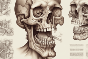
Oral Mucosa
- The oral cavity is lined with oral mucosa, also known as the oral mucous membrane
- It is a stratified squamous epithelial lining
- This extends from the margins of the lips to the tonsils
Categories of Oral Mucosa
- Specialized Mucosa: On the dorsum of the tongue adapted for taste
- Masticatory Mucosa: Comprises the gingiva and hard palatal tissue, endures trauma during mastication
- Lining Mucosa: All other areas of oral mucosa
Specialized Mucosa
- It covers the dorsal surface of the tongue
- It accommodates the sensation of taste
Masticatory Mucosa
- The gingiva and hard palate are masticatory mucosa
- It is firmly attached to the underlying structures
- Exhibits parakeratinized or keratinized epithelium
- The pressure from food leads to parakeratinization or keratinization of the tissue
Lining Mucosa
- Exhibits non-keratinized epithelium
- Consists of the mucosa covering the lips, cheeks, floor of the mouth, inferior surface of the tongue, soft palate, uvula, and alveolar mucosa
Keratinization
- The process by which epithelial cells differentiate or mature
- Begins with basal cells moving through epithelial layers
- More keratinized cells are tougher and more resistant
Types of Mucosa
- Keratinized: A layer of dead cells without nuclei on the surface (e.g., hard palate)
- Parakeratinized: Features some dead cells without nuclei and some dying cells with flattened nuclei on the surface
- Nonkeratinized: Cells on the surface tend to have nuclei that appear fairly healthy and normal (e.g., alveolar mucosa)
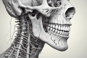
The Periodontium
- Periodontium consists of the tissues supporting and surrounding the teeth
- The periodontium is divided into two units: Gingival Unit and Attachment Unit
Gingival Unit
- The gingival unit comprises Free gingiva (Gingival margin), Interdental papilla, Attached gingiva, and Alveolar mucosa
Free Gingiva
- It extends from the gingival margin to the base of the gingival sulcus
- It is light pink in color
- Averages 0.5 to 2mm in depth
- Is located next to the enamel approximately 0.5 – 2mm coronal to the CEJ
- Forms collar separated from the tooth via the gingival sulcus
- Can be keratinized or parakeratinized
- It closely fits around the tooth without direct attachment
- Being unattached, it can be stretched away from the tooth with a periodontal probe
Gingival Papilla (Interdental Papilla)
- The free gingiva is located in the triangular interdental spaces
- The apex is sharp in anterior teeth, blunt in posterior teeth
- There are facial and lingual interdental papilla
- The lateral borders and tip are formed by free gingiva
- The center is formed by attached gingiva
- When inflamed, it is redder than normal, puffy, and has a blunted apex
Gingival Sulcus
- A gingival groove that often occurs is on the outside of the free gingiva, corresponding to the base of the sulcus
- Health has sulcus termed
- Inflammation has "pocket" term
- Healthy depth is 1-3 mm when measured with a probe
- The inner portion is nonkeratinized
- The tooth surface and free gingiva have space between
- Outer portion is the free tissue which is keratinized
- Attached tissue starts at the bottom of the pocket
Junctional Epithelium
- It attaches the gingiva to the tooth
- Its length is ~1-2 mm .
- It forms a collar around the tooth's neck
- It is a nonkeratinized stratified squamous epithelium
- The fact that it is a nonkeratinized stratified sqaumous epithelium makes it more permeable to fluids, bacteria and toxins
Attached Gingiva
- Extends apically from the base of the sulcus
- It is attached to the bone by collagenous fibers
- Has a stippled texture, resembling dimpled skin (orange peel)
- Is keratinized
- Covered by stratified squamous epithelium
- Color ranges from light to dark pink
- May contain pigment (Melanin pigmentation)
Alveolar Mucosa
- It joins the attached gingiva at the mucogingival junction
- It is continuous with the tissues to the vestibule
- Tissue is thin and soft
- It's loosely attached to the underlying bone
- Consists of lining mucosa
- Is smooth, thin, and nonkeratinized
Attachment Unit
- The attachment unit comprises cementum, alveolar bone, and the periodontal ligament
Cementum
- A hard mineralized tissue layer covers the root of the tooth
- Without Cementum, a tooth would fall out
- Cementum provides an attachment for periodontal ligament fibers to the tooth
- The outer cementum layer safeguards dentin and prevents the exposure and possible damage from open dentinal tubules
Types of Cementum
- Cellular or acellular
- Acellular always covers the cervical 1/3 with possible root coverage outside of the apical section
- Cellular will cover the apical part and form over acellular when needed
More About Cementum
- Similar qualities as bones
- Can have periods of growth or absorbency
- Grows by apposition (addition)
- Appears with collagen or tissue made of connecting tissue from periodontal area
Alveolar Bone
- Alveolar bone proper lines the sockets holding tooth roots
- Thin, compact
- It lines the sockets where the teeth lie
- It allows blood vessels/nerves to pass through
- The socket is called the alveolus, which supports and protects the roots
- The alveolar process contains both proper bone and alveolus components that surround and support teeth in the maxilla and mandible
Compact & Spongy bone
- Compact (cortical) bone is the exterior portion of the alveolar
- Spongy bone is the interior
Alveolar crest
- The alveolar crest portion is the alveolar bone, varies in size and shape based on the tooth position
- Alveolar bone's existence is dependent on teeth
- Alveolar bone resorbs if teeth are extracted
Periodontal Ligaments
- A connective tissue surrounds/ adheres teeth's root with alveolar bones
- Collagen fibers connect the tissue to cementum (which anchor its teeth in bone socket and prevent movement)
- Anchors with Sharpey's collagen fibers
- Forms/ keeps tissue
- Provides nutrition + sensory messages like pain
Arrangement of Periodontal Ligament Fibers
- Alveolar crest group (cervical to alveolar crest)
- Horizontal group (runs horizontally from tooth to alveolar bone)
- Oblique group (fibers that run diagonally from bone to cementum)
- Apical group (fibers by apex)
- Interradicular group- between roots of multi-rooted teeth
Function of PDL Fibers
- The fiber arrangement gives support
- Prevents teeth being pulled out
- Also serves to protect bone and tissues
- Minimizing damage in pushing
- Constant change/ shift based on the pressures
Pressure exerted that leads to functional change includes the following:
- Active eruption: Eruption to compensate for abrasion
- Mesial drift: Movement of molars to compensate for abrasion
- Masticatory occlusal forces: Tooth movement while chewing
- Orthodontic corrective forces: Dentist placing pressure
- Traumatic occlusal forces: Premature contact during occlusion
Periodontal ligament and tooth Position
- A tooth suspends because of PDL, there is movement w/ a bony cavity
- Expansion allows tipping/ compressing
Studying That Suits You
Use AI to generate personalized quizzes and flashcards to suit your learning preferences.