Podcast
Questions and Answers
Which of the following is NOT a protective structure of the brain?
Which of the following is NOT a protective structure of the brain?
- Cerebrospinal fluid
- Vertebral arteries (correct)
- Cranial meninges
- Cranial bones
The cranial dura mater is composed of three layers.
The cranial dura mater is composed of three layers.
False (B)
What is the name of the structure that attaches to the crista galli?
What is the name of the structure that attaches to the crista galli?
falx cerebri
The development of the nervous system begins with a thickening of the ectoderm called the ________.
The development of the nervous system begins with a thickening of the ectoderm called the ________.
Match the following meninges layers with their descriptions:
Match the following meninges layers with their descriptions:
Blood flows to the brain via which arteries?
Blood flows to the brain via which arteries?
The circle of Willis is a network of veins that helps to circulate blood to the brain.
The circle of Willis is a network of veins that helps to circulate blood to the brain.
What are the two layers of the cranial dura mater?
What are the two layers of the cranial dura mater?
Which cranial nerve is responsible for monitoring blood pressure and oxygen levels?
Which cranial nerve is responsible for monitoring blood pressure and oxygen levels?
The Vestibulocochlear nerve (VIII) is primarily responsible for taste.
The Vestibulocochlear nerve (VIII) is primarily responsible for taste.
Which cranial nerve is associated with the secretion of saliva?
Which cranial nerve is associated with the secretion of saliva?
The Vagus nerve (X) is involved in the motility and secretion of the organs of the _________ canal.
The Vagus nerve (X) is involved in the motility and secretion of the organs of the _________ canal.
Which of the following cranial nerves has a sensory function related to taste from the epiglottis?
Which of the following cranial nerves has a sensory function related to taste from the epiglottis?
Match the cranial nerves with their primary motor functions:
Match the cranial nerves with their primary motor functions:
Which cranial nerve is NOT associated with monitoring blood pressure?
Which cranial nerve is NOT associated with monitoring blood pressure?
The Accessory nerve (XI) has autonomic functions related to the digestive canal.
The Accessory nerve (XI) has autonomic functions related to the digestive canal.
Which cranial nerve is responsible for the sense of smell?
Which cranial nerve is responsible for the sense of smell?
The oculomotor nerve (III) is solely responsible for the movement of the eyeballs.
The oculomotor nerve (III) is solely responsible for the movement of the eyeballs.
What is the primary function of the optic nerve (II)?
What is the primary function of the optic nerve (II)?
The trigeminal nerve (V) is responsible for sensations from the face, oral cavity and the anterior two-thirds of the ______.
The trigeminal nerve (V) is responsible for sensations from the face, oral cavity and the anterior two-thirds of the ______.
Match the following cranial nerves with their primary function:
Match the following cranial nerves with their primary function:
Which cranial nerve is responsible for chewing?
Which cranial nerve is responsible for chewing?
The facial nerve (VII) is only responsible for controlling facial expression muscles.
The facial nerve (VII) is only responsible for controlling facial expression muscles.
Which cranial nerve controls the movement of the eyeballs and is not the oculomotor nerve?
Which cranial nerve controls the movement of the eyeballs and is not the oculomotor nerve?
Which of the following is NOT a function of the hypothalamus?
Which of the following is NOT a function of the hypothalamus?
The corpus callosum is a bundle of gray matter tracts that connects the right and left hemispheres of the cerebrum.
The corpus callosum is a bundle of gray matter tracts that connects the right and left hemispheres of the cerebrum.
What is the primary function of the basal nuclei (corpus striatum)?
What is the primary function of the basal nuclei (corpus striatum)?
The _ regulates circadian rhythms.
The _ regulates circadian rhythms.
Which lobe of the cerebrum is responsible for motor functions?
Which lobe of the cerebrum is responsible for motor functions?
The cerebral cortex is composed of white matter.
The cerebral cortex is composed of white matter.
Match the following terms with their description:
Match the following terms with their description:
Which of the following best describes the function of the limbic system?
Which of the following best describes the function of the limbic system?
Which cranial nerve is associated with the manipulation of food?
Which cranial nerve is associated with the manipulation of food?
The Trochlear nerve (IV) is primarily responsible for the sense of smell.
The Trochlear nerve (IV) is primarily responsible for the sense of smell.
What is the Roman numeral designation for the Abducens nerve, based on the provided mnemonic?
What is the Roman numeral designation for the Abducens nerve, based on the provided mnemonic?
The _____ nerve is responsible for vision.
The _____ nerve is responsible for vision.
Which cranial nerve is considered 'somatic'?
Which cranial nerve is considered 'somatic'?
The Oculomotor nerve (III) controls the superior oblique muscle of the eye.
The Oculomotor nerve (III) controls the superior oblique muscle of the eye.
According to the provided mnemonic, what does 'Faci' refer to?
According to the provided mnemonic, what does 'Faci' refer to?
The spinal accessory nerve (CN XI) provides motor innervation to which muscles?
The spinal accessory nerve (CN XI) provides motor innervation to which muscles?
The Hypoglossal nerve (CN XII) is primarily responsible for sensory functions of the tongue.
The Hypoglossal nerve (CN XII) is primarily responsible for sensory functions of the tongue.
What is the primary cause of an ischemic stroke?
What is the primary cause of an ischemic stroke?
A transient ischemic attack (TIA) is characterized by temporary cerebral dysfunction due to impaired blood flow, typically lasting for ______ minutes.
A transient ischemic attack (TIA) is characterized by temporary cerebral dysfunction due to impaired blood flow, typically lasting for ______ minutes.
Which of the following is a result of aging on the nervous system?
Which of the following is a result of aging on the nervous system?
Hemorrhagic strokes are caused by a blood clot obstructing blood flow to the brain.
Hemorrhagic strokes are caused by a blood clot obstructing blood flow to the brain.
What are the main hallmarks of Alzheimer's disease?
What are the main hallmarks of Alzheimer's disease?
Flashcards
Cerebrospinal Fluid
Cerebrospinal Fluid
The fluid that surrounds the brain and spinal cord providing cushioning and protection.
Dura Mater
Dura Mater
The outer layer of the meninges, a tough membrane that helps protect the brain and spinal cord.
Arachnoid Mater
Arachnoid Mater
The middle layer of the meninges, a delicate membrane that surrounds the brain and spinal cord.
Pia Mater
Pia Mater
Signup and view all the flashcards
Development of the Nervous System
Development of the Nervous System
Signup and view all the flashcards
Circle of Willis
Circle of Willis
Signup and view all the flashcards
Brain Organization
Brain Organization
Signup and view all the flashcards
Brain Protection
Brain Protection
Signup and view all the flashcards
What is the function of the thalamus?
What is the function of the thalamus?
Signup and view all the flashcards
What is the function of the hypothalamus?
What is the function of the hypothalamus?
Signup and view all the flashcards
What is the epithalamus?
What is the epithalamus?
Signup and view all the flashcards
What is the cerebral cortex?
What is the cerebral cortex?
Signup and view all the flashcards
What are gyri?
What are gyri?
Signup and view all the flashcards
What are sulci?
What are sulci?
Signup and view all the flashcards
What is the corpus callosum?
What is the corpus callosum?
Signup and view all the flashcards
What are the basal nuclei?
What are the basal nuclei?
Signup and view all the flashcards
Olfactory Nerve (I)
Olfactory Nerve (I)
Signup and view all the flashcards
Optic Nerve (II)
Optic Nerve (II)
Signup and view all the flashcards
Oculomotor Nerve (III)
Oculomotor Nerve (III)
Signup and view all the flashcards
Trochlear Nerve (IV)
Trochlear Nerve (IV)
Signup and view all the flashcards
Trigeminal Nerve (V)
Trigeminal Nerve (V)
Signup and view all the flashcards
Abducens Nerve (VI)
Abducens Nerve (VI)
Signup and view all the flashcards
Facial Nerve (VII)
Facial Nerve (VII)
Signup and view all the flashcards
Vestibulocochlear Nerve (VIII)
Vestibulocochlear Nerve (VIII)
Signup and view all the flashcards
Glossopharyngeal Nerve (IX)
Glossopharyngeal Nerve (IX)
Signup and view all the flashcards
Vagus Nerve (X)
Vagus Nerve (X)
Signup and view all the flashcards
Accessory Nerve (XI)
Accessory Nerve (XI)
Signup and view all the flashcards
What is the primary function of the Accessory Nerve (CN XI)?
What is the primary function of the Accessory Nerve (CN XI)?
Signup and view all the flashcards
What is the primary function of the Hypoglossal Nerve (CN XII)?
What is the primary function of the Hypoglossal Nerve (CN XII)?
Signup and view all the flashcards
What are some key changes that occur in the nervous system during aging?
What are some key changes that occur in the nervous system during aging?
Signup and view all the flashcards
Explain what a stroke is and what causes it.
Explain what a stroke is and what causes it.
Signup and view all the flashcards
What is a Transient Ischemic Attack (TIA) and how is it different from a stroke?
What is a Transient Ischemic Attack (TIA) and how is it different from a stroke?
Signup and view all the flashcards
What is Alzheimer's disease and what are its defining features?
What is Alzheimer's disease and what are its defining features?
Signup and view all the flashcards
What does the Hypoglossal nerve control?
What does the Hypoglossal nerve control?
Signup and view all the flashcards
How can you remember the names of the cranial nerves?
How can you remember the names of the cranial nerves?
Signup and view all the flashcards
What is the function of the Olfactory nerve?
What is the function of the Olfactory nerve?
Signup and view all the flashcards
What is the function of the Optic nerve?
What is the function of the Optic nerve?
Signup and view all the flashcards
What is the function of the Oculomotor nerve?
What is the function of the Oculomotor nerve?
Signup and view all the flashcards
What does the Trochlear nerve control?
What does the Trochlear nerve control?
Signup and view all the flashcards
What is the Trigeminal nerve responsible for?
What is the Trigeminal nerve responsible for?
Signup and view all the flashcards
What does the Facial nerve control?
What does the Facial nerve control?
Signup and view all the flashcards
Study Notes
Introduction
- The chapter aims to explain the organization, protection, and blood supply of the brain.
- It will compare and contrast brain areas and their functions.
- It will discuss the functional organization of the brain.
- It will cover cranial nerve structure and function.
Brain Organization, Protection, and Blood Supply
- The nervous system begins with a thickening of the ectoderm called the neural tube.
- The brain's development follows a sequence of primary and secondary vesicles.
- The adult brain develops from these initial structures.
Development of the Nervous System
- Table 14.1 details the development of the brain, showing primary and secondary vesicles and their adult derivatives. Three primary vesicles (prosencephalon, mesencephalon, rhombencephalon) initially form, then develop into five secondary vesicles.
- Each of those secondary vesicles is involved in the eventual formation of certain structures of the adult brain.
Brain Organization
- The cerebral cortex processes sensory information, sends messages to skeletal muscles, integrates incoming and outgoing nerve impulses, and performs tasks such as thinking, learning, remembering.
- The basal ganglia coordinate slow, sustained movements and suppress useless movement patterns.
- The thalamus relays most sensory information from the spinal cord and certain brain parts to the cerebral cortex, and interprets sensory messages such as pain, temperature, and pressure.
- The hypothalamus controls body temperature, respiration, and heartbeat; it also directs hormone secretions.
- The cerebellum coordinates subconscious movements, contributes to muscle tone, posture, and balance.
- The brain stem controls many cranial nerves, regulates heartbeat and breathing, is involved in consciousness, and transmits impulses between the brain and spinal cord.
Protection
- The brain is protected by cranial bones, cranial meninges (pia, arachnoid, and dura mater).
- Cranial dura mater is composed of two layers (external periosteal/internal meningeal – only spinal has one).
- Cerebrospinal fluid further protects the brain.
Cranial Bones and Meninges
- Cranial meninges consist of dura mater (periosteal and meningeal layers [spinal has one]), arachnoid, and pia mater.
- The falx cerebri is a type of dura matter.
Extensions of the Dura Mater
- Dura matter extensions include the falx cerebri, and the tentorium cerebelli.
- The falx cerebri attaches to the crista galli.
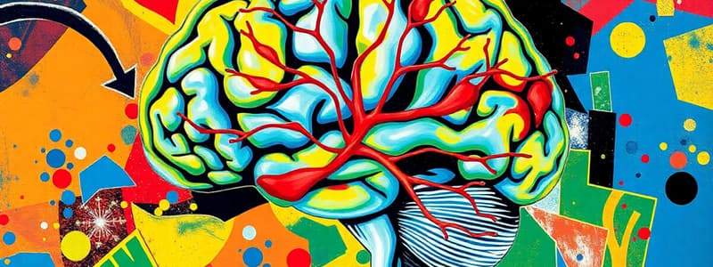
Blood Flow to the Brain
- Blood flows to the brain via the vertebral and carotid arteries, and returns to the heart via venous sinuses.
- The Circle of Willis is a circulatory anastomosis that ensures reliable blood supply to the brain.
Importance of Blood Flow to the Brain
- The brain uses about 20% of the body's oxygen supply.
- Interrupted oxygen supply can lead to brain cell damage or death.
- Glucose deficiency can cause mental confusion, dizziness, convulsions, and unconsciousness.
Brain Blood Flow
- The blood-brain barrier is a result of tight junctions between brain capillaries and nearby astrocytes.
- The barrier isolates brain cells from many substances in the blood.
Blood - Brain Barrier (BBB)
- The BBB protects the brain from harmful substances by preventing their passage into brain tissue.
- The BBB can sometimes prevent the entry of therapeutic drugs.
- Brain injury can lead to impairment of the BBB, potentially allowing substances to enter brain tissue.
Brain Blood Flow
- The BBB can be helpful in certain pharmacological situations.
- Older antihistamine drugs cross the BBB and induce sedation, making them unsuitable for allergy relief but sometimes useful as sleep aids.
- Newer antihistamines do not cross the BBB.
Cerebrospinal Fluid (CSF)
- CSF protects the brain and spinal cord from chemical and physical injury.
- It carries oxygen, glucose, and other vital substances to nerve tissue.
Ventricles and the Choroid Plexus
- The ventricles of the brain contain CSF.
- The choroid plexuses produce CSF.
CSF Flow
- CSF flows through the ventricles and into the subarachnoid space.
- Arachnoid granulations absorb CSF into venous sinuses.
- The flow of CSF is crucial for maintaining normal brain function.
Frontal Section of the Brain and Spinal Cord
- Cranial subarachnoid space holds about 110mL of CSF.
- Spinal subarachnoid space holds about 25mL of CSF.
- Lateral ventricle horn can hold 25-30mL of CSF.
- Fourth ventricle can hold 2-3mL of CSF.
- Third ventricle can hold 2-3mL of CSF.
Production and Flow of CSF
- CSF production and drainage imbalance creates hydrocephalus.
- Hydrocephalus arises from congenital abnormalities, head injury, bleeding into the brain.
Regions of the Brain (Anatomy Overview)
- This section presents an overview of the regions of the brain and their importance.
The Brainstem and Reticular Formation
- The brainstem comprises the medulla oblongata, pons, and midbrain.
The Brainstem (1 of 2)
- These components of the brainstem are associated with regions of brain and spinal cord for motor and sensory functions.
The Brainstem (2 of 2)
- The brainstem controls various functions associated with cranial nerves, including sensory regions such as the Vestibulocochlear (VIII) and hypoglossal (XII) nerves.
- The medulla oblongata controls various functions including heart rate, respiratory rate, vasoconstriction, swallowing, coughing, vomiting, sneezing, hiccupping.
Medulla Oblongata (1 of 2)
- Medulla oblongata structure and its connections to the superior aspect of the spinal cord with tracts.
Medulla Oblongata (2 of 2)
- Cranial nerves such as the Vestibulocochlear (VIII) and hypoglossal (XII) nerves are associated.
- The medulla controls functions including heart rate, respiratory rate, vasoconstriction, swallowing, coughing, vomiting, sneezing, hiccupping.
Pons (1 of 2)
- The pons is located superior to the medulla.
- It links brain parts through tracts.
- Visual and auditory impulses are processed via the pons.
Pons (2 of 2)
- Cranial nerves, such as the trigeminal, abducens, facial, and vestibular branches of the vestibulocochlear nerve are associated with the pons.
- The pons controls voluntary skeletal muscle movements through a pathway from the cerebrum to the cerebellum.
- The pons regulates respiration.
Midbrain (1 of 3)
- The midbrain is located superior to the medulla oblongata.
- It extends from the pons to the diencephalon.
Midbrain (2 of 3)
- The midbrain contains the cranial nerves: oculomotor (III) and trochlear (IV).
- The midbrain relates to regions including the cerebral peduncles, corpora quadrigemina, substantia nigra & red nuclei, and medial lemniscus.
Midbrain (3 of 3)
- The midbrain connects the cerebrum to the cerebellum and spinal cord, and relates to the processing of visual and auditory stimuli.
The Cerebellum (1 of 3)
- The cerebellum is located in the inferior and posterior aspects of the cranial cavity.
- It consists of two hemispheres and a central vermis.
- Sections of the cerebellum are identified by their location (superior v. inferior).
The Cerebellum (2 of 3)
- The cerebellum controls various motor functions; its structure includes cerebellar hemispheres and the vermis.
The Cerebellum (3 of 3)
- The cerebellum coordinates contractions of skeletal muscles, and maintains fine tune motor functions for normal muscle tone, posture, and balance.
The Diencephalon
- The diencephalon is situated superior and posterior to the midbrain.
The Diencephalon (1 of 2)
- The diencephalon includes three major parts. These are the thalamus, hypothalamus, and epithalamus.
- The diencephalon is involved in controlling many activities of the body.
The Diencephalon (2 of 2)
- This section presents an overview of the diencephalon.
Thalamus (1 of 2)
- The thalamus is positioned superior to the midbrain.
- It contains nuclei that serve as relay stations for all sensory inputs (except smell).
Thalamus (2 of 2)
- The structure of the thalamus is described.
Hypothalamus
- The hypothalamus lies inferior to the thalamus.
Epithalamus
- The epithalamus lies superior and posterior to the thalamus.
- It contains the pineal gland and habenular nuclei, involved in olfactory functions.
Circumventricular Organs (CVOs) of the Diencephalon
- Parts of the diencephalon, the CVOs, sense chemical changes in the blood.
- CVOs lack a blood-brain barrier, allowing direct monitoring.
- The pineal gland and parts of the hypothalamus, including the pituitary gland are associated.
- They regulate homeostatic activities of the endocrine and nervous systems.
Summary of Functions of the Diencephalon
- The thalamus relays information to the cerebral cortex. The hypothalamus controls autonomic nervous system activity, hormone production, and regulates emotional and behavioral patterns.
- The epithalamus consists of the pineal gland.
The Cerebrum
- The cerebral cortex composes the gray matter of the cerebrum.
The Cerebrum (1 of 3)
- Gyri, fissures, and sulci are aspects of the structure of the cerebral cortex.
- Deeper to the cortex, white matter tracts connect parts of the brain and spinal cord.
- The corpus callosum connects the right and left hemispheres.
The Cerebrum (2 of 3)
- Anatomical details of the lobes of the cerebral cortex, including the gyri, sulci, and fissures are described.
The Cerebrum (3 of 3)
- Lobes include the frontal, parietal, temporal, and occipital lobes.
- The insula is also described.
Cerebrum White Matter Tracts
- The cerebral white matter mainly consists of association, commissural, and projection tracts.
Basal Nuclei (Corpus Striatum) of the Cerebrum
- Basal nuclei (corpus striatum) are paired masses deep in the cerebral hemispheres.
- These nuclei initiate and terminate movements, suppress unwanted movements and regulate muscle tone.
The Limbic System
- The limbic system involves emotional and behavioral functions, including smell/odor memory and emotional aspects.
Functional Organization of the Cerebral Cortex (1 of 2)
- The cerebral cortex has sensory, motor, and association areas.
Functional Organization of the Cerebral Cortex (2 of 2)
- This section overviews the functional areas of the cerebral cortex, including sensory, motor, and association areas.
Functional Areas of the Cerebrum
- Descriptions of the primary somatosensory, primary motor, visual, auditory, and other cortical areas.
The Cerebrum
- The sensory homunculus depicts the areas of the body represented in the somatosensory cortex, reflecting the sensory information they receive.
- The motor cortex section is outlined, and the motor homunculus describes the areas that control various body parts, with extensive representation for skilled hand and face movements, demonstrating the brain's control over these actions.
The Cerebrum (1 of 2)
- These anatomical sections are described, focusing on white matter tracts, associated with function and hemispheric lateralization.
The Cerebrum (2 of 2)
- The corpus callosum unites the left and right cerebral hemispheres across the white matter.
The Cerebrum
- The basal nuclei have a function in regulating muscle contractions for automatic movements like walking and spontaneous laughter.
Summary of Functions of the Cerebrum
- The sensory and motor areas of the cerebral cortex are described, and the role of regions in the cerebral cortex is described, such as the corpus striatum and the limbic system.
Hemispheric Lateralization (1 of 3)
- The brain's hemispheres exhibit subtle differences, despite general symmetry.
Hemispheric Lateralization (2 of 3)
- Each hemisphere specializes in certain functions, a feature known as hemispheric lateralization.
Hemispheric Lateralization (3 of 3)
- Lateralization differences are less pronounced in females.
- The left hemisphere is generally associated with logic, language, and numerical and scientific skills.
- The right hemisphere is associated with musical and artistic awareness, spatial perception, face and emotion recognition, and creating mental images.
Functional Differences between Right and Left Hemispheres
- Right and left hemisphere functions are delineated.
Brain Waves (1 of 2)
- Brain waves are electrical signals generated by neurons in the cortex.
- Electrodes measuring brain waves on the scalp are called an electroencephalogram (EEG).
Brain Waves (2 of 2)
- Various types of brain waves (alpha, beta, theta, delta) reflect different states of consciousness.
- EEG shows the electrical activity of the cerebral cortex.
Brain Waves
- EEG measurements are valuable for studying normal brain functions (such as sleep patterns) and diagnosing brain disorders like epilepsy or trauma.
- An EEG also determines if “brain death” has occurred.
Cranial Nerves
- Cranial nerves are named and numbered
The Twelve Cranial Nerves
- The twelve cranial nerves are listed, with descriptions of their fibers.
Cranial Nerves
- This table lists cranial nerves by designation, number, origin, roots, contents, and target. Spinal and cranial nerves are compared.
Cranial Nerves (1 of 8)
- Table 14.4 summarizes cranial nerves by component (sensory, motor, autonomic) and function.
Cranial Nerves (2 of 8)
- A more detailed look at component and function is presented for trigeminal (V) and abducens (VI) nerves.
Cranial Nerves (3 of 8)
- This table details components and functions of the facial (VII) and vestibulocochlear (VIII) nerves.
Cranial Nerves (4 of 8)
- Components and functions for the glossopharyngeal (IX) nerve are further detailed. Glossopharyngeal (IX) nerve functions include taste, proprioception in swallowing muscles and autonomic function.
Cranial Nerves (5 of 8)
- The Vagus (X) nerve components and functions are more extensively described in the sensory and motor/ branchial and autonomic functions.
Cranial Nerves (6 of 8)
- The accessory (XI) and hypoglossal (XII) cranial nerves are covered by the components and functions.
Cranial Nerves (7 of 8)
Mnemonic device for remembering cranial nerves by number and function with corresponding letters in the mnemonic.
Cranial Nerves (8 of 8)
- This is an overview of the cranial nerves.
Olfactory, I (1 of 2)
- The olfactory nerve (I) structure and its pathways.
Olfactory, I (2 of 2)
- This section overviews the olfactory (I) nerve..
Optic, II (1 of 2)
- Details of the optic nerve (II) structure and its pathway.
Optic, II (2 of 2)
- This section details the optic nerve (II).
Oculomotor, III (1 of 2)
- The structure and pathways of Oculomotor (III) nerve are shown.
Oculomotor, III (2 of 2)
- The oculomotor (III) nerve function is covered.
Trochlear, IV (1 of 2)
- The trochlear nerve (IV) structure and related pathways are presented.
Trochlear, IV (2 of 2)
- Trochlear (IV) nerve function is discussed.
Trigeminal, V (1 of 2)
- Structure and pathways of the trigeminal nerve (V).
Trigeminal, V (2 of 2)
- This section details the trigeminal (V) nerve.
Abducens, VI (1 of 2)
- The abducens nerve (VI) structure and related pathways.
Abducens, VI (2 of 2)
- Abducens (VI) nerve function is discussed.
Facial, VII (1 of 2)
- The facial nerve (VII) structure and related pathways.
Facial, VII (2 of 2)
- Facial nerve (VII) function covered.
Vestibulocochlear, VIII (1 of 2)
- The vestibulocochlear nerve (VIII) structure and pathways related to auditory and balance functions.
Vestibulocochlear, VIII (2 of 2)
- Vestibulocochlear (VIII) function is covered.
Glossopharyngeal, IX (1 of 2)
- The glossopharyngeal nerve (IX) structure and related pathways.
Glossopharyngeal, IX (2 of 2)
- Function of the glossopharyngeal (IX) nerve.
Vagus, X (1 of 2)
- The vagus nerve (X) structure and related pathways.
Vagus, X (2 of 2)
- The vagus nerve (X) function is described.
Accessory, XI (1 of 2)
- The accessory nerve (XI) structure and related pathways.
Accessory, XI (2 of 2)
- Accessory nerve (XI) function covered.
Hypoglossal, XII (1 of 2)
- The hypoglossal nerve (XII) structure and related pathways are discussed.
Hypoglossal, XII (2 of 2)
- Hypoglossal nerve (XII) function covered.
Aging and the Nervous System
- Aging impacts nervous tissue, resulting in neuron loss, reduced nerve impulse capacity, impaired information processing, slowing motor function, increased reflex time, and eventual degeneration in vision, hearing, taste, smell, touch, and balance.
Cerebrovascular Accident (CVA) or Stroke
- Stroke is caused by lack of oxygen to brain cells due to blood clot (ischemic stroke; 85%) or blood vessel rupture (hemorrhagic stroke; 15%).
Transient Ischemic Attack (TIA)
- A TIA is temporary cerebral dysfunction caused by temporary impaired blood flow.
- Symptoms include dizziness, weakness, and numbness.
Alzheimer's Disease
- Alzheimer's is a progressive dementia characterized by loss of reasoning and self-care abilities.
- Hallmarks are neuronal degeneration, beta-amyloid plaques, and neurofibrillary tangles (Tau).
Disorders (3 of 3)
- Brain tumors can be malignant or benign and present in the brain.
- ADHD is a learning disorder characterized by inattention, hyperactivity, and impulsivity; Likely has strong genetic influences.
Chapter 14
- A summary of the chapter's content covered.
Studying That Suits You
Use AI to generate personalized quizzes and flashcards to suit your learning preferences.


