Podcast
Questions and Answers
What role do microfilaments primarily play in neurons?
What role do microfilaments primarily play in neurons?
- Maintaining membrane integrity and cell morphology (correct)
- Myelination of axons
- Facilitation of neurotransmitter release
- Transporting organelles along the axon
What is the difference between fast and slow axonal transport?
What is the difference between fast and slow axonal transport?
- Fast transport is slower than slow transport.
- Fast transport is mediated exclusively by microfilaments.
- Fast transport occurs at approximately 400 mm/day. (correct)
- Slow transport involves a known recognition mechanism.
Which factor stimulates myelination in neurons?
Which factor stimulates myelination in neurons?
- Decreased ATP availability in axons
- Increased oxidative stress in oligodendrocytes
- Inhibition of action potential generation
- Release of Leukaemia Inducing Factor (LIF) (correct)
Which of the following is a symptom of multiple sclerosis?
Which of the following is a symptom of multiple sclerosis?
What makes oligodendrocytes particularly vulnerable in the central nervous system?
What makes oligodendrocytes particularly vulnerable in the central nervous system?
Which type of neuronal firing pattern consistently produces action potentials?
Which type of neuronal firing pattern consistently produces action potentials?
What is the primary function of the soma in a neuron?
What is the primary function of the soma in a neuron?
Which component of the neuronal cytoskeleton is primarily involved in cellular transport?
Which component of the neuronal cytoskeleton is primarily involved in cellular transport?
Neurofilaments are most commonly found in which part of a neuron?
Neurofilaments are most commonly found in which part of a neuron?
What neurological disorders are associated with the dysfunction of microtubules?
What neurological disorders are associated with the dysfunction of microtubules?
What is the primary function of GABAergic interneurones?
What is the primary function of GABAergic interneurones?
Which neuronal type typically has long axons that project to other parts of the CNS?
Which neuronal type typically has long axons that project to other parts of the CNS?
What is the diameter range of proximal dendrites?
What is the diameter range of proximal dendrites?
How are neurotransmitters classified in neurons?
How are neurotransmitters classified in neurons?
What type of neurones are typically found in the central nervous system with a cell soma and multiple processes?
What type of neurones are typically found in the central nervous system with a cell soma and multiple processes?
Which neurotransmitter is primarily associated with inhibitory neurotransmission?
Which neurotransmitter is primarily associated with inhibitory neurotransmission?
What distinguishes relay neurones from projection neurones?
What distinguishes relay neurones from projection neurones?
Which of the following is a non-classical neurotransmitter?
Which of the following is a non-classical neurotransmitter?
What is one of the key roles of ependymal cells in the central nervous system?
What is one of the key roles of ependymal cells in the central nervous system?
Which type of glial cell is specifically associated with the myelination of peripheral nerves?
Which type of glial cell is specifically associated with the myelination of peripheral nerves?
What role do radial cells play in neural development?
What role do radial cells play in neural development?
What major function do astrocytes provide in the central nervous system?
What major function do astrocytes provide in the central nervous system?
Which glial cells are considered the main immune cells of the central nervous system?
Which glial cells are considered the main immune cells of the central nervous system?
What are the primary types of cells in the brain?
What are the primary types of cells in the brain?
What key components make up a neurone?
What key components make up a neurone?
Which of the following classification methods for neurones is NOT mentioned?
Which of the following classification methods for neurones is NOT mentioned?
Which staining technique identifies the entirety of a cell?
Which staining technique identifies the entirety of a cell?
What type of neurone relays signals between other neurones?
What type of neurone relays signals between other neurones?
What is the approximate somatic diameter range of a neurone?
What is the approximate somatic diameter range of a neurone?
Which of the following pairs of neurones is correctly matched with their function?
Which of the following pairs of neurones is correctly matched with their function?
Which theory of neuronal organization was proposed by Ramon y Cajal?
Which theory of neuronal organization was proposed by Ramon y Cajal?
Flashcards
Sensory neurones
Sensory neurones
These cells receive information from the sensory organs and transmit it to the CNS.
Motor neurones
Motor neurones
These cells transmit signals from the CNS to muscles and glands, controlling movement and responses.
Interneurones
Interneurones
These cells connect different neurones within the CNS, facilitating communication and complex processing.
Soma
Soma
Signup and view all the flashcards
Dendrites
Dendrites
Signup and view all the flashcards
Axon
Axon
Signup and view all the flashcards
Axonal transport
Axonal transport
Signup and view all the flashcards
Dendritic transport
Dendritic transport
Signup and view all the flashcards
Tonic Firing
Tonic Firing
Signup and view all the flashcards
Phasic/Bursting Firing
Phasic/Bursting Firing
Signup and view all the flashcards
Fast Spiking
Fast Spiking
Signup and view all the flashcards
Soma (Perikaryon)
Soma (Perikaryon)
Signup and view all the flashcards
Neuronal Cytoskeleton
Neuronal Cytoskeleton
Signup and view all the flashcards
Multipolar neurons
Multipolar neurons
Signup and view all the flashcards
Projection neurons
Projection neurons
Signup and view all the flashcards
Relay neurons
Relay neurons
Signup and view all the flashcards
Major Neurotransmitters
Major Neurotransmitters
Signup and view all the flashcards
Electrical properties of neurons
Electrical properties of neurons
Signup and view all the flashcards
Ependymal cells
Ependymal cells
Signup and view all the flashcards
Radial cells
Radial cells
Signup and view all the flashcards
Bergmann glia
Bergmann glia
Signup and view all the flashcards
Muller glia
Muller glia
Signup and view all the flashcards
Schwann Cells
Schwann Cells
Signup and view all the flashcards
Microfilaments
Microfilaments
Signup and view all the flashcards
Oligodendrocytes
Oligodendrocytes
Signup and view all the flashcards
Multiple sclerosis
Multiple sclerosis
Signup and view all the flashcards
Study Notes
Cellular Organisation of the CNS: Neurones & Glia
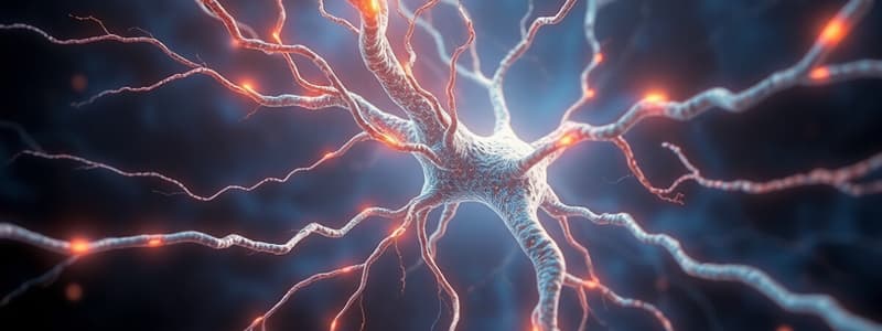
- The brain primarily comprises two cell types: neurones and glia.
- Neurones carry signals; glia provide support.
- Neurones exhibit diverse morphologies, functions, neurotransmitter types, and electrical activity.
- Neuronal classification is complex. Factors to consider include function, morphology, electrical activity, neurotransmitters, and gene expression.
Neurones
- Neuronal size: somatic diameter ranges from 5 to 50 µm.
- Dendrites: typically 1-10, originating from the soma, ranging in diameter from 1-5 µm (proximal) and 0.2-1 µm (distal); extending ~10-500μm.
- Axons: generally thicker and longer than dendrites, typically 1-10 µm in diameter (up to 20 µm); lengths vary significantly (micrometers to millimeters to meters).
Neuronal Classification
-
Sensory neurones: afferents to the CNS.
-
Motor neurones: efferents from the CNS.
-
Interneurones: relay signals between neurones.
- This classification primarily applies to the peripheral nervous system, not the CNS.
-
Neuronal classification is multifaceted, considering function, morphology, electrical properties, neurotransmitters, and gene expression.
Neurone Doctrine
- Historical debate between the reticular and neurone theories of brain structure. Reticular theory proposed continuous network; neurone theory favoured discrete neurones.
- Camillo Golgi championed the reticular theory.
- Santiago Ramón y Cajal championed the neurone theory, highlighting synaptic junctions.
- The neurone doctrine, supporting the concept of discrete neurons communicating via synapses, became firmly established in the 1950s with the discovery of synapses in the brain.
Advances in Histology & Microscopy
- Nissl stain (1885): highlighting nucleic acid-rich areas, predominantly endoplasmic reticulum (ER), for cellular identification.
- Golgi stain (1873): silver staining that visualizes the entire neuronal structure.
Fluorescence Microscopy
- Conventional fluorescence microscopy (late 1970s): utilized for visualizing a wide variety of aspects in biological tissues and cells to improve resolution.
- Multiphoton microscopy (1990s): advanced fluorescence techniques revealing enhanced resolution and depth penetration.
Electron Microscopy
- High-resolution visualization of cellular structures (e.g., neurons and glia) down to the nanometer region.
Neuronal Structures
- Multipolar neurones: the most common type in the CNS; possess numerous dendrites and a single axon.
- Bipolar neurones: have two processes, 1 dendrite and 1 axon.
- Pseudo-unipolar neurones: single axon that divides into two branches, one extending to the periphery and the other to the CNS.
- Unipolar neurones: single process that splits into two branches.
Projection & Local Neurones
- Projection neurones: transmit signals across long distances within the CNS; exhibit long axons.
- Local/relay neurones: transmit signals within a confined brain region; characterized by short axons.
- Projection and relay cells often use neurotransmitters like glutamate, GABA, serotonin, and dopamine.
- Interneurones are a type of relay neurone predominantly GABAergic; occasionally are cholinergic or dopaminergic.
GABAergic Interneurones
- Inhibitory neurones contributing to the majority of interneurones in the CNS.
- Crucial for preventing excessive neuronal activity (e.g., epilepsy).
- Regulate signal precision (e.g., visual acuity) and coordinate activity across multiple neurones (e.g., oscillations).
Neuronal Circuit Example
- Simple illustration of a projection neurone, a relay neurone (both sending signals in the same direction), and an inhibitory interneurone (sending signals in the opposite direction).
Classification by Neurotransmitter
- Major neurotransmitters include glutamate, GABA, acetylcholine, dopamine, serotonin, glycine, noradrenaline, and histamine (collectively, "classical" primary neurotransmitters). Other are comparatively less prevalent.
Classification by Electrical Properties
- Firing patterns: categorization by neuronal firing frequency or regularity; include tonic (regular), phasic (burst), and fast spiking.
Neuronal Compartments
- Neurones are highly compartmentalized, having different functions in specific regions (e.g., soma for protein synthesis, dendrites for signal reception, axons for signal transmission).
Soma ("Perikaryon")
- The cell body containing standard organelles such as the nucleus, Golgi apparatus, and lysosomes.
- Unique protein synthesis and degradation site in neurones.
Neuronal Cytoskeleton
- Similar to other eukaryotic cells; composed of microtubules, neurofilaments, and microfilaments.
- Essential for cell structure, growth, and transport.
Microtubules
- Contribute to neuronal transport by facilitating kinesin- and dynein-mediated movement of vesicles and other components.
Neurofilaments
- Significant role in structural maintenance and growth of axonal structure.
Microfilaments
- Formed from actin; present in dendrites, axons, and growth cones.
- Essential for the maintenance of cell shape, membrane integrity, organization of membrane proteins and interaction with the extracellular environment.
Neuronal Cytoskeleton & Transport
- Illustrates cellular components of neurones.
Axonal Transport
- Transportation of molecules to different parts within the axon and synapse.
- Fast axonal transport: high speed, primarily used for organelles and vesicles to move along microtubules, using kinesin and dynein motors.
- Slow axonal transport: slower speeds, mainly used for components (e.g., proteins) that move continuously.
More Histology
- Stains are carried along axonal transport, for example horseradish peroxidase.
- Reverse transport of stains (retrograde transport) enables mapping of neuronal circuits.
Cytoskeleton & Transport in Nervous Pathologies
- Key cytoskeletal proteins involved in neurodegenerative diseases (e.g., tau protein in Alzheimer's disease, neurofilaments in Charcot-Marie-Tooth disease).
Dendrites
- Start as extensions of the perikaryon, tapering and branching as they extend.
- Primary receptive area of the neuron.
- Dendritic spines are numerous, small protrusions on dendrites, important areas of synapse formation.
Dendritic Spines
- Membranous extensions of dendrites forming postsynaptic parts of synapses.
- Essential for proper synaptic function.
- Structure and contents crucial for synaptic function.
- Compartmentalisation (the spine neck) restricts movement between the spine and dendrite.
- Contain organelles (e.g., ribosomes, some ER)
Dendritic Spine Structure
- Contain abundant microfilaments (actin).
- Lack microtubules and intermediate filaments.
- Vary in shape, including thin, stubby, and mushroom-like, influencing synapse efficacy.
Dendritic Transport
- Dendritic transport shares similarities with axonal transport; motor proteins (e.g., kinesin, dynein) along microtubules propel various components.
- Dendrites are rich in microtubules.
- Components such as receptors, lysosomes, etc., undergo anterograde and retrograde transport within the dendrites.
Summary
- Neurones and brain cells exhibit significant diversity in their size, shape, function, neurotransmitter usage, and electrical activity.
- Neuronal diversity reflects multifaceted control of communication.
- Soma are a key site for protein synthesis and cellular processes.
Glia
- Glial cells are the supporting cells of the Central Nervous System (CNS).
- Glial cells comprise multiple types, including astrocytes, oligodendrocytes (Schwann cells within the periphery), microglia, ependymal, and radial glia.
- A key ratio between glia and neurons is approximately 1-to-1.
Astrocytes
- Form a substantial proportion (20-40%) in the brain; crucial for structural and metabolic functions.
- Regulate ion homeostasis (primarily potassium).
- Contribute to transmitter uptake and metabolism (glutamate, GABA, dopamine, etc.).
- Release nutrients/lactate to provide energy (e.g., glycogenolysis & gluconeogenesis).
- Interact with neurones and blood vessels extensively.
- Play a role in neuroprotection; astrogliosis (reactive astrocytosis) is the adaptive cellular response to CNS damage.
- Participate in signalling; associated with calcium waves.
- Intertwined with blood vessels; critical to maintaining function.
- Astrogliosis (neuronal dysfunction) arises from CNS issues.
Astrocytic Domains
- Three dimensional reconstruction of astrocytes in a specific brain region (e.g., the dentate gyrus).
- Reveals overlap between astrocytes and neurons (visual distinction from glial cells).
Metabolic Support (Astrocytes)
- Participate in ion homeostasis and transmitter metabolism.
- Support energy requirements of neurons (e.g., release of lactate from astrocytes to meet energy demands).
- Crucial in energy homeostasis; release of neurotransmitter precursors into extracellular space.
Astrocytes & CNS Disorder
- Proliferation and modification of astrocytes are associated with CNS damage.
- Astrocytoma, a glioma, is the most frequent type of brain cancer in adults.
Astrogliosis
- A protective reaction, initially beneficial, that may become maladaptive and contribute to neurological disorders if prolonged.
- Triggered by many factors, including injuries such as trauma, strokes, diseases, and infections.
Astrocytes & Blood Vessels
- Extensive contact between astrocytes (in particular, astrocytic end-feet) and blood vessels.
- These contacts facilitate the exchange of nutrients and waste products between blood and brain tissue.
- The end-feet wrap around blood vessels and maintain the integrity of the blood-brain barrier; this separation prevents immune cells and proteins from the blood from entering the brain unnecessarily.
Functional Hyperemia
- Mechanisms in response to the brain's elevated energy demands.
- Brain's ability to increase blood flow in response to elevated activity.
- Signaling from neurons (via nitric oxide, NO), and astrocytes (e.g., phospholipase A2, PLA2) plays an important role in regulating blood flow.
Mechanisms of Vasomodulation
- Illustration of the physiological processes involved in regulating blood flow in the brain. Mechanisms involve an astrocyte response triggered by changes in neuronal activity and oxygen availability, initiating signalling cascade to adjacent blood vessels, ultimately regulating their dilation and constriction.
Just Support Cells?
- Astrocytes and other glial cells, in response to neurotransmitters, may regulate or conduct signals alongside neurons.
Astrocyte Signalling
- Astrocytes communicate via connexin hemichannels; although not like a clear syncytium, astroglial cells may function independently in the brain.
- Ca2+ waves (initially implicated by IP3 receptors) accompany neurotransmitter release and subsequent neuronal activation.
Astrocytic Calcium/Sodium Waves
- Illustration of the calcium (Ca2+) and sodium (Na+) waves observed in astrocytes in response to a specific stimulus (e.g., glutamate).
Gliotransmission
- Transmission between astrocytes and neurons involve the release of neurotransmitters, such as glutamate, D-serine, ATP, and adenosine from astrocytes.
- ATP may trigger astrocytic Ca2+ wave responses in the brain.
Astrocytes & Synapses
- Interactions between astrocytes and synapses play a critical role in synaptic function from their origin (in the retina).
Extrasynaptic Signalling
- Slow inward and outward currents (SICs and SOCs) likely arise from astrocyte-mediated release of neurotransmitters (Glutamate, GABA) and result in neuron activation.
- Astrocytic signals may influence neuronal synchronicity — the simultaneous activation of multiple neurons.
SICs & Neuronal Synchronisation
- Large, slow inward currents in neurons, potentially driven by astrocytic gliotransmitter release.
- Activation of NMDA receptors may involve these neuronal signals or currents.
- Neuronal activation may occur across multiple neurons, even distant ones, influenced by glial signalling.
CNS Immune System
- The unique "privileged" immune system in the CNS involves the separation from the peripheral system.
- Separated by the blood-brain barrier (BBB).
- Immune cells (e.g., microglia) play a crucial role in the CNS's immune response.
Microglia
- Microglia are resident immune cells in the CNS.
- Normally found in the ramified state, surveying the CNS for signs of damage, and in an activated state, exhibiting proliferation and an amoeboid form in response to activation.
- Non-phagocytic phase characterised by soma enlargement and process shrinkage; phagocytic phase involves complete amoeboid transformation.
Microglia Activation
- Microglia transform from a ramified shape to an activated ameboid morphology in response to various cues (e.g., damage).
- Activated microglia can recruit additional immune cells across the blood-brain barrier (BBB) if necessary.
Microglial Immune Signalling
- Activating triggers involve infection, injury, and neurodegenerative disorders.
- Similar signalling molecules are used across diverse CNS immune cells.
- Microglia respond to potentially damaging molecules like LPS, amyloid-beta, and others.
- Inflammatory mediators (e.g., IL-1, IL-6, TNFα) are released.
Pathological Immune Responses
- Chronic immune responses in the brain can become detrimental.
- Associated with neurodegenerative disorders and epilepsy.
- Release of cytotoxic molecules (e.g., ROS, glutamate) may lead to neuronal damage; exacerbated by recruitment from the peripheral immune system.
Oligodendrocytes
- Responsible for myelin formation in axons of the CNS.
- Pathology: multiple sclerosis leads to oligodendrocyte damage and demyelination.
Oligodendrocyte Myelination
- Myelination process is regulated by electrical activity and other molecules.
- Myelination is impacted by factors like axonal ATP release and various components of axonal membrane proteins (e.g., adhesion molecules).
Oligodendrocyte Pathologies
- Oligodendrocytes are particularly susceptible to detrimental factors such as oxidative stress, high metabolic demand for myelination, iron concentration buildup, and low expression of antioxidant and associated receptors.
- This vulnerability contributes to neurodegenerative/inflammatory damage.
- Death from surrounding neuronal or astrocytic cells can contribute to damage and degeneration in oligodendrocytes.
Multiple Sclerosis
- An immune-mediated CNS disorder characterized by inflammation and demyelination, culminating in neuronal damage.
- Symptoms include muscle weakness, decreased coordination, sensory deficits, and autonomic disturbances.
Ependymal Cells
- Line the ventricles of the brain and are rod-shaped and ciliated.
- Crucial in the production and regulation of cerebrospinal fluid (CSF).
Radial Glia
- Crucial during development; their role is in directing neuronal migration.
- The progenitor cells facilitate proper architecture of the developing brain.
Other Glial Cells
- Bergmann glia: cerebellar radial glia found in adults; act similarly to astrocytes.
- Muller glia: retinal radial glia; perform similar roles as astrocytes.
- Schwann cells: peripheral myelinating glia analogous to oligodendrocytes.
- Satellite glia: support neurons in the autonomic ganglia.
Summary (Glia)
- Astrocytes: provide metabolic and structural support, protection, and signalling.
- Microglia: participate in the CNS's immune response.
- Oligodendrocytes: essential for myelination.
Studying That Suits You
Use AI to generate personalized quizzes and flashcards to suit your learning preferences.


