Podcast
Questions and Answers
Neurons are cells that make up the ______ system.
Neurons are cells that make up the ______ system.
nervous
The ______ of a neuron contains the nucleus and organelles.
The ______ of a neuron contains the nucleus and organelles.
cell body
Neurons receive information through ______.
Neurons receive information through ______.
dendrites
The ______ is the information giver in neural communication.
The ______ is the information giver in neural communication.
The ______ matter consists of cell bodies.
The ______ matter consists of cell bodies.
The ______ is a folded sheet of gray matter.
The ______ is a folded sheet of gray matter.
The basal ganglia is involved in ______ control and skill learning.
The basal ganglia is involved in ______ control and skill learning.
The ______ system is involved in emotional and motivational responses.
The ______ system is involved in emotional and motivational responses.
Sensation involves the effects of a stimulus on sensory ______.
Sensation involves the effects of a stimulus on sensory ______.
Perception involves the interpretation of a stimulus based on prior ______.
Perception involves the interpretation of a stimulus based on prior ______.
Rod cells are specialized for low ______ of light.
Rod cells are specialized for low ______ of light.
The fovea is entirely made of ______ → visual acuity.
The fovea is entirely made of ______ → visual acuity.
The optic nerve leaves the eye at the ______ spot.
The optic nerve leaves the eye at the ______ spot.
Simple cells in V1 respond to particular ______ and single points of light.
Simple cells in V1 respond to particular ______ and single points of light.
Complex cells in V1 respond to the ______ of orientation and movement.
Complex cells in V1 respond to the ______ of orientation and movement.
The geniculostriate pathway is the best understood and makes the largest contribution to human ______ perception.
The geniculostriate pathway is the best understood and makes the largest contribution to human ______ perception.
Hemianopia is cortical blindness restricted to one half of the visual ______ (associated with damage to the primary visual cortex in one hemisphere).
Hemianopia is cortical blindness restricted to one half of the visual ______ (associated with damage to the primary visual cortex in one hemisphere).
Quadrantanopia is cortical blindness restricted to a ______ of the visual field.
Quadrantanopia is cortical blindness restricted to a ______ of the visual field.
A ______ is a small region of cortical blindness.
A ______ is a small region of cortical blindness.
Blindsight is the inability to report perceiving visual ______ even though performance suggests otherwise.
Blindsight is the inability to report perceiving visual ______ even though performance suggests otherwise.
V4 is a region of extrastriate cortex associated with ______ perception and color constancy.
V4 is a region of extrastriate cortex associated with ______ perception and color constancy.
Akinetopsia is a failure to perceive visual ______ due to damage to V5.
Akinetopsia is a failure to perceive visual ______ due to damage to V5.
The case of LM involves the inability to detect direction of movement but can detect ______ motion.
The case of LM involves the inability to detect direction of movement but can detect ______ motion.
Object recognition involves the perception of ______ elements such as edges of various lengths and contrasts.
Object recognition involves the perception of ______ elements such as edges of various lengths and contrasts.
The structural descriptions refer to the memory of the ______ representation of an object.
The structural descriptions refer to the memory of the ______ representation of an object.
Apperceptive agnosia is a failure to recognize objects due to a deficit at the level of ______ perception.
Apperceptive agnosia is a failure to recognize objects due to a deficit at the level of ______ perception.
The case of HJA is an example of ______ agnosia, where the patient can see the parts but not the whole.
The case of HJA is an example of ______ agnosia, where the patient can see the parts but not the whole.
The parahippocampal place area (PPA) is an area of the brain that responds more to ______ than objects.
The parahippocampal place area (PPA) is an area of the brain that responds more to ______ than objects.
The fusiform face area (FFA) is an area in the inferior temporal lobes that responds more to ______ than other visual objects.
The fusiform face area (FFA) is an area in the inferior temporal lobes that responds more to ______ than other visual objects.
Prosopagnosia is the inability to recognize ______ familiar faces.
Prosopagnosia is the inability to recognize ______ familiar faces.
Flashcards are hidden until you start studying
Study Notes
Introducing the Brain
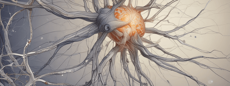
- Neurons are cells that make up the nervous system, consisting of a cell body (soma), dendrites, and axon.
- The cell body contains the nucleus and organelles.
- Dendrites receive information, while the axon sends information.
Neural Communication
- Terminal buttons of a neuron and dendrites of another neuron communicate through a small gap called a synapse.
- Presynaptic neuron is the information giver, while postsynaptic neuron is the information receiver.
- Action potential electrically charges the presynaptic neuron, inducing the release of informative chemicals called neurotransmitters.
Terms of Directional References
- Lateral: refers to the outer regions of the brain.
- Medial: refers to the central regions of the brain.
Gross Organization of the Brain
- Gray matter consists of cell bodies.
- White matter consists of axons and glia (support cells involved in tissue repair and myelin formation).
Cerebral Cortex
- Folded sheets of gray matter.
- Raised surfaces are called gyri (gyrus).
- Folds are called sulci (sulcus).
- The folded structure helps to increase area/volume ratio (efficiency in packaging of the brain).
4 Main Parts of the Cerebral Cortex
- No specific information provided about the 4 main parts.
Subcortex
- Lies under the white matter.
- Consists of a gray matter collection.
- Includes basal ganglia, limbic system, thalamus, and hypothalamus.
Basal Ganglia
- Responsible for motor control and skill learning.
- Disorders associated with poverty or excess of movement (e.g., Parkinson's and Huntington's).
Limbic System
- No specific information provided about the limbic system.
Diencephalon
- Includes thalamus and hypothalamus.
Midbrain and Hindbrain
- No specific information provided about midbrain and hindbrain.
Sensation & Perception
- Sensation: Effects of stimulus on sensory organs
- Perception: Interpretation of stimulus based on prior experience
- The brain actively constructs the visual representation of the world
From Eye to Brain
- Rod cells: Specialized for low intensity of light and movement
- Cone cells: Specialized for high intensity of light and color information
- Fovea: Entirely made of cones, responsible for visual acuity
- Blind spot: Where the optic nerve leaves the eye
Geniculostriate Pathway
- Optic nerve → Optic chiasm → Optic tract → Lateral Geniculate Nucleus (LGN) → Primary visual cortex (V1)
Primary Visual Cortex (V1)
- Located in the occipital lobe, responsible for visual processing
- V1 is the first stage of processing in the cortex
- Simple cells: Respond to particular orientation and single points of light
- Complex cells: Combination of simple cells, larger receptive fields, respond to movement of orientation, do not respond to single points of light
- Hypercomplex cells: Just outside V1, built from responses of complex cells, respond to orientation and length
Cortical & Non-Cortical Routes
- Geniculostriate pathway is the best understood and makes the largest contribution to human visual perception
- Other routes are evolutionary older
- Pathway to suprachiasmatic nucleus in the hypothalamus provides information about time of day
- Pathways via superior colliculus and inferior pulvinar are important for orienting stimuli
Problems with Primary Visual Cortex
- Retinotopic organization: Layout of the receptive fields of neurons in V1 reflect the spatial organization of the retina
- Hemianopia: Cortical blindness restricted to one half of the visual field
- Quadrantanopia: Cortical blindness restricted to a quarter of the visual field
- Scotoma: A small region of cortical blindness
Blindsight
- Inability to report perceiving visual stimulus even though performance suggests otherwise
- Case of DB: Reported not seeing stimuli but oriented his eyes correctly toward stimuli
Extrastriate Areas in Vision
- V4: Associated with color perception and color constancy
- Achromatopsia: A failure to perceive color due to damage to V4
- V5 (or MT): Associated with motion perception
- Akinetopsia: A failure to perceive visual motion due to damage to V5
Dual Stream Visual Processing
- No specific information provided
Object Recognition
-
- Perception of basic elements (e.g., edges of various lengths, contrasts & orientations)
-
- Grouping physical elements (depth cues and divide surfaces)
-
- The viewer-centered description is then matched onto stored 3D descriptions of the structure of objects
-
- Meaning is attributed to the stimulus
Agnosia
- Failure in object recognition
- Apperceptive agnosia: A failure to recognize objects due to a deficit at the level of object perception
- Associative agnosia: A failure to recognize objects due to a deficit at the level of semantic memory
- Case of HJA: Seeing the part but not the whole, impaired at deciding if objects are real or made up and naming objects
Categorical Perception
- Category specificity: The brain represents different categories in different ways (and/or different regions)
- Parahippocampal place area (PPA): Area of the brain that responds to scenes more than objects
- The extrastriate body area (EBA): Area of the brain that responds to the human body more than to faces, scenes or objects
Face Recognition
- Fusiform face area (FFA): An area in the inferior temporal lobes that responds more to faces than other visual objects
- Prosopagnosia: Inability to recognize previously familiar faces
Studying That Suits You
Use AI to generate personalized quizzes and flashcards to suit your learning preferences.


