Podcast
Questions and Answers
How do nodes of Ranvier contribute to faster nerve impulse transmission?
How do nodes of Ranvier contribute to faster nerve impulse transmission?
They allow the action potential to 'jump' from node to node, a process called saltatory conduction, greatly increasing the speed of electrical impulses.
What is the role of neurotransmitters in synaptic transmission?
What is the role of neurotransmitters in synaptic transmission?
Neurotransmitters carry the signal across the synapse by diffusing across the gap and binding to receptors on the postsynaptic neuron, initiating a new electrical impulse.
How does the sodium-potassium pump contribute to maintaining the resting membrane potential?
How does the sodium-potassium pump contribute to maintaining the resting membrane potential?
It actively transports sodium ions ($Na^+$) out of the cell and potassium ions ($K^+$) into the cell, against their concentration gradients, which helps establish and maintain the negative resting membrane potential.
Describe the key difference between sensory and motor neurons in terms of their function and direction of impulse transmission.
Describe the key difference between sensory and motor neurons in terms of their function and direction of impulse transmission.
In a reflex arc, what is the role of the interneuron (also called association or relay neuron)?
In a reflex arc, what is the role of the interneuron (also called association or relay neuron)?
Explain how the presence of myelin sheath affects the speed of nerve impulse transmission.
Explain how the presence of myelin sheath affects the speed of nerve impulse transmission.
What is the difference between white matter and gray matter in the central nervous system, in terms of composition and location?
What is the difference between white matter and gray matter in the central nervous system, in terms of composition and location?
Describe how a stimulus's strength can be coded into a nerve impulse.
Describe how a stimulus's strength can be coded into a nerve impulse.
How does an action potential propagate along an unmyelinated axon?
How does an action potential propagate along an unmyelinated axon?
Explain how the action of stimulants like caffeine and depressants like anesthetics affect the transmission of nerve impulses at the synapse.
Explain how the action of stimulants like caffeine and depressants like anesthetics affect the transmission of nerve impulses at the synapse.
What is the role of the cerebral cortex in sensory perception?
What is the role of the cerebral cortex in sensory perception?
How does the corpus callosum facilitate communication between the two cerebral hemispheres?
How does the corpus callosum facilitate communication between the two cerebral hemispheres?
Explain the roles of the dura mater, arachnoid mater, and pia mater in protecting the central nervous system.
Explain the roles of the dura mater, arachnoid mater, and pia mater in protecting the central nervous system.
What factors are controlled by the medulla oblongata, and why is damage to this area often fatal?
What factors are controlled by the medulla oblongata, and why is damage to this area often fatal?
How do the symptoms of Parkinson's disease result in low dopamine levels in the brain?
How do the symptoms of Parkinson's disease result in low dopamine levels in the brain?
Describe the difference between the sympathetic and parasympathetic divisions of the autonomic nervous system.
Describe the difference between the sympathetic and parasympathetic divisions of the autonomic nervous system.
What is the function of ascending and descending tracts in the spinal cord?
What is the function of ascending and descending tracts in the spinal cord?
Describe the sequence of events that occurs when a nerve impulse reaches the axon terminal and crosses a synapse.
Describe the sequence of events that occurs when a nerve impulse reaches the axon terminal and crosses a synapse.
Identify the types of neurons that carry signals towards and away from the CNS in the efferent division of the PNS?
Identify the types of neurons that carry signals towards and away from the CNS in the efferent division of the PNS?
What are the structural components of the brainstem?
What are the structural components of the brainstem?
What are some of the advantages of stem cell therapy for nuerological conditions such as Alzheimer's and Parkinson's
What are some of the advantages of stem cell therapy for nuerological conditions such as Alzheimer's and Parkinson's
What is the difference between alzheimer's disease plaques, and neurofibrillary tangles?
What is the difference between alzheimer's disease plaques, and neurofibrillary tangles?
What brain features are responsible for enabling the communication between the left and right hemispheres?
What brain features are responsible for enabling the communication between the left and right hemispheres?
Identify some stimulis and corresponding receptors that are stimulated in the body
Identify some stimulis and corresponding receptors that are stimulated in the body
What is the effect of stimulation of the paraysmpathetic nerves on the lungs?
What is the effect of stimulation of the paraysmpathetic nerves on the lungs?
Flashcards
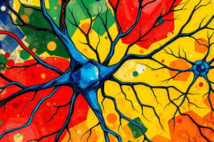
What are neurons?
What are neurons?
Cells that make up the nervous system; consist of a cell body, dendrites, and an axon.
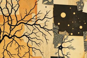
What are dendrites?
What are dendrites?
Extensions of the cytoplasm that carry impulses toward the cell body; they are highly branched.
What is an axon?
What is an axon?
Extension of the cytoplasm that carries impulses away from the cell body; can be quite long.
What is the myelin sheath?
What is the myelin sheath?
Signup and view all the flashcards
What is the neurilemma?
What is the neurilemma?
Signup and view all the flashcards
What are Nodes of Ranvier?
What are Nodes of Ranvier?
Signup and view all the flashcards
What are axon terminals?
What are axon terminals?
Signup and view all the flashcards
What are neurotransmitters?
What are neurotransmitters?
Signup and view all the flashcards
What is a synapse?
What is a synapse?
Signup and view all the flashcards
What are sensory neurons?
What are sensory neurons?
Signup and view all the flashcards
What are motor neurons?
What are motor neurons?
Signup and view all the flashcards
What are interneurons?
What are interneurons?
Signup and view all the flashcards
What are multipolar neurons?
What are multipolar neurons?
Signup and view all the flashcards
What are bipolar neurons?
What are bipolar neurons?
Signup and view all the flashcards
What are pseudo-unipolar neurons?
What are pseudo-unipolar neurons?
Signup and view all the flashcards
What is a nerve fiber?
What is a nerve fiber?
Signup and view all the flashcards
What is a nerve impulse?
What is a nerve impulse?
Signup and view all the flashcards
What is membrane potential?
What is membrane potential?
Signup and view all the flashcards
What is resting membrane potential?
What is resting membrane potential?
Signup and view all the flashcards
What is the sodium-potassium pump?
What is the sodium-potassium pump?
Signup and view all the flashcards
What is an action potential?
What is an action potential?
Signup and view all the flashcards
What is Depolarisation?
What is Depolarisation?
Signup and view all the flashcards
What is Repolarisation?
What is Repolarisation?
Signup and view all the flashcards
What is the refractory period?
What is the refractory period?
Signup and view all the flashcards
Saltatory conduction
Saltatory conduction
Signup and view all the flashcards
Study Notes
- Neurons are cells that constitute the nervous system
- Neurons vary in shape and size, but all contain a cell body, dendrites, and an axon
Cell Body
- Spherical in shape
- Approximately 0.025 mm in diameter
- Contains a nucleus and other organelles
- Most cell bodies are located in the central nervous system (CNS)
- Outside the CNS, cell bodies cluster into ganglia
Nucleus
- Contains genetic information
Dendrites
- Extensions of the cytoplasm
- Highly branched structures
- Carry impulses towards the cell body
Axon
- Extension of the cytoplasm
- Length ranges from less than 1 mm to between 75-100 cm
- Conducts impulses away from the cell body
- Nerve fibers are bundles of axons
- Nerve fibers with myelin sheaths are classified as myelinated
Myelin Sheath
- Fatty, white substance
- Insulates and protects the axon
- Increases the speed of nerve impulse transmission
- Forms white matter in the nervous system
- Created by Schwann cells in the peripheral nervous system (PNS) or by glial cells in the CNS
- Acts as an electrical insulator, protects the axon, and speeds up nerve impulses
Neurilemma
- Membrane that covers the myelin sheath
- Provides protective functions
- Assists in the regrowth of damaged axons and dendrites
Schwann Cell
- Produces myelin covering some axons
Nodes of Ranvier
- Gaps located in the myelin sheath
- Facilitates impulse "jumping" to increase electrical impulse speed via saltatory conduction
Axon Terminals
- Branched ends that enable the axon to contact up to 1000 other neurons
- Enlarged ends contain neurotransmitter-filled vesicles
- Neurotransmitters are released into the synapse to transmit impulses
- The point where a motor neuron meets a muscle is called the motor end plate
Synapses
- The location where an axon terminal joins a dendrite or the cell body of another neuron
- Neurons don't physically touch
- The space between neurons is bridged by neurotransmitters
- A neuromuscular junction is a synapse where a neuron meets skeletal muscle
Types of Neurons - Sensory
- Also known as Afferent or receptor neurons
- Transmit messages from receptors in the sense organs or skin to the brain and spinal cord within the central nervous system
Types of Neurons - Motor
- Also known as efferent or effector neurons
- Carry messages from the central nervous system to effectors, such as muscles and glands
Types of Neurons - Interneurons
- Also known as Association neurons, connector neurons, or relay neurons
- Located in the central nervous system
- Link sensory and motor neurons
Neuron Structure - Multipolar
- Features one axon with multiple dendrites extending from the cell body
- Example: Motor neuron, Interneuron
Neuron Structure - Bipolar
- Possesses one axon and one dendrite
- Both the axon and dendrite may have many branches at their ends
- Example: Neurons in the eye, ear and nose
Neuron Structure - Pseudo-unipolar
- Has a single axon from the cell body that splits into two extensions
- One extension connects to dendrites, while the other ends in axon terminals
- Example: Sensory neurons
Neuron Structure - Unipolar
- Characterized by only one extension, an axon
- Not found in humans or other vertebrates
Nerve Fiber
- Term for any long extension of cytoplasm of a nerve cell body
- Usually refers to an axon
Nerve
- Bundles of nerve fibers held together by connective tissue
Nerve Impulses
- Nerve impulses are messages conveyed along a nerve fiber
- Transmission speed is rapid
- Involves electrochemical processes
- Changes in electrical voltage caused by changes in ion concentrations on either side of the cell membrane
Electrical Potential
- Opposite electrical charges attract, measured in volts or millivolts
- Extracellular fluid has a more positive charge due to sodium (Na+) and chloride (Cl-) ions
- Intracellular fluid has more K+ ions and negatively charged organic ions resulting in a negative charge
Membrane potential
- Differences in ion concentration creates potential between inside and outside of the cell membrane
Resting Membrane Potential
- Resting membrane potential of an unstimulated nerve fibre is -70 mV
- The inside's potential is 70 mV less than the outside
- The membrane is polarized in this state
Ion Movement
- Ions move through protein channels since they cannot move directly through the cell membrane
- Leakage channels remain open continuously
- Voltage-gated channels open only when the nerve is stimulated
Resting Membrane Potential Conditions
- Extracellular fluid is more positively charged than intracellular fluid at resting membrane potential
- Sodium ion concentration is 10x higher outside, but limited leakage channels limit facilitated diffusion
- Potassium ion concentration is 30x higher inside the neuron, thus it is highly permeable to potassium
Sodium-Potassium Pump
- Sodium and potassium ions move via a carrier protein
- Moves two potassium ions into the cell for every three sodium ions removed
- Reduction of positive ions inside the cell is achieved with this process
- Movement of ions is against the concentration gradient with active transport and utilizes ATP
Action Potential
- Sufficient stimulation of neuron will passes the signal
- Rapid depolarization and then repolarization of the membrane results from opening and closing of voltage-gated channels
- This last 1 millisecond and is called an action potential
Depolarization
- Voltage-gated sodium channels open, allowing sodium ions to flow into the axon
- Results in the axon becoming positively charged
Repolarization
- Sodium channels close, voltage-gated potassium channels open
- Potassium ions move out of the axon makes the inside negatively charged again
Refractory Period
- Axon becomes slightly hyperpolarized for a short period
- Potassium ions leave the fibre and charges settle back to the original conditions
- The period when the membrane reaches -55mV until it returns to -70mV resting membrane potential
All or Nothing Response
- Stimulation must exceed electrical change threshold to cause an electrical response
Hyperpolarization
- Excess potassium channels remain open during repolarization
- Decreases membrane potential below normal resting potential
Impulse Transmission
- Action potential in one membrane section triggers another in adjacent sections
- This continues along the neuron's length and is called a nerve impulse
Unmyelinated Nerve Fiber
- Electrical current spreads along the fibre
- Membrane depolarization in one area causes depolarization in the immediately adjacent area
- The speed and amplitude is constant
- The impulse strength is undiminished as it travels
- Action potential does not travel, successive action potentials are generated down the impulse
Myelinated Nerve Fiber
- In myelinated fibres, the insulating myelin sheath prevents action potential formation, the impulse "jumps" between Nodes of Ranvier where myelin sheath is absent
- Impulse travels faster along a myelinated sheath with around 140 m/s vice 2 m/s in an unmyelinated fibre
Size and Strength
- Strong stimuli depolarizes more nerve fibers than weak ones
- Strong stimuli produce more nerve impulses in given time periods
Transmission Across Synapse
- Synapse is a small gap between neurons, so electrical impulses (action potential) cannot cross
- Changes to a chemical messenger is made, then diffuses across the synapse and back to an electrical impulse on the other side:
- When the nerve impulse reaches axon terminals, voltage-gated calcium ion channels open and stimulate depolarization
- Synaptic vesicles fuse with the membrane, releasing special chemicals called neurotransmitters by exocytosis
- The neurotransmitters diffuse across the synaptic gap and attach to receptors on membrane
- Sodium ions enter, causing it to depolarize
- Receptors and neurotransmitters are specific and create a lock and key
- The neuron depolarizes and an impulse forms
Neurotransmitters After Excitation
- Returns to the axon terminal
- Reabsorbed
- Recycled or broken down by enzymes
- Moved away by diffusion
Common Neurotransmitters
- Acetylcholine: Muscle movement, memory, learning and attention
- Dopamine: "Feel good", pleasure, satisfaction, movement and motivation
- Serotonin: "Happy", plays a role in mood, sleeping and digestion
- Histamine: Keeps the brain awake with arousal
Stimulants and Depressants
- Influence nerve impulse transmission mostly at the synapse and the neuromuscular junction
- Stimulants increase transmission, like caffeine and Benzedrine
- Anaesthetics and hypnotics, such as Benzodiazepines, are examples of depressants
Receptors
- Structure able to detect/signal changes in the human body
- Some receptors are located in sense organs, others being located near nerve endings and other body parts
- Receptors detect stimuli and allow for body responses
Thermoreceptors
- Located in the skin and hypothalamus
- Respond to heat and cold
Osmoreceptors
- Located In the hypothalamus
- Sensitive to very small osmotic pressure changes
Chemoreceptors
- Located in the nose and mouth
- Stimulated by certain chemicals
- Sensitive to smell and taste
Touch Receptor (mechanoreceptors/pressure receptors)
- Located in the skin
- Sensitive to touch
Pain Receptors (nociceptors)
- Located in Most organs, not in the brain, but highly concentrated in the skin
- Stimulated by tissue damage
Reflexes
- Rapid, automatic responses to internal/external environmental changes
- Four properties:
- Requires stimulus
- Involuntary
- Rapid
- Stereotyped
Spinal Reflexes
- Coordinated by spinal cord
- A nerve impulse travels from the receptor to the effector, also known as a reflex arc
Reflex Arc Components
- Receptor reacts to an internal/external change via initiating a sensory neuron nerve impulse
- Sensory neurons carry impulses to spinal cord or brain
- At least one synapse connects and directs motor neuron impulses
- Motor neuron carries the impulse to an effector
- Effector receives the nerve impulse and causes reaction
Common Spinal Reflexes
- Blinking when touching the cornea; sneezing or coughing when the nose/trachea is irritated; constriction of the pupil given intense light
Hormonal and Nervous System Coordination
Nervous System
- The nature of message is electrical impulses and neurotransmitters
- Transports the message along neurons where it has its affects
- The type of response is usually local and specific
- The process is very fast, it takes milliseconds
- Duration is brief and it stops when the stimulus stops
Endocrine System
- The nature of message is electrical a Hormones
- Transports the message by bloodstream
- The affects are all-body cells
- Affects can be very general and widespread
- The transmission is slower and can takes seconds to days
- Can continuously last even after the stimulus has stopped
Central Nervous System
- Brain and spinal cord make up the central nervous system
- It is the control center and integrates, processes, and distributes information
- Contains over 1000 million neurons and gives us the ability to think and feel
Spinal Cord
- Links the brain with peripheral nerves to the rest of the human body
- There are 31 pairs of spinal nerves that branch off the spinal cord
- Houses 1000 million neurons, being the largest part of the CNS
- Sensory and motor nerves are both situated inside the spinal cord
Spinal Cord - White Matter
- Bundles of myelinated nerve fibers (axons and dendrites) make up this, carrying messages to and from the brain
Spinal Cord - Grey Matter
- Nerve Cell Bodies, dendrites and unmyelinated neurons( ie: connector) make up this
Spinal Cord - Tracts
- Bundle of nerve fibers within, only a tract if in the spinal cord or brain
Spinal Cord - Hemispheres
- White matter forms tracts, conveying impulses between hemispheres as well as forming connections between the cortex and brain or spinal cord regions
Cerebrum (Largest Part of Brain)
- Largest part of brain, making up 85% of brain's mass
Cerebral Cortex
- The deeply convoluted and wrinkled surface that contains grey matter
White Matter
- Lies deep within beneath the cortex, with deep additional grey matter called basal ganglia
Brain Lobes
- Divided into 2 Hemispheres and has 4 Lobes:
- Left: Language, analytic thinking, math, etc
- Right: thought, intuition, creativity, art and music, etc
- This accounts for speech, thoughts, learning, emotion, senses, vision, hearing, memory and voluntary movements
Brain Symmetry
- Divided into 2 symmetrical Hemispheres (L + R) and is covered by large, convoluted surface area where the
- Hemispheres are divided by longitudinal fissures
- The thin CEREBRAL CORTEX on the exterior is made of grey matter Gyrus > ridges( high points) Sulcus > grooves (low points)
Brain Lateralisation
- Right sensory cortex receives sensory stimuli from the LEFT side of the body.
- If the right cortex area is damaged then the person is not aware of sensation to certain parts on the opposite side of body
Frontal Lobe Function
- Deals with memory, thought, personality decision making, emotions and imagination
- Primary Motor Cortex: -Sends impulses to voluntary muscles, used for initiation
Temporal Lobe Function
- Interprets/processes auditory stimulus, which enables hearing
Parietal Lobe Function
- Deals with sensation and perception stimuli for the user
- Primary Sensory Cortex:
- Receives pain/touch impulses
- Primary Sensory Cortex:
Occipital Lobe Function
- Interprets/processes visual stimulus for the user to see.
Cerebellum Structure
- 2nd largest part of the brain and accounts for 1/8th the Cerebrum's size sitting below it
- Resembles a tree or Cauliflower where it consists of both grey and white matter
Cerebellum Function
-Coordinates skeletal muscles to produce smooth, efficient movements, balance and posture while being connected to semi-circular canals for maintaining assistance
Medulla Oblongata Structure
- Is at the bottom of the brain and sits in front of cerebellum, connecting the spinal cord
Medulla Oblongata Function
- Controls unconscious/involuntary reactions, cardiac function, diameter of bloods vessels, breathing and swallowing
- Deals with facial reflexes: Vomiting, sneezing, coughing(fatal if damaged)
Hypothalamus Structure
- Lies just above the pituitary gland between the thalamus and brainstem at the top of the brain
Hypothalamus Function
-Controls regulation of body temperature, blood pressure, food, water, heart and urinary control, pupil diameter for controlling homeostasis
Corpus Callosum Structure
- Underneath the cerebrum is where the myelinated nerve connects left & right hemisphere by structure
Corpus Callosum Function
- Nerve impulses travels along, communication in the brain is made possible
Protecting the Brain
- Made of 3 parts vertebrae bones, Skull Meninges and cerebrospinal fluid which acts as a physical protective barrier
Brain - Cranium
- Hard boney case protects complete brain
Meninges
- 3 layer connective tissues over CNS
- Dura mater(outer): tough fibrous layer below bones -Arachnoid(middle)- filmy web containing cerebrospinal fluid(spiderweb) – Pia mater(inner)- innmost with blood vessels
Meninges 1-3
- Gives the physical protection protective barrier of three layer to protect;
- Provides anatomical structural Support and holds everything together
- Enclosed Fluid inside the spinal cord and brain is made to cushion damage.
Cerebrospinal Fluid
- Clear watery fluid found in between arachnoid and pia mater
- Tissue fluids contain a distinct feature from cerebrospinal as the capillaries reduce substances from moving into the CSF -Functions as a shock absorber
Cerebrospinal Fluid - Process
- The process starts supporting the spinal cord which floats the spine
- Transport happens where the bloodstream makes and re-enters capillaries
- This happens by feeding nutrients into the cord making it possible to carry waste
Spinal Cord
- An extension of the spinal cord with vertebrae
- Carry’s spinal nerves through the body(31 pairs).
- There are 12 Cranial nerves coming form the brain and its components
Spinal Cord Structure
- Consists of white(nerve fibres) vs grey matter(unmyelinated):
- Consists of the hollow canal which make up CSF which also have ascending tracts where nerve make way to the brain
Spinal Cord Roots - Ascending and Descending Tracts
 -ASCENDING: mylenated fibres of sensory neuron travels the brain
 -DESCENDING:  melyenated fibres of  motor neuron transmits through brain
Spinal Cord - Dorsal and Ventral Roots
 - Dorsal Root: Contains sensory neurons with Dorsal Root Ganglions being at the same surface
      - Ventral Root  - Contain motor neurons for muscle movements both roots combine and travels through mixed nerves
Spinal Cord - Function
- To allow muscles reflexes.
- To deliver nerve traffic through brain
Nervous Pathway
- If sensory receptors detect the stimulus, a transmission to brain happens for an impulse to take place
Nervous Pathway: The Process
- The process starts from the sensory receptors that transmit to the brain through a stimulus signal.
- The action is then carried along the sensory neurons to spinal cord
- The signal impulse gets crossed that make the sensory neurons
- They transmit the signal to ascending and descending parts
- The signals carry themselves from brain stream for impulse with response.
- They deliver information from sensory receptors, area that receives and interprets information
- Impulse travel is transmitted after moving towards parts of the body 8 At the base of the brain, the nerve carrying the impulse crosses to the opposite side of the body. This is cross-over.
- Then its time to down the spinal. 10 Impulse get crossed between areas in the cord and area gets impulses to the motor for function
- The is ready to cause impulses towards the motor neurons
- At neuromuscular, stimulates the whole muscle with signal.
The Cerebellum: Coordinated Movements
- Receives the cerebrum with the movements to give the inputs for smooth and connected output
Division of Nervous Systems Comparison
Central
- spinal cord or brain
- Primarily: processing and integrative signals with responses with acts site of control processing with info within itself
Peripheral
- Everywhere
- Gathers for sensory and movements with signal relays for responding with signal
Somatic System
- Controls the muscles that provide movement
- The nerve carries through to the muscle
- Releases signals that provide signal
Autonomic
-
Self governing and is autonomous
-
ANS works muscles organs breathing heart rate and controls things to breath
ANS Function
- Controls and keeps blood pressure constant
- Keeps a equal air flow 3)Keeps rate for body to constant.
Unlike somatic Involves 2 nerons for pre signals and the transmitter of choice Nerve system gets either transmitted back with signals
Similarities and Differences Comparison
Both are the same when it comes to functions and parts/motor neurons
The difference is as show with the function for autonomic vs somatic.
### Automomic and differences between two
Effects - Muscles glands and heart control and invoulentty Functions - Environment control of all systems Nerves - Sends 2 fiber with neurons that get nerves by autonomic
Transmitter - Transmitter or nerves Control - Normally does that involuntary and 1 nerve Effects - Muscles that are used
Controlling Nerve System
- Nerves connect and controls brains to eyes skins heart and many functions
- Integrated signals where it can process as its function
- Autonomic makes sure of all actions Comparison of Afferent and Effect Division AFFECT: Gets signal in and with muscles
2 Division and Structures
-
- Muscles and skins
- Internal such as organ
Structure Function
- Is responsible and sensitive
- System with spinal or brain helps through sensory touch organs and stretch where Nerve goes through afferent
Types
Sensory receptors and nueron for temp and press
Efferent- 2 types, 4 with it when auto:
-
- Makes the skeletal movement which is voulentey
-
- In to involentey
Spinal Cord Structure and functions.
- They brain sends signal for bodily movement for changed functions with functions
- It brings the motor system with movements Types:
- Uses receptors
The structure provides
- Gives the two structure of autonomic and somatic
-The stimulaits, the autonomic makes a involuntary while somatic gets it a vountlry
A. - Afect nerves sends nerve B.. So with muscles with movements that is somatic and auto keeps hypothalamus
Fight or flight
Is know and swets goes to activate
heart goes up and gets more blood flow. Air goes out and increases breath Vessels Increase to all with function and restricts with constant. All happens from sympathetic.
Paragraphatic
Controls where the muscles back which also does help with motors neurons And all comes with nerves such cranial actions and also gets often detected They come to those regions and comes down the motor and help neurons and
Both are similar through this the structures has
- Decrement of heart and lung decrease
Parkisions.
Gets to the muscle and helps with neurons where these calls produce more cordination. Help and symptoms are through chemical physical etc
Alizhemors
Progress where get connected to the brian and the cells create and do. They help the synaptic tranmistters Is a neuron which They help and give the to and help by reducing with anti help. Stem symphonies help cells with the divisons
So Alzheimer and perkinson
Alizemers.
The signals deposit protien
Perkinson
Help and has the neurons that has impairments, helps to the dopamine production.
Studying That Suits You
Use AI to generate personalized quizzes and flashcards to suit your learning preferences.