Podcast
Questions and Answers
What structural components form the protofilaments of microtubules?
What structural components form the protofilaments of microtubules?
Alpha and beta tubulin heterodimers form the protofilaments of microtubules.
How do microfilaments contribute to synaptic plasticity in neurons?
How do microfilaments contribute to synaptic plasticity in neurons?
Microfilaments allow for changes in neuronal shape and motility, which is crucial for synaptic plasticity.
Why are neurofilaments considered the 'bones' of the cytoskeleton?
Why are neurofilaments considered the 'bones' of the cytoskeleton?
Neurofilaments are referred to as the 'bones' of the cytoskeleton because they are strong and stable, providing structural support.
Describe the role of capping proteins in microtubule dynamics.
Describe the role of capping proteins in microtubule dynamics.
What is the diameter of microfilaments, and how do they form?
What is the diameter of microfilaments, and how do they form?
What components make up the ultrastructure of a neuron?
What components make up the ultrastructure of a neuron?
Describe the role of the nucleus in a neuron's soma.
Describe the role of the nucleus in a neuron's soma.
What are Nissl bodies and where are they found?
What are Nissl bodies and where are they found?
What is the main function of mitochondria in neurons?
What is the main function of mitochondria in neurons?
Explain the significance of the neuron doctrine.
Explain the significance of the neuron doctrine.
Identify the three components of the neuronal cytoskeleton.
Identify the three components of the neuronal cytoskeleton.
What is the function of lipofuscin bodies in neurons?
What is the function of lipofuscin bodies in neurons?
How do microtubules contribute to the structure of neurons?
How do microtubules contribute to the structure of neurons?
What role do synapses play in neuronal communication?
What role do synapses play in neuronal communication?
Describe the types of axonal transport found in neurons.
Describe the types of axonal transport found in neurons.
What are neurofilaments composed of and what is their significance in neurodegenerative diseases?
What are neurofilaments composed of and what is their significance in neurodegenerative diseases?
Explain the role of the sodium-potassium pump in maintaining the neuronal membrane potential.
Explain the role of the sodium-potassium pump in maintaining the neuronal membrane potential.
Describe what happens during depolarization of the neuronal membrane.
Describe what happens during depolarization of the neuronal membrane.
What occurs during hyperpolarization in a neuron?
What occurs during hyperpolarization in a neuron?
What is the diameter of the neuronal membrane and its significance?
What is the diameter of the neuronal membrane and its significance?
What are neurofibrillary tangles and their association with Alzheimer's disease?
What are neurofibrillary tangles and their association with Alzheimer's disease?
How does the action potential begin in a neuron?
How does the action potential begin in a neuron?
What is the impact of voltage-gated channels during the action potential cycle?
What is the impact of voltage-gated channels during the action potential cycle?
What is the significance of dendritic spines in neuronal connectivity?
What is the significance of dendritic spines in neuronal connectivity?
Describe the role of the axon hillock in generating an action potential.
Describe the role of the axon hillock in generating an action potential.
What distinguishes myelinated axons from unmyelinated axons in terms of conduction velocity?
What distinguishes myelinated axons from unmyelinated axons in terms of conduction velocity?
How does synaptic plasticity relate to the shape of dendritic spines?
How does synaptic plasticity relate to the shape of dendritic spines?
Explain the mechanism of summation in the context of EPSPs and IPSPs.
Explain the mechanism of summation in the context of EPSPs and IPSPs.
What is the role of microtubules in axonal transport?
What is the role of microtubules in axonal transport?
How many types of dendritic spine shapes are identified and what distinguishes them?
How many types of dendritic spine shapes are identified and what distinguishes them?
What factors influence action potential conduction velocity in axons?
What factors influence action potential conduction velocity in axons?
Study Notes
The Neuron
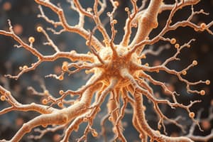
- Neurons are the fundamental units of the nervous system, responsible for receiving, processing, and transmitting information via electrochemical signaling.
- Camillo Golgi developed a silver staining method that made the entire neuron (soma, axon, and dendrites) visible under a microscope. He believed neurons were interconnected, forming a continuous network.
- Santiago Ramon y Cajal used Golgi's staining method to visualize brain circuitry, highlighting the distinct structures of neurons. He proposed the Neuron Doctrine, stating that neurons are discrete units that communicate via synapses but are not physically connected.
- The soma (cell body) contains the nucleus and surrounding cytoplasm (perikaryon). It is responsible for protein synthesis and other cellular functions.
- Organelles of the soma:
- Nucleus: involved in protein synthesis but not replication in adult neurons.
- Ribosomes: sites of protein translation, either free or associated with the endoplasmic reticulum (ER).
- ER: present in both smooth and rough forms (Nissl bodies). Nissl staining reveals the abundance of ER in neurons, indicative of their high metabolic activity.
- Golgi apparatus: involved in post-translational processing of proteins.
- Mitochondria: responsible for ATP generation, crucial for maintaining the membrane potential.
- Lysosomes: contain enzymes that break down cellular organelles.
- Lipofuscin bodies: accumulate lysosomal waste, appearing yellow/brown and increasing with age.
- The cytoskeleton provides structural support and shape to neurons, but it’s dynamic and constantly reorganizing.
- Microtubules: composed of alpha and beta tubulin proteins, they are dynamic structures that can grow or shrink. They are organized in a longitudinal manner and can branch to form new microtubules.
- Microfilaments: the thinnest cytoskeletal fibers, composed of actin polymers. They are responsible for changing neuronal shape and motility, especially important in dendrites.
- Neurofilaments: intermediate filaments made up of three protofibrils. They are less dynamic than microtubules and microfilaments, providing strong structural support, often referred to as the ‘bones’ of the cytoskeleton.
- Neurofibrillary tangles: abnormal accumulations of neurofilaments, associated with Alzheimer's disease.
The Neuronal Membrane
- The neuronal membrane is a phospholipid bilayer that forms a barrier, isolating the cytosol from the extracellular fluid. It contains transmembrane proteins that control the passage of ions in and out of the neuron.
- Transmembrane proteins:
- Receptor proteins: bind to signaling molecules.
- Channel proteins: allow the passage of specific ions.
- NA-K pump: uses ATP to actively move 3 sodium ions out of the cell and 2 potassium ions in, contributing to maintaining the membrane potential.
- Voltage-gated channels: open or close based on changes in membrane potential, allowing the passage of specific ions.
- The neuronal membrane is semi-permeable, allowing controlled movement of ions to maintain the resting membrane potential, which is approximately -70mV.
- Depolarization: Stimulus causes the membrane potential to become less negative; voltage-gated sodium channels open, and sodium ions move into the cell, making it more positive.
- Repolarization/Hyperpolarization: Sodium channels close, and potassium channels open. Potassium ions move out of the cell, driving the membrane potential back down to negative values. Hyperpolarization refers to the potential becoming even more negative than the resting potential.
The Action Potential
- The action potential is a rapid, short-lasting change in membrane potential that propagates down the axon. It is triggered when the membrane potential reaches a threshold of -55mV.
- The axon hillock is the site where EPSPs and IPSPs summate, determining whether an action potential is generated.
- The initial segment is where the action potential is generated.
- Conduction velocity: The speed at which an action potential travels down the axon, determined by the axon diameter and myelination.
Dendrites
- Dendrites are branching extensions of the neuron that receive synaptic input from other neurons.
- Dendritic spines: small projections on dendrites, typically forming one synapse each. A high number of spines indicates higher connectivity.
- Synaptic plasticity: the ability of dendrites to modify their shape and structure over time, altering the strength of synapses.
The Axon
- The axon is a specialized structure for transmitting electrical impulses (action potential) from the cell body to other neurons or target cells.
- Myelination: insulation of the axon by a sheath of myelin, greatly increasing conduction velocity (Group A and B fibers).
- Unmyelinated axons: lack myelin, conduction is slower (Group C fibers, e.g., pain signals).
- Axon collaterals: branches of the axon that allow a single neuron to communicate with multiple other neurons.
Axonal Transport
- Axonal transport: the movement of materials between the soma and the axon terminal, relying on microtubules as “tracks”.
- Fast transport: transports materials at speeds of 50-400 mm/day.
- Slow transport: transports materials at slower speeds.
Studying That Suits You
Use AI to generate personalized quizzes and flashcards to suit your learning preferences.
Related Documents
Description
This quiz covers the fundamental aspects of neurons, including their structure, functions, and key contributions from scientists like Camillo Golgi and Santiago Ramon y Cajal. Explore the roles of different neuron components and their importance in the nervous system.


