Podcast
Questions and Answers
What role do Schwann cells play in nerve fiber regeneration after Wallerian degeneration?
What role do Schwann cells play in nerve fiber regeneration after Wallerian degeneration?
- They assist in the degradation of myelin debris.
- They promote inflammation at the injury site.
- They completely replace the damaged axon.
- They form a regeneration tube around the remaining axon. (correct)
Which type of nerve fibers is responsible for transmitting sharp, pricking pain sensations?
Which type of nerve fibers is responsible for transmitting sharp, pricking pain sensations?
- Alpha fibers
- C fibers
- A-delta fibers (correct)
- Beta fibers
What is the primary function of the non-adapting nature of pain receptors?
What is the primary function of the non-adapting nature of pain receptors?
- To distinguish between different types of pain.
- To provide a constant warning signal about ongoing damage. (correct)
- To limit the intensity of pain signals.
- To help with the adaptation to chronic pain.
Which chemical is NOT associated with exciting pain nerve fibers?
Which chemical is NOT associated with exciting pain nerve fibers?
What primary effect does tissue ischemia have on pain sensation?
What primary effect does tissue ischemia have on pain sensation?
What type of pain is characterized by a dull, aching sensation and is transmitted by C fibers?
What type of pain is characterized by a dull, aching sensation and is transmitted by C fibers?
Muscle spasms can lead to pain primarily due to which of the following mechanisms?
Muscle spasms can lead to pain primarily due to which of the following mechanisms?
Which statement about fast and slow pain pathways is correct?
Which statement about fast and slow pain pathways is correct?
What type of cells are responsible for myelination in the Central Nervous System (CNS)?
What type of cells are responsible for myelination in the Central Nervous System (CNS)?
Which process describes the degeneration of the axon distal to the site of nerve injury?
Which process describes the degeneration of the axon distal to the site of nerve injury?
Which characteristic differentiates slow pain pathways from fast pain pathways?
Which characteristic differentiates slow pain pathways from fast pain pathways?
What can contribute to the experience of pain during tissue ischemia?
What can contribute to the experience of pain during tissue ischemia?
What is the primary role of the analgesia system in the body?
What is the primary role of the analgesia system in the body?
What does the gate control theory of pain suggest?
What does the gate control theory of pain suggest?
Which of the following functions is NOT associated with the hypothalamus?
Which of the following functions is NOT associated with the hypothalamus?
Which structure is formed by the extensions of the dura mater?
Which structure is formed by the extensions of the dura mater?
What is the primary role of the analgesia system in pain suppression?
What is the primary role of the analgesia system in pain suppression?
Which neurotransmitter is primarily inhibited by the analgesia system to reduce pain signals?
Which neurotransmitter is primarily inhibited by the analgesia system to reduce pain signals?
What phenomenon describes the pattern where pain from internal organs is perceived as occurring in a different part of the body?
What phenomenon describes the pattern where pain from internal organs is perceived as occurring in a different part of the body?
According to the Gate Control Theory, non-painful stimuli can reduce pain perception by:
According to the Gate Control Theory, non-painful stimuli can reduce pain perception by:
Which layer of the meninges is the outermost and most durable?
Which layer of the meninges is the outermost and most durable?
What is the role of dural venous sinuses?
What is the role of dural venous sinuses?
Which component is NOT typically found in the cerebrospinal fluid (CSF)?
Which component is NOT typically found in the cerebrospinal fluid (CSF)?
What structure separates the two cerebral hemispheres?
What structure separates the two cerebral hemispheres?
What is the primary role of the arachnoid villi in the drainage of cerebrospinal fluid (CSF)?
What is the primary role of the arachnoid villi in the drainage of cerebrospinal fluid (CSF)?
Which structure is located between the brainstem and the cerebellum?
Which structure is located between the brainstem and the cerebellum?
Which function is primarily regulated by the medulla oblongata?
Which function is primarily regulated by the medulla oblongata?
What is a key function of the pons?
What is a key function of the pons?
Which part of the brainstem is responsible for the most basic life functions such as consciousness?
Which part of the brainstem is responsible for the most basic life functions such as consciousness?
Which of the following is NOT a function of the hypothalamus?
Which of the following is NOT a function of the hypothalamus?
The midbrain is involved in which of the following functions?
The midbrain is involved in which of the following functions?
Which ventricle is considered a narrow, midline cavity located between the two halves of the thalamus?
Which ventricle is considered a narrow, midline cavity located between the two halves of the thalamus?
Study Notes
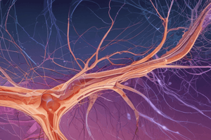
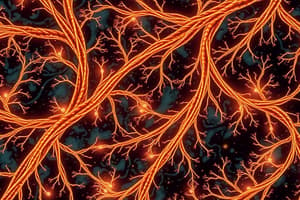
Myelination of Nerve Fibers
- In the CNS, oligodendrocytes myelinate multiple axons, forming an insulating myelin sheath that speeds up impulse conduction.
- In the PNS, Schwann cells myelinate a single segment of one axon, also enhancing impulse velocity.
Wallerian Degeneration and Nerve Fiber Regeneration
- Wallerian degeneration occurs after nerve injury, leading to degeneration of the axon segment distal to the injury site and breakdown of the myelin sheath.
- Regeneration in the PNS involves surviving Schwann cells forming a regeneration tube, guiding the regrowth of the axon towards its target.
Non-Adapting Nature of Pain Receptors
- Pain receptors (nociceptors) do not adapt, continually responding to the presence of harmful stimuli.
- This characteristic ensures pain serves as a persistent warning signal for ongoing tissue damage.
Slow vs. Fast Pain Pathways
- Fast pain pathways are transmitted by A-delta fibers, causing sharp, immediate, localized pain.
- Slow pain pathways utilize C fibers, resulting in dull, aching pain with a slower onset and more diffuse sensation.
Chemicals Exciting Pain Nerve Fibers
- Pain nerve fibers can be excited by bradykinin, histamine, prostaglandins, substance P, and ATP, all of which are related to tissue damage or inflammation.
Tissue Ischemia and Pain
- Ischemia results in a lack of oxygen, leading to metabolic waste accumulation like lactic acid, which stimulates pain receptors.
Muscle Spasm and Pain
- Muscle spasms create sustained muscle contraction, compressing blood vessels and leading to ischemia and pain due to reduced oxygen flow.
Analgesia System in Pain Suppression
- The analgesia system includes brain and spinal cord networks that modulate pain perception through neurotransmitters like endorphins, inhibiting pain signal transmission.
Concept of Referred Pain
- Referred pain occurs when pain from an internal organ is perceived in a different body area due to convergence of sensory nerves at the spinal cord level.
Gate Control Theory of Pain
- Gate Control Theory posits that non-painful input can inhibit pain signals in the spinal cord, effectively "closing the gate" to pain perception.
Meninges and Dural Venous Sinuses
- The meninges consist of three layers: dura mater (outermost), arachnoid mater (middle), and pia mater (innermost).
- Dural venous sinuses, formed between dura mater layers, drain venous blood from the brain to the internal jugular veins.
- Dural extensions include falx cerebri (separating cerebral hemispheres), tentorium cerebelli (separating cerebrum from cerebellum), and falx cerebelli (separating cerebellar hemispheres).
Cerebrospinal Fluid (CSF)
- CSF is secreted by choroid plexuses, primarily in brain ventricles, and is a clear fluid containing water, glucose, proteins, ions, and some white blood cells.
- It drains through ventricles into the subarachnoid space, absorbed by arachnoid villi into venous sinuses.
- Functions include cushioning the brain, maintaining a stable environment, and removing metabolic waste.
Ventricles of the Brain
- Lateral ventricles are two large cavities in the cerebral hemispheres.
- Third ventricle lies between the thalamic halves.
- Fourth ventricle is situated between the brainstem and cerebellum, connecting with the spinal canal.
Brainstem and Medulla Oblongata
- The brainstem regulates essential life functions, including heart rate, breathing, and consciousness.
- The medulla oblongata, at the brainstem's lower end, controls cardiovascular and respiratory functions and reflexes like coughing.
Structure and Functions of the Pons
- The pons lies above the medulla and connects different brain areas with its nerve tracts.
- It is involved in breathing, sleep regulation, sensory information relay, and movement coordination.
Structure and Functions of the Midbrain
- The midbrain, above the pons, consists of the tectum and tegmentum, influencing sensory processing and motor control.
- It regulates visual and auditory info processing and sleep-wake cycles.
Functions of the Hypothalamus
- Key roles include regulating body temperature, hunger/thirst, circadian rhythms, autonomic nervous system responses, and endocrine system control via the pituitary gland.
Studying That Suits You
Use AI to generate personalized quizzes and flashcards to suit your learning preferences.
Description
Explore the intricate processes of myelination in nerve fibers, covering the roles of oligodendrocytes and Schwann cells. Understand Wallerian degeneration and the regenerative capabilities of the PNS. Additionally, learn about the pain receptors' behavior and the distinctions between slow and fast pain pathways.