Podcast
Questions and Answers
Where are voltage-gated Ca2+ channels mainly present?
Where are voltage-gated Ca2+ channels mainly present?
- At the cell body
- At the axon terminals (correct)
- At the nodes of Ranvier
- At dendritic spines
Abnormalities of ion channels are known as ionopathies.
Abnormalities of ion channels are known as ionopathies.
False (B)
In the resting state, negatively charged ions are lined along the ______ of the membrane and positively charged ions are lined along the exterior of the membrane.
In the resting state, negatively charged ions are lined along the ______ of the membrane and positively charged ions are lined along the exterior of the membrane.
interior
What term is used to describe the change in membrane potential toward the resting value after depolarization?
What term is used to describe the change in membrane potential toward the resting value after depolarization?
Which type of synapse occurs through the release of neurotransmitters?
Which type of synapse occurs through the release of neurotransmitters?
What is the role of the presynaptic neuron in a synapse?
What is the role of the presynaptic neuron in a synapse?
What is the space between the presynaptic and postsynaptic membranes called?
What is the space between the presynaptic and postsynaptic membranes called?
___________ contain neurotransmitters and are released into the synaptic cleft for communication.
___________ contain neurotransmitters and are released into the synaptic cleft for communication.
EPSP leads to the inhibition of the postsynaptic neuron.
EPSP leads to the inhibition of the postsynaptic neuron.
What are the three main divisions of the nervous system?
What are the three main divisions of the nervous system?
What are the two principal cell types that make up the Central Nervous System?
What are the two principal cell types that make up the Central Nervous System?
Astrocytes are named for their star shape and are primarily found in the spinal cord.
Astrocytes are named for their star shape and are primarily found in the spinal cord.
Neurons transmit impulses in the form of ________.
Neurons transmit impulses in the form of ________.
Match the neuron types with their descriptions:
Match the neuron types with their descriptions:
Which component of the nerve Na+ channel is responsible for the upstroke of the action potential in nerve and skeletal muscle?
Which component of the nerve Na+ channel is responsible for the upstroke of the action potential in nerve and skeletal muscle?
What is the major regulator of erythropoiesis secreted from the kidney?
What is the major regulator of erythropoiesis secreted from the kidney?
At rest, the activation gate of the Na+ channel is open.
At rest, the activation gate of the Na+ channel is open.
What is the thick capsule covering the kidneys called?
What is the thick capsule covering the kidneys called?
What are the areas in the axon where the action potential is first initiated?
What are the areas in the axon where the action potential is first initiated?
Each kidney in an adult is roughly about the size of one's _____ (11 cm in length and 6 cm in width).
Each kidney in an adult is roughly about the size of one's _____ (11 cm in length and 6 cm in width).
In myelinated axons, the mode of propagation of action potential is known as _____ conduction.
In myelinated axons, the mode of propagation of action potential is known as _____ conduction.
The glomerulus is part of the nephron responsible for reabsorption of substances.
The glomerulus is part of the nephron responsible for reabsorption of substances.
Match the following functions of the kidney with their descriptions:
Match the following functions of the kidney with their descriptions:
Match the following cell types with their functions in the kidney:
Match the following cell types with their functions in the kidney:
What are some important functions of the flow of blood through the kidneys? (Select all that apply)
What are some important functions of the flow of blood through the kidneys? (Select all that apply)
What is the main determinant of Glomerular Filtration Rate (GFR)?
What is the main determinant of Glomerular Filtration Rate (GFR)?
Renal plasma flow (RPF) can be estimated from the clearance of an organic acid para-aminohippuric acid (PAH). Renal blood flow (RBF) is calculated from the RPF and the __________.
Renal plasma flow (RPF) can be estimated from the clearance of an organic acid para-aminohippuric acid (PAH). Renal blood flow (RBF) is calculated from the RPF and the __________.
The Fick principle states that the amount of a substance entering an organ equals the amount of the substance leaving the organ.
The Fick principle states that the amount of a substance entering an organ equals the amount of the substance leaving the organ.
Match the following substances with their effects on arterioles:
Match the following substances with their effects on arterioles:
What are the three layers of the basement membrane?
What are the three layers of the basement membrane?
What type of cells make up the epithelial cell layer?
What type of cells make up the epithelial cell layer?
Increasing renal blood flow decreases glomerular filtration rate.
Increasing renal blood flow decreases glomerular filtration rate.
The major function of the tubule is to reabsorb ___ and solutes from the tubular fluid.
The major function of the tubule is to reabsorb ___ and solutes from the tubular fluid.
Match the passive transport mechanism with its description:
Match the passive transport mechanism with its description:
What role do peritubular capillaries play in the absorption process?
What role do peritubular capillaries play in the absorption process?
Blood in peritubular capillaries has low oncotic pressure.
Blood in peritubular capillaries has low oncotic pressure.
How does the combination of high oncotic and low hydrostatic pressures in peritubular capillaries affect water uptake?
How does the combination of high oncotic and low hydrostatic pressures in peritubular capillaries affect water uptake?
Glucose is reabsorbed completely from tubular fluid in the ______.
Glucose is reabsorbed completely from tubular fluid in the ______.
What are the two mechanisms of coupled transport described in the content?
What are the two mechanisms of coupled transport described in the content?
What is the primary focus of reabsorption process in the proximal tubule?
What is the primary focus of reabsorption process in the proximal tubule?
Na+ reabsorption in the proximal tubule is a passive transport mechanism.
Na+ reabsorption in the proximal tubule is a passive transport mechanism.
About 67% of filtered _ is reabsorbed along with 67% of water in the proximal tubule.
About 67% of filtered _ is reabsorbed along with 67% of water in the proximal tubule.
Match the following terms with their descriptions:
Match the following terms with their descriptions:
Flashcards are hidden until you start studying
Study Notes
Nervous System
- The nervous system is a system of communication that allows individuals to interact with their external environment and integrates functions of various internal organ systems.
- It provides the basis for anatomical and physiological communications that help in the smooth execution of systemic functions.
Division of Nervous System
- The nervous system is divided into:
- Central Nervous System (CNS)
- Peripheral Nervous System (PNS)
- Autonomic Nervous System (ANS)
Central Nervous System (CNS)
- The CNS includes:
- Brain
- Spinal cord
- Brain is divided into three parts:
- Prosencephalon (forebrain)
- Telencephalon: includes cerebral hemisphere, amygdala, basal ganglia, hippocampus
- Functions: perception of sensations, cognition, learning, and memory, and planning and programming of responses
- Mesencephalon (midbrain)
- Functions: central pattern generator for locomotion, and nuclei for righting reflexes
- Rhombencephalon (hindbrain)
- Metencephalon: includes pons and cerebellum
- Myelencephalon: includes medulla oblongata
- Functions: control of cardiovascular and respiratory functions, motor activities, sleep-wakefulness, and visceral functions
- Prosencephalon (forebrain)
Spinal Cord
- The spinal cord starts from the base of the skull as the extension of medulla and continues till the body of the first lumbar vertebra.
- The spinal cord has 31 segments, each having a motor and a sensory nerve root.
- The main function of the spinal cord is to receive sensory inputs from peripheral structures and transmit them to the brain and convey signals originating from the brain motor and autonomic areas to the target structures.
Cellular Component of Central Nervous System
- The CNS consists of two principal cell types:
- Glial cells (neuroglia)
- Functions: support and protect neurons, maintain homeostasis of fluids that bath the neurons
- Neurons
- Functions: transmit impulses in the form of action potentials
- Glial cells (neuroglia)
Glial Cells
- Types of glial cells:
- Astrocytes
- Functions: provide mechanical matrix, serve metabolic and nutritive roles, help regulate blood flow, and phagocytose neural debris
- Oligodendrocytes
- Functions: form myelin sheath, insulate axons, and limit current flow
- Ependymal cells
- Functions: unclear
- Microglia
- Functions: scavenger cells, eliminate debris and organisms
- Astrocytes
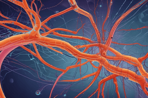
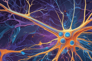
Neuron
- A neuron consists of:
- Cell body (soma)
- Contains: nucleus, cytoplasm, organelles, and Nissl bodies
- Dendrites
- Functions: receive information, conduct impulses toward the cell body
- Axon
- Functions: transmit impulses away from the cell body
- Cell body (soma)
- Types of neurons:
- Unipolar neurons
- Pseudounipolar neurons
- Bipolar neurons
- Multipolar neurons
- Golgi type 1 and 2
- Sensory neurons
- Motor neurons
- Pyramidal cells
- Stellate cells
Synapses in CNS
- Communication between neurons occurs through synaptic transmission.
- Synapses are the neuro-neuronal junctions through which information from one neuron passes to the other.
- Types of synapses:
- Axodendritic synapse
- Axosomatic synapse
- Axoaxonic synapse
- Dendrodendritic synapse
- Mechanism of synaptic transmission:
- Presynaptic mechanisms: vesicle release, calcium influx, and exocytosis
- Postsynaptic mechanisms: neurotransmitter binding, receptor activation, and generation of postsynaptic potential
Mechanism of Synaptic Transmission
- Presynaptic mechanisms:
- Vesicle release
- Calcium influx
- Exocytosis
- Postsynaptic mechanisms:
- Neurotransmitter binding
- Receptor activation
- Generation of postsynaptic potential### Neurotransmitter Binding and Synaptic Potential
- Neurotransmitter binds to receptors in the postsynaptic membrane, causing a conformational change that opens ion channels or triggers a cascade of biochemical reactions.
- This binding generates a change in ionic permeability of the cell, leading to a synaptic potential.
Synaptic Potential
- Synaptic potential is a change in membrane potential of the postsynaptic neuron, lasting longer than an action potential.
- Can be either excitatory (depolarizing) or inhibitory (hyperpolarizing).
Types of Synaptic Potentials
- Excitatory Postsynaptic Potential (EPSP)
- Occurs when the postsynaptic membrane becomes depolarized (less negative).
- Can cause repeated firing of the postsynaptic neuron if the EPSP is large enough.
- Caused by opening of Na+ or Ca2+ ion channels, or closure of K+ channels.
- Inhibitory Postsynaptic Potential (IPSP)
- Occurs when the postsynaptic membrane becomes hyperpolarized (more negative).
- Decreases the excitability of the postsynaptic neuron.
- Caused by opening of Cl- channels, or closure of Na+ or Ca2+ channels.
Ion Channels
- Neuronal Ion Channels
- Nongated (leaky) channels: Na+, K+, Cl- channels present throughout the neuronal membrane.
- Gated channels: Voltage-gated (Na+, Ca2+), Ligand-gated (at synaptic sites), Mechanical-gated (sensory nerve endings).
- ATP-driven pumps: Na+-K+ ATPase helps maintain ionic balance.
Distribution of Na+ Channels
- Highest concentration in:
- Initial segment of the axon (350-500 channels/μm²)
- Nodes of Ranvier (2000-12,000 channels/μm²)
Membrane Potentials
- Resting Membrane Potential (RMP): -70 mV
- Depolarization: a decrease in membrane potential (becomes less negative).
- Hyperpolarization: an increase in membrane potential (becomes more negative).
- Repolarization: return of the membrane potential to RMP after depolarization.
Genesis of Nerve Potential
- Excitability: ability of cells to generate action potentials.
- Stimulus: can be electrical, chemical, or mechanical.
- Action Potential: a transient change in membrane potential of about 100 mV, conducted along the axon in an all-or-none fashion.
Graded Potentials
- Electrotonic Potentials: local, nonpropagated potentials of small magnitude, in response to a depolarizing or hyperpolarizing stimulus of lesser strength.
- Types: Catelectrotonic potential (depolarizing), Anelectrotonic potential (hyperpolarizing).
- Properties:
- Graded in nature
- Decremental conduction
- Depolarizing or hyperpolarizing
- Summation of potentials
Action Potential
- Phases:
- Depolarization: gradual depolarization to threshold, rapid ascent, overshoot, and peak.
- Repolarization: rapid falling phase and slower terminal part.
- Ionic Basis:
- Resting membrane potential: K+ conductance is high, Na+ conductance is low.
- Upstroke: rapid opening of Na+ channels, inward Na+ current.
- Repolarization: inactivation of Na+ channels, increased K+ conductance, outward K+ current.
Nerve Na+ Channel
- Voltage-Gated Na+ Channel: responsible for the upstroke of the action potential.
- Gates: activation gate opens quickly, inactivation gate closes after a time delay.
- Function: regulates Na+ flow into the cell during the action potential.### Action Potential Generation and Conduction
- The potential of the adjacent membrane decreases as Na+ ions move to the negative area, bringing the membrane to the firing level.
- This opens voltage-gated Na+ channels, firing an action potential.
- The process is repeated, with positive charges flowing to the adjacent resting membrane, decreasing its potential, and activating voltage-gated Na+ channels.
- The action potential does not move on its own, but generates a new action potential in the membrane ahead.
- Sodium and potassium channels open and close sequentially along the axonal membrane, allowing the action potential to travel.
Myelinated Axon (Saltatory Conduction)
- Myelin acts as an insulator, preventing ion flow across the membrane.
- Voltage-gated Na+ channels are present in large numbers at the nodes of Ranvier.
- The local current travels like a graded potential, decreasing by 37% over 3 mm, and arrives at the adjacent node of Ranvier.
- The nodal membrane is depolarized to the threshold level, firing an action potential.
- The action potential rapidly proceeds from one node to the next, pausing to regenerate at each node, in a process called saltatory conduction.
Advantages of Myelinated Axon
- Faster velocity of conduction.
- Conservation of energy, as ionic flux occurs only at the nodes of Ranvier.
Direction of Propagation of Action Potential
- In motor neurons, the action potential is conducted from the axon hillock towards the axon terminal.
- In sensory neurons, it propagates from the first node of Ranvier towards the CNS (anterograde conduction of impulse).
- The action potential does not travel back towards the trigger zone due to the refractory period.
Renal System
- The kidneys play a vital role in maintaining homeostatic functions, including regulating ECF volume and composition.
- Kidneys form urine and excrete waste products, regulate blood pressure, electrolyte composition, acid-base balance, and plasma osmolality.
Functions of Kidney
- Urine formation and excretion of waste products.
- Regulation of ECF volume.
- Regulation of blood pressure.
- Regulation of electrolyte composition.
- Acid-base balance.
- Regulation of plasma osmolality.
- Regulation of erythropoiesis.
- Endocrine functions.
Anatomy and Blood Supply
- The kidneys are bean-shaped, paired organs located in the posterior part of the abdomen.
- Each kidney is about 11 cm in length, 6 cm in width, and weighs around 150 g.
- The kidneys are covered by a thick capsule, and have three main regions: cortex, medulla, and papilla.
Structure of the Nephron
- A nephron is the basic structural and functional unit of the kidney.
- Each nephron consists of a glomerulus, a renal tubule, and a collecting duct.
- The glomerulus is a glomerular capillary network surrounded by Bowman's capsule.
- The renal tubule consists of the proximal convoluted tubule, proximal straight tubule, loop of Henle, distal convoluted tubule, and collecting duct.
Secretory Cells of Kidney
- Juxtaglomerular (JG) cells secrete renin, which activates the renin-angiotensin system.
- Interstitial cells (IS cells) secrete erythropoietin and prostaglandins.
Renal Vasculature
- Blood enters the kidney via the renal artery, which branches into interlobar arteries, arcuate arteries, and cortical radial arteries.
- The afferent arterioles deliver blood to the glomerular capillaries, and the efferent arterioles deliver blood to the peritubular capillaries.
Functions of Vasa Recta
- Provides oxygen and nutrients to the nephron segments.
- Delivers substances to the nephron for secretion into the tubular lumen.
- Serves as a pathway for the return of reabsorbed water and solutes to the circulatory system.
- Participates in concentration and dilution of urine.
Juxtaglomerular Apparatus
- Consists of the macula densa, juxtaglomerular cells, and lacis cells.
- The macula densa acts as a sensor, monitoring the change in ionic composition and rate of flow in the tubular fluid.
- Juxtaglomerular cells secrete renin, which activates the renin-angiotensin system.
- Lacis cells secrete renin and erythropoietin.
Renal Clearance
- Renal clearance is the volume of plasma completely cleared of a substance by the kidneys per unit time.
- Calculated using the equation: C = [U]x × V / [P]x.
- Substances with high renal clearance are completely removed on a single pass of blood through the kidneys.
Studying That Suits You
Use AI to generate personalized quizzes and flashcards to suit your learning preferences.