Podcast
Questions and Answers
What space separates the arachnoid mater from the dura mater?
What space separates the arachnoid mater from the dura mater?
- Subdural space (correct)
- Epidural space
- Intrathecal space
- Subarachnoid space
Which of the following structures is contained within the subarachnoid space?
Which of the following structures is contained within the subarachnoid space?
- Epidural fat
- Venous sinuses
- Cerebrospinal fluid (CSF) (correct)
- Pia mater
In which demographic group is the incidence of epidural hemorrhage higher?
In which demographic group is the incidence of epidural hemorrhage higher?
- Children under 12
- Adolescents and younger adults (correct)
- Elderly individuals
- Women over 60
What is a non-traumatic cause of potential issues in the dural venous sinus area?
What is a non-traumatic cause of potential issues in the dural venous sinus area?
How does aging affect the dura mater's attachment to the overlying bone?
How does aging affect the dura mater's attachment to the overlying bone?
What shape is the lentiform nucleus described as resembling?
What shape is the lentiform nucleus described as resembling?
Which of the following is NOT a common cause of epidural hemorrhage?
Which of the following is NOT a common cause of epidural hemorrhage?
Which structure can pass through the subarachnoid space?
Which structure can pass through the subarachnoid space?
What is the primary location of an epidural hemorrhage?
What is the primary location of an epidural hemorrhage?
Which artery is most commonly implicated in an epidural hemorrhage?
Which artery is most commonly implicated in an epidural hemorrhage?
Under what circumstances is non-surgical management of an epidural hematoma appropriate?
Under what circumstances is non-surgical management of an epidural hematoma appropriate?
What defines prompt surgical intervention in cases of epidural hemorrhage?
What defines prompt surgical intervention in cases of epidural hemorrhage?
What is the significance of the pterion in relation to epidural hemorrhages?
What is the significance of the pterion in relation to epidural hemorrhages?
What CT scan finding is characteristic of an epidural hemorrhage?
What CT scan finding is characteristic of an epidural hemorrhage?
Which of the following conditions indicates a more severe case of epidural hemorrhage requiring surgical treatment?
Which of the following conditions indicates a more severe case of epidural hemorrhage requiring surgical treatment?
What is the importance of repeat neuroimaging 6-8 hours after brain injury in cases of epidural hemorrhage?
What is the importance of repeat neuroimaging 6-8 hours after brain injury in cases of epidural hemorrhage?
What type of blood is primarily received from the brain by cerebral veins?
What type of blood is primarily received from the brain by cerebral veins?
Which structure is responsible for the drainage of cerebrospinal fluid?
Which structure is responsible for the drainage of cerebrospinal fluid?
Which of the following sinuses is NOT a venous sinus that drains blood from the brain?
Which of the following sinuses is NOT a venous sinus that drains blood from the brain?
What is a unique characteristic of the walls of cerebral veins?
What is a unique characteristic of the walls of cerebral veins?
Which layer of the meninges closely invests the brain surface?
Which layer of the meninges closely invests the brain surface?
Which of the following statements about the arachnoid granulations is true?
Which of the following statements about the arachnoid granulations is true?
What happens to the blood after it drains into the internal jugular vein?
What happens to the blood after it drains into the internal jugular vein?
Which term describes the characteristics of the sinuses mentioned in the content?
Which term describes the characteristics of the sinuses mentioned in the content?
What is the primary function of the diaphragma sellae?
What is the primary function of the diaphragma sellae?
Which artery is primarily responsible for supplying blood to the dura mater?
Which artery is primarily responsible for supplying blood to the dura mater?
What condition is indicated by the involvement of the posterior branch of the meningeal artery?
What condition is indicated by the involvement of the posterior branch of the meningeal artery?
What anatomical structure passes through the small opening at the center of the diaphragma sellae?
What anatomical structure passes through the small opening at the center of the diaphragma sellae?
Which clinical scenario is most likely to cause damage to the middle meningeal artery?
Which clinical scenario is most likely to cause damage to the middle meningeal artery?
What is the primary role of the tentorium cerebelli?
What is the primary role of the tentorium cerebelli?
Which structure runs alongside the attachment of the tentorium cerebelli to the petrous bone?
Which structure runs alongside the attachment of the tentorium cerebelli to the petrous bone?
What shape is the falx cerebelli described as?
What shape is the falx cerebelli described as?
Where is the occipital sinus located in relation to the falx cerebelli?
Where is the occipital sinus located in relation to the falx cerebelli?
Which layer of the dura mater is represented by the endosteal layer?
Which layer of the dura mater is represented by the endosteal layer?
The transverse sinus is associated with which bone?
The transverse sinus is associated with which bone?
What is the anatomical significance of the falx cerebelli?
What is the anatomical significance of the falx cerebelli?
Which structure does not run along the tentorium cerebelli's attachments?
Which structure does not run along the tentorium cerebelli's attachments?
Study Notes
Dural Venous Sinuses
- Dural venous sinuses collect blood from the brain and drain into the internal jugular vein, which returns blood to the lungs for oxygenation.
- Notable sinuses: superior sagittal sinus, transverse sinus, confluence of sinuses, occipital sinus, inferior sagittal sinus, straight sinus, cavernous sinus, superior and inferior petrosal sinuses, and sigmoid sinus.
Arachnoid Mater
- Positioned between the pia mater and dura mater, creating spaces for cerebrospinal fluid (CSF).
- Subdural space lies below the dura mater; subarachnoid space contains CSF and serves as a pathway for cerebral arteries, veins, and cranial nerves.
- Arachnoid granulations, formed from arachnoid villi, facilitate the drainage of unoxygenated blood and CSF from the subarachnoid space.
Epidural Hemorrhage
- Occurs between the dura mater and skull, resulting from trauma such as vehicle collisions or falls.
- Characterized by clear borders and confined by lateral sutures, appearing dense or white on CT scans.
- Anterior division of the middle meningeal artery is the most frequently damaged vessel, with the pterion being the common injury site.
- Surgical intervention is required for hematomas over 30ml or if GCS is below 9 with pupillary abnormalities.
Non-Traumatic Causes of Hemorrhage
- Infections or abscesses, coagulopathy, hemorrhagic tumors, and vascular malformations (e.g., dural arteriovenous fistula) may lead to hemorrhagic conditions.
Pia Mater
- The innermost meningeal layer, difficult to separate from brain tissue during dissection.
- Composed of a vascular membrane covered by mesothelial cells, closely adheres to the brain and descends into sulci.
- Extends around cranial nerves, fusing with their epineurium.
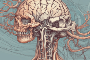
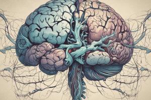
Dura Mater and Its Features
- Comprised of an endosteal layer and a meningeal layer.
- The tentorium cerebelli is a crescent-shaped fold of dura that covers the cerebellum, with the superior petrosal sinus and transverse sinus running along its borders.
- The falx cerebelli and diaphragma sellae are additional folds providing structural support within the cranial cavity.
Blood Supply
- Mainly supplied by the middle meningeal artery, which branches from the external carotid artery to the internal maxillary artery.
- The dura mater has extensive blood supply due to its widespread presence throughout the brain.
Studying That Suits You
Use AI to generate personalized quizzes and flashcards to suit your learning preferences.
Description
This quiz explores the structure and function of dural venous sinuses, arachnoid mater, and the occurrence of epidural hemorrhage. Test your knowledge on notable sinuses, cerebrospinal fluid pathways, and the implications of trauma on the brain. Ideal for students studying neuroanatomy.