Podcast
Questions and Answers
What do nervous tissues receive, transmit, and integrate?
What do nervous tissues receive, transmit, and integrate?
Information from outside and inside the body
What are the two primary cell types in the nervous system?
What are the two primary cell types in the nervous system?
Neurons and Glial cells
What are neurons?
What are neurons?
Excitatory cells that transmit electrical signals
What is the Nissl Method used for?
What is the Nissl Method used for?
What type of cell is a neuron?
What type of cell is a neuron?
What cells are collectively known as neuroglia?
What cells are collectively known as neuroglia?
Name the four principal types of neuroglia cells that are recognized in the CNS.
Name the four principal types of neuroglia cells that are recognized in the CNS.
What is the function of oligodendrocytes?
What is the function of oligodendrocytes?
Name the layers of cerebral cortex.
Name the layers of cerebral cortex.
What are the layers that the cerebellum comprises?
What are the layers that the cerebellum comprises?
In cross section, what does the spinal cord consist of?
In cross section, what does the spinal cord consist of?
Name the connective tissue layers of nerve fibers.
Name the connective tissue layers of nerve fibers.
The human eye has three coats or layers. They are...
The human eye has three coats or layers. They are...
What is the middle vascular coat of the eye also called?
What is the middle vascular coat of the eye also called?
Name the five layers that the cornea consists of.
Name the five layers that the cornea consists of.
Name the two parts that the retina is principally composed of.
Name the two parts that the retina is principally composed of.
The retina is subdivided into how many recognizable layers?
The retina is subdivided into how many recognizable layers?
Name the layers of the retina (from the outside to the inside of the eye).
Name the layers of the retina (from the outside to the inside of the eye).
The eyelid consists of a dense fibro-elastic plate known as the _____ plate.
The eyelid consists of a dense fibro-elastic plate known as the _____ plate.
It consists of the bony labyrinth and _____ labyrinth filled with perilymph and endolymph respectively
It consists of the bony labyrinth and _____ labyrinth filled with perilymph and endolymph respectively
Name parts of bony labyrinth.
Name parts of bony labyrinth.
What are the functions of the semicircular canals and cochlea?
What are the functions of the semicircular canals and cochlea?
The lumen of the cochlea is divided into three chambers. Name the chambers.
The lumen of the cochlea is divided into three chambers. Name the chambers.
The _____ of Corti is located in the scala media where it is surrounded by the endolymph.
The _____ of Corti is located in the scala media where it is surrounded by the endolymph.
The vestibule contains...
The vestibule contains...
The cristae ampullaris of the semicircular canals contains hair cells with _____ and kinocilium, embedded in the cupula, supported by supporting cells, and innervated by vestibular nerve fibers.
The cristae ampullaris of the semicircular canals contains hair cells with _____ and kinocilium, embedded in the cupula, supported by supporting cells, and innervated by vestibular nerve fibers.
Flashcards
Nervous Tissue Definition
Nervous Tissue Definition
Tissues that receive, transmit, and integrate information to control body activities.
Central Nervous System (CNS)
Central Nervous System (CNS)
Brain and spinal cord; the control center.
Peripheral Nervous System (PNS)
Peripheral Nervous System (PNS)
Nerves outside the brain and spinal cord, spinal and cranial.
Somatic Nervous System
Somatic Nervous System
Signup and view all the flashcards
Autonomic Nervous System
Autonomic Nervous System
Signup and view all the flashcards
Neurons
Neurons
Signup and view all the flashcards
Glial Cells/Neuroglia
Glial Cells/Neuroglia
Signup and view all the flashcards
Hematoxylin and Eosin (H+E)
Hematoxylin and Eosin (H+E)
Signup and view all the flashcards
Heavy Metal Impregnation Technique
Heavy Metal Impregnation Technique
Signup and view all the flashcards
Immunohistochemistry
Immunohistochemistry
Signup and view all the flashcards
Nissl Method
Nissl Method
Signup and view all the flashcards
Neuron Structure: Perikaryon
Neuron Structure: Perikaryon
Signup and view all the flashcards
Astrocytes
Astrocytes
Signup and view all the flashcards
Oligodendrocytes
Oligodendrocytes
Signup and view all the flashcards
Microglia
Microglia
Signup and view all the flashcards
Ependymal cell
Ependymal cell
Signup and view all the flashcards
Oligodendrocytes Function
Oligodendrocytes Function
Signup and view all the flashcards
Schwann cells Function
Schwann cells Function
Signup and view all the flashcards
Cerebral Cortex
Cerebral Cortex
Signup and view all the flashcards
Brain Nuclei
Brain Nuclei
Signup and view all the flashcards
Inner White Matter
Inner White Matter
Signup and view all the flashcards
Cerebellum Folia
Cerebellum Folia
Signup and view all the flashcards
Outer Molecular Layer
Outer Molecular Layer
Signup and view all the flashcards
Middle Purkinje cell layer
Middle Purkinje cell layer
Signup and view all the flashcards
Inner Granular Cell Layer
Inner Granular Cell Layer
Signup and view all the flashcards
Spinal Cord
Spinal Cord
Signup and view all the flashcards
Spinal Cord Gray Matter
Spinal Cord Gray Matter
Signup and view all the flashcards
Spinal Cord White Matter
Spinal Cord White Matter
Signup and view all the flashcards
Nerve Fiber
Nerve Fiber
Signup and view all the flashcards
Endoneurium
Endoneurium
Signup and view all the flashcards
Study Notes
- Nervous tissues receive, transmit, and integrate information to control the body's activities.
- Highly specialized, interconnected neurons process information and generate response signals.
Central Nervous System (CNS)
- Consists of the brain and spinal cord.
- Protected within the skull and vertebral canal.
- Covered by meninges.
- Bathed by cerebrospinal fluid (CSF).
Peripheral Nervous System (PNS)
- Includes paired spinal and cranial nerves, and ganglia.
Functional Divisions
- Somatic Nervous System: Supplies the body wall and limbs, involved in voluntary functions.
- Autonomic Nervous System: Supplies viscera and glands, controls involuntary organ functions.
Cells of the Nervous System
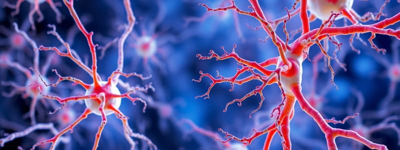
- Neurons: Excitable cells that transmit electrical signals.
- Glial Cells (Neuroglia): Supporting cells including astrocytes, oligodendrocytes, ependymal cells, microglia, and Schwann cells.
Stains Used
- Hematoxylin and Eosin (H+E).
- Immunohistochemistry: Used to study neuron-specific proteins like neurofilament protein.
- Heavy Metal Impregnation Technique: Gold and silver are used to study neuron morphology.
- Nissl Method: Stains RNA for rough endoplasmic reticulum (Nissl Substance); the axon lacks Nissl Substance.
Neuron Structure
- Excitatory cell with a cell body (perikaryon) containing the nucleus, Nissl bodies, neurofilaments, and microtubules.
- Has a single axon and one or more dendrites.
Glial Cells (Neuroglia in CNS)
- Non-neuronal support cells in the nervous tissue.
- Oligodendrocytes
- Astrocytes
- Microglia
- Ependymal cells
Astrocytes
- Exhibit a small cell body, a large oval nucleus, and a dark-stained nucleolus.
- Have long, thin, radiating processes located between neurons and blood vessels.
- Perivascular fibrous astrocytes surround capillaries.
- Long processes extend to and terminate on the capillary as perivascular end feet.
Microglia
- Smallest neuroglial cells with elongated nuclei and fine branched processes.
- Originate from bone marrow precursors.
- Found throughout the CNS (white and gray matter).
- Act like macrophages, migrating to injury sites.
- Proliferate, become phagocytic, and remove dead or foreign tissue.
Oligodendrocytes and Schwann Cells
- Oligodendrocytes are smaller than astrocytes with fewer cytoplasmic processes.
- Oligodendrocytes myelinate axons in the CNS.
- A single oligodendrocyte can surround and myelinate several axons.
- Schwann cells myelinate axons in the PNS, forming one myelin sheath around the internode of a single myelinated axon.
Ependymal Cells
- Typically cuboidal-columnar.
- Often have cilia, originating from neuroepithelial cells of the neural plate.
- Line the ventricles of the brain and spinal canal.
- Play a role in the production of cerebrospinal fluid (CSF).
- Tumors of ependymal cells are called ependymomas.
Cerebrum
- Layers of neurons are arranged closely to form the gray matter or cerebral cortex.
- Neurons grouped deeply form nuclei (thalamus, basal ganglia).
- Inner white matter conveys axons of neurons (bundles of nerve fibers).
- The cerebral cortex has six layers: Molecular (plexiform), External granular, External pyramidal, Internal granular, Internal pyramidal, Multiform (fusiform).
Cerebellum
- Surface layer of gray matter (cortex) is arranged into folds (folia).
- Central core of nerve fibers forms the white matter.
- Outer molecular layer (ML) consists of unmyelinated nerve fibers.
- Middle Purkinje cell layer (PL) is made of Purkinje cells with large cell bodies in a single layer.
- Inner granular cell layer (GL) consists of small neurons.
Spinal Cord
- Neurons form the central core (gray matter). Fibers form the outer section (white matter).
- Gray matter is butterfly-shaped. It contains neurons and extends to form ventral, dorsal & lateral horns.
- White matter has nerve fibers forming ascending & descending tracts surrounding the gray matter.
Connective Tissue Layers of Nerve Fibers
- Nerves have nerve fibers in nerve fascicles.
- Endoneurium: Delicate connective tissue around each nerve fiber with Schwann cells.
- Perineurium: Connective tissue covering each nerve fascicle.
- Epineurium: Connective tissue covering the entire nerve.
Eye Layers
- Fibro-elastic outer coat (corneo-scleral coat): Includes sclera and cornea.
- Sclera: White posterior part of dense collagen fibers.
- Cornea: Anterior transparent part.
- Middle vascular coat (Uveal tract): Consists of the iris, ciliary body, and choroid.
- Choroid lies between the sclera and retina.
- Iris regulates light via the pupil aperture.
- Inner photosensitive nervous coat:The Retina.
Cornea Layers
- Corneal epithelium: Stratified squamous non-keratinized epithelium (5-6 layers).
- Bowman’s membrane or anterior limiting membrane: Compactly packed collagen fibrils.
- Stroma: 200 layers of collagen fibrils with fibroblasts in an organized manner.
- Descemet’s membrane: Compactly packed collagen fibrils.
- Endothelium: Simple squamous epithelium of the anterior chamber.
Retina Parts
- Pigmented cuboidal epithelium: Contains melanin to absorb light after photoreceptor stimulation, preventing back reflection.
- Multilayered nervous part: Contains photoreceptors (rods and cones), bipolar neurons, and ganglionic multipolar neurons. Association neurons and glial cells (horizontal cells, amacrine cells, and Müller’s cell) are present. Has 10 recognizable layers.
Retina Layers (Outside to Inside)
- Pigmented layer: Layer of pigmented simple cuboidal epithelium.
- Photoreceptor layer: Outer and inner segments of rods and cones.
- External limiting membrane.
- Outer nuclear layer.
- Outer plexiform layer.
- Inner nuclear layer.
- Inner plexiform layer.
- Ganglionic cell layer.
- Nerve fiber layer.
- Internal limiting membrane.
Eyelid
- Dense fibro-elastic plate (tarsal plate) covered externally by thin skin and lined internally by the conjunctiva.
- Orbicularis oculi muscle can be seen between the skin and tarsal plate.
- Contains 12-30 tarsal (Meibomian) glands within the tarsal plate.
- Ciliary sweat glands are associated with the eyelashes.
- Conjunctiva covers the sclera's exposed part and the inner eyelid surface. It contains non-keratinized, stratified columnar or cuboidal epithelium, and goblet cells.
Inner Ear
- Bony labyrinth and membranous labyrinth filled with perilymph and endolymph.
- Parts of bony labyrinth:
- Vestibule: Contains maculae in the utricle and saccule for balance.
- Semicircular canals: Crista ampullaris for balance.
- Cochlea: Organ of Corti for hearing.
Cochlea
- Divided into three chambers: scala vestibuli, scala media (cochlear duct), and scala tympani.
- Scala vestibuli and scala tympani are filled with perilymph.
- Scala media is filled with endolymph.
- The organ of Corti is in the scala media, surrounded by endolymph.
Organ of Corti
- Rests on the basilar membrane
- Has inner and outer hair cells, as well as supporting cells.
- Apical part of hair cell show multiple cilia that attach to the tectorial membrane.
- Basal part of hair cells relate directly to the dendritic process of afferent sensory neuron.
- The fibrous tectorial membrane rests on top of the stereo cilia of the hair cells.
Vestibule
- Contains the utricle and the saccule (otolith organs).
- Each has a macula with sensory and supporting cells.
- Within each macula, stereocilia are embedded in the gelatinous otolithic membrane.
- Head movements bend the cilia due to gravity and position information is sent to the central nervous system.
Semicircular Canals
- Arranged in different planes; each contains a tube filled with endolymph.
- Each canal has an expanded end called the ampulla, which contains the crista ampullaris with hair cells, stereocilia, and kinocilium.
- Stereocilia and kinocilium are embedded in the cupula held by supporting cells, and innervated by vestibular nerve fibers.
- Head movements make the cupula and cilia in the cilia move, which stimulates or inhibits nerve fibers
Studying That Suits You
Use AI to generate personalized quizzes and flashcards to suit your learning preferences.


