Podcast
Questions and Answers
What is the primary imbalance that defines ischemia?
What is the primary imbalance that defines ischemia?
- Compromised myocardial perfusion due to an imbalance between oxygen supply and demand. (correct)
- Increased ventricular wall compressibility.
- Reduced diastolic time affecting ventricular relaxation.
- Excessive myocardial oxygen supply relative to demand.
Which of the following factors directly influences myocardial oxygen demand?
Which of the following factors directly influences myocardial oxygen demand?
- Myocardial contractility (correct)
- Diastolic time
- Ventricular wall compressibility
- Coronary vessel lumen size
What is the potential outcome if ischemia is not treated promptly?
What is the potential outcome if ischemia is not treated promptly?
- Reversible cell damage with immediate restoration of blood flow.
- Injury, apoptosis, or necrosis (cell death). (correct)
- Increased aerobic metabolism in myocardial cells.
- Efficient waste removal (CO2, H2O) in affected cells.
What is the primary result of anaerobic metabolism during ischemia?
What is the primary result of anaerobic metabolism during ischemia?
What is a key early consequence of ischemia on myocardial cells?
What is a key early consequence of ischemia on myocardial cells?
What is a key feature of Non-ST Elevation Myocardial Infarction (NSTEMI)?
What is a key feature of Non-ST Elevation Myocardial Infarction (NSTEMI)?
What ECG change is typically observed during unstable angina?
What ECG change is typically observed during unstable angina?
Which cardiac biomarker is considered the preferred biomarker for MI diagnosis?
Which cardiac biomarker is considered the preferred biomarker for MI diagnosis?
What ECG finding is indicative of myocardial ischemia?
What ECG finding is indicative of myocardial ischemia?
Which zone of myocardial tissue is characterized by decreased perfusion but cells remain viable if blood flow is restored?
Which zone of myocardial tissue is characterized by decreased perfusion but cells remain viable if blood flow is restored?
What is the primary purpose of angiogenesis in the context of myocardial ischemia?
What is the primary purpose of angiogenesis in the context of myocardial ischemia?
What role does the sympathetic nervous system play in response to decreased cardiac output?
What role does the sympathetic nervous system play in response to decreased cardiac output?
What is a potential consequence of long-term activation of the renin-angiotensin-aldosterone system (RAAS) in heart failure?
What is a potential consequence of long-term activation of the renin-angiotensin-aldosterone system (RAAS) in heart failure?
Which diagnostic tool is used to assess myocardial damage and identify hypokinetic or akinetic areas?
Which diagnostic tool is used to assess myocardial damage and identify hypokinetic or akinetic areas?
Which clinical manifestation is characteristic of left-sided heart failure?
Which clinical manifestation is characteristic of left-sided heart failure?
What is a primary goal when decreasing preload in the treatment of heart failure?
What is a primary goal when decreasing preload in the treatment of heart failure?
What is the mechanism of action of ACE inhibitors in the treatment of heart failure?
What is the mechanism of action of ACE inhibitors in the treatment of heart failure?
Which medication class increases contractility and acts as a negative chronotrope (reducing hear rate)?
Which medication class increases contractility and acts as a negative chronotrope (reducing hear rate)?
What is the primary purpose of Cardiac Resynchronization Therapy (CRT)?
What is the primary purpose of Cardiac Resynchronization Therapy (CRT)?
Which of the Beta-Blocker Generations is better tolerated and less bradycardia?
Which of the Beta-Blocker Generations is better tolerated and less bradycardia?
Flashcards
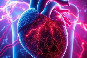
Ischemia
Ischemia
Myocardial perfusion is compromised due to an imbalance between myocardial oxygen supply and demand
Oxygen Supply
Oxygen Supply
Regulated by coronary vessel lumen size, ventricular wall compressibility, and diastolic time
Oxygen Demand
Oxygen Demand
Dependent on myocardial contractility, heart rate, and ventricular wall stress (preload and afterload)
Causes of Ischemia
Causes of Ischemia
Signup and view all the flashcards
Pathophysiology of Ischemia
Pathophysiology of Ischemia
Signup and view all the flashcards
Consequences of Inadequate ATP
Consequences of Inadequate ATP
Signup and view all the flashcards
Impact of Cell Death
Impact of Cell Death
Signup and view all the flashcards
Acute Coronary Syndrome (ACS)
Acute Coronary Syndrome (ACS)
Signup and view all the flashcards
Atherosclerosis
Atherosclerosis
Signup and view all the flashcards
Unstable Angina - Pathophysiology
Unstable Angina - Pathophysiology
Signup and view all the flashcards
Non-ST Elevation Myocardial Infarction (NSTEMI) - Pathophysiology
Non-ST Elevation Myocardial Infarction (NSTEMI) - Pathophysiology
Signup and view all the flashcards
ST Elevation Myocardial Infarction (STEMI) - Pathophysiology
ST Elevation Myocardial Infarction (STEMI) - Pathophysiology
Signup and view all the flashcards
Myocardial Injury
Myocardial Injury
Signup and view all the flashcards
Zone of Infarct
Zone of Infarct
Signup and view all the flashcards
Zone of Injury
Zone of Injury
Signup and view all the flashcards
Zone of Ischemia
Zone of Ischemia
Signup and view all the flashcards
Angiogenesis
Angiogenesis
Signup and view all the flashcards
Ventricular Remodeling
Ventricular Remodeling
Signup and view all the flashcards
What is Heart Failure?
What is Heart Failure?
Signup and view all the flashcards
Compensatory Mechanisms - Heart Failure
Compensatory Mechanisms - Heart Failure
Signup and view all the flashcards
Study Notes
- Ischemia occurs when myocardial perfusion is compromised due to an imbalance between myocardial oxygen supply and demand.
Oxygen Supply and Demand
- Oxygen supply depends on coronary vessel lumen size, ventricular wall compressibility, and diastolic time.
- Oxygen demand depends on myocardial contractility, heart rate, and ventricular wall stress (influenced by preload and afterload).
Causes and Pathophysiology of Ischemia
- Blockage in the coronary artery and coronary artery spasm can cause ischemia.
- Increased demand not met by increased supply results in supply ischemia.
- Ischemia can occur even at rest if the supply is insufficient due to blockage or spasm.
- Ischemia is reversible if blood flow is restored before permanent damage.
- Untreated ischemia leads to injury, apoptosis, or necrosis (cell death).
Effects and Metabolic Shift of Ischemia
- Ischemia occurs within 10 seconds of blood flow interruption.
- Depressed contractility occurs within 1 minute.
- Necrosis occurs within 20 minutes if blood flow is not restored.
- Aerobic metabolism produces 36 ATP molecules and efficient waste removal (CO2, H2O).
- Anaerobic metabolism produces 2 ATP molecules and toxic pyruvic and lactic acids.
Consequences of Inadequate ATP
- Inefficient sodium-potassium pump
- Cell membrane alterations
- Can lead to edema, arrhythmias, cell death, and impaired myocardial contraction.
Impact of Cell Death
- Reduced ventricular muscle available for contraction (hypokinesis or akinesis).
- Decreased ventricular compliance and elasticity.
- Reduced cardiac output.
- Incomplete ventricular emptying.
- Activation of the sympathetic nervous system.
Acute Coronary Syndrome (ACS)
- ACS starts with plaque rupture in a coronary artery, leading to myocardial infarction if perfusion isn't restored quickly.
- Includes unstable angina, non-ST elevation MI, and ST elevation MI.
Atherosclerosis
- Atherosclerosis involves plaque buildup in arteries, leading to ischemia.
- Key steps: endothelial injury, inflammation, fatty streak formation, plaque development, and plaque rupture leading to thrombosis.
Myocardial Infarction (MI) Symptoms
- Shortness of breath and chest tightness.
- Intense and prolonged chest pain.
- Nausea and Fainting.
- Intense sweating and pain in left shoulder, arm, jaw, and back.
Unstable Angina
- Disrupts a vulnerable or unstable atherosclerotic plaque.
- This leads to transient episodes of vessel occlusion
- Symptoms are unpredictable and cannot be controlled by medication or rest.
- Often shows ST segment depression and T wave inversion during pain and usually negative cardiac enzymes.
Non-ST Elevation Myocardial Infarction (Non-STEMI)
- Necrosis of myocardial tissue, but it doesn't involve the full thickness.
- A thrombus forms, partially occluding the lumen.
- Symptoms: Severe and abrupt pain; difficulty obtaining pain relief.
- Often shows ST segment depression and T wave inversion
- Positive for cardiac biomarkers.
ST Elevation Myocardial Infarction (STEMI)
- Complete arterial occlusion results in necrosis of the full thickness of the ventricle.
- Symptoms: Severe and abrupt pain; difficulty obtaining pain relief.
- Shows ST elevation and Elevated cardiac biomarkers.
Cardiac Biomarkers
- Creatinine Kinase (CK) shows heart, brain, skeletal muscles rises 3-6 hours post-infarction, peaks 12-24 hours.
- CKMB, troponin T & Troponin I are used to detect specificity in the heart to identify MI diagnosis.
ECG Changes
- Normal ECG: Shows a p wave, a q wave, an r wave, an s wave, and a t wave.
- The QRS complex represents ventricular depolarization.
- Ischemia: Depicted by ST depression or inverted T waves.
- Injury: Depicted by ST elevation.
- Infarct: Depicted by pathological Q waves.
Zones of Myocardial Infarction
- Zone of Infarct: Cell death and necrosis eventually replaced by scar tissue.
- Zone of Injury: Myocardial cells are damaged but still viable.
- Zone of Ischemia: Perfusion is decreased, but cells remain viable.
Collateral Circulation and Ventricular Remodeling
- Angiogenesis: Formation of new blood vessels to bypass blockages.
- Collateral Circulation: New blood vessels restore flow to the myocardial tissue.
- Ventricular Remodeling: Regeneration of myocardial tissue.
Myocardial Infarction (MI) Study Guide
- Pain: Lactic acid buildup or myocardial stretching.
- Sympathetic stimulation and catecholamine release.
- Dysrhythmias: Anaerobic metabolism during ischemia leads to insufficient ATP.
- Nausea and vomiting: Reflex stimulation of vomiting centers by pain fibers.
Diagnosis and Assessment
- Diagnosis based on thorough history, risk factor identification
- Physical assessment, 12-lead or 15-lead ECG, cardiac enzyme detection, echocardiogram.
- ECG: Identifies changes indicating myocardial infarction.
- Cardiac Enzymes: Detect cellular injury and Echocardiogram: Assesses myocardial damage.
Fourth Universal Definition of MI
- Myocardial injury: Detectable elevation in cardiac troponin
- Myocardial infarction: Acute myocardial injury with acute myocardial ischemia.
- Pathophysiological MI definition: Myocardial cell death due to prolonged ischemia.
Immediate Non-Pharmacological Interventions
- Immediate Transfer: To an acute care facility via EMS (911) and supplemental oxygen.
- 12-Lead ECG: Identifies infarcted ventricle area and potential culprit coronary artery.
- Continuous Cardiac Rhythm Monitoring: Observes changes in rhythm.
- Vital Signs Monitoring: Assesses cardiac output.
- Bed Rest: Conserves energy, reduces cardiac demand and Blood Tests: Cardiac biomarkers.
Medications for Heart Failure
- Morphine: Pain management and vasodilation.
- Oxygen: Supplements oxygen supply.
- Nitroglycerin: Vasodilation, decreased afterload/myocardial oxygen demand.
- Aspirin: Prevents platelet aggregation.
- Beta Blockers: Reduced myocardial oxygen demand.
- Heparin: Accelerates antithrombin action.
- Glycoprotein IIb/IIIa Inhibitors: Decreases thrombus formation.
- ACE Inhibitors: Prevent angiotensin I to II conversion and ARBs: Prevent angiotensin from binding to receptors.
- Statins: Inhibit cholesterol synthesis.
Consequences of MI and Healing
- Consequences of MI and healing depend on infarct location and size.
- Enzyme Release: Damaged myocardial cells release cardiac enzymes.
- Leukocyte and Neutrophil Invasion: Enzyme release breaks down necrotic tissue.
- Scar Formation (Week 3): Fibrous connective tissue replaces necrotic tissue.
- Wall Motion Abnormalities: Hypokinetic.
What is Heart Failure?
- Heart failure is a complex clinical syndrome.
- The heart's inability to pump blood adequately.
- Heart failure is a syndrome with symptoms of cardiac abnormality.
Causes of Heart Failure
- Valve disease and inflammatory processes.
- Cardiomyopathy and uncontrolled hypertension.
Epidemiology of Heart Failure
- Approximately 500,000 people live with heart failure in Canada.
- 50,000 new cases are identified annually.
- Annual mortality rate: 5-50%.
Myocardial Changes and Systemic Effects
- Decreased cardiac output causing reduced stroke volume.
- Fluid leakage from capillaries increases hydrostatic pressure.
Left-Sided Heart Failure
- Fluid leaks into lung tissue (pulmonary edema).
- Right-Sided Heart Failure: Fluid leaks into the liver, jugular vein, and peripheral tissues.
- Altered tissue perfusion and pulmonary congestion.
- Gas exchange impairment (hypoxia) and dyspnea.
- Cough with frothy pink sputum and paroxysmal nocturnal dyspnea.
Right-Sided Heart Failure
- Blood isn't effectively ejected, increasing pressure on the right side of the heart.
- Peripheral tissue congestion (liver, gastrointestinal tract).
Summary Table: Left vs. Right Heart Failure
| Feature | Left-Sided Heart Failure | Right-Sided Heart Failure |
|---|---|---|
| Affected Ventricle | Left | Right |
| Primary Issue | Decreased cardiac output/Pul congestion | Decreased cardiac output/Peripheral congestion |
| Fluid Leakage | Pulmonary tissues | Peripheral tissues |
| Clinical Signs | Dyspnea/Cough/Pul edema/ Hypoxia | Peripheral edema/Jugular venous distention |
Types of Heart Failure
- Left-sided failure affects the left ventricle and right-sided failure affects the right ventricle.
Systolic vs. Diastolic Heart Failure
| Feature | Systolic Heart Failure | Diastolic Heart Failure |
|---|---|---|
| Cause | CAD/ Ventricular infarction | Hypertension/Neoplasm/Aging |
| Ejection Fraction | Decreased | Normal |
| Left Ventricular End Diastolic Volume | Increased | Low |
| Left Ventricular End Diastolic Pressure | Increased | Abnormally High |
| Ventricular Function | Impaired pumping ability | Impaired relaxation |
Compensatory Mechanisms
- Activation of the sympathetic nervous system causes peripheral vasoconstriction.
- Renin-Angiotensin-Aldosterone System (RAAS) preserve fluid volume.
- Endothelin causes vasoconstriction.
- Atrial Natriuretic Peptide releases stretch receptors.
Diagnosis of Heart Failure
- History of risk factors and physical exam.
- Lab Tests: Reduce glomerular filtration rate, elevated liver enzymes.
- Chest X-Ray: Detects enlarged heart.
- Echocardiography: Assesses heart structure and function.
- Ventricularography: Assesses left ventricular systolic dysfunction.
Clinical Manifestations of Heart Failure
- Left-Sided Heart Failure: Pulmonary congestion and reduced organ perfusion.
- Right-Sided Heart Failure: Peripheral congestion.
- Chronic Heart Failure: Fatigue, dyspnea, and edema.
Treatment of Heart Failure aims to:
- Decrease preload (intravascular volume).
- Decrease afterload also reduce heart rate and blood pressure.
Medications for Heart Failure
- Afterload Reduction: Nasirotide and Spironolactone.
- Improving Gas Exchange: Morphine and Supplemental Oxygen.
- Increasing Cardiac Contractility: Digoxin/Dobutamine/Milrinone/Aminone.
- Reducing Anxiety: Morphine and Non-Pharmacological Measures.
Beta-Blocker Generations
- First Generation: Propranolol
- Second Generation (ẞ1 selective): Metoprolol, Atenolol, Bisoprolol
- Third Generation (ẞ and a blockers): Carvedilol
Studying That Suits You
Use AI to generate personalized quizzes and flashcards to suit your learning preferences.