Podcast
Questions and Answers
What is the primary function of myocytes?
What is the primary function of myocytes?
- Support structural integrity
- Transport of materials
- Contraction (correct)
- Storage of nutrients
Which component forms the thin filaments in muscle tissue?
Which component forms the thin filaments in muscle tissue?
- Fibrous actin (correct)
- Globular actin (G-actin)
- Collagen
- Myosin II
How are striated muscle fibers characterized?
How are striated muscle fibers characterized?
- Uniform diameter
- Presence of cross-striations (correct)
- Non-elastic fibers
- Absence of myofilaments
What type of muscle is responsible for producing skeletal movement and maintaining posture?
What type of muscle is responsible for producing skeletal movement and maintaining posture?
What is the diameter of thick filaments in muscle tissue?
What is the diameter of thick filaments in muscle tissue?
What is the primary function of the granules containing ANP and BNP in cardiac myocytes?
What is the primary function of the granules containing ANP and BNP in cardiac myocytes?
Where are the nuclei of cardiac myocytes typically located within the cell?
Where are the nuclei of cardiac myocytes typically located within the cell?
What components primarily make up the transverse portion of the intercalated disc?
What components primarily make up the transverse portion of the intercalated disc?
Which structural feature of cardiac myocytes is primarily responsible for their energy production?
Which structural feature of cardiac myocytes is primarily responsible for their energy production?
What distinguishes the lateral component of the intercalated disc under transmission electron microscopy?
What distinguishes the lateral component of the intercalated disc under transmission electron microscopy?
What type of muscle fibers are primarily adapted for long, slow contractions and maintaining posture?
What type of muscle fibers are primarily adapted for long, slow contractions and maintaining posture?
Which connective tissue layer surrounds groups of muscle fibers to form fascicles?
Which connective tissue layer surrounds groups of muscle fibers to form fascicles?
Which of the following accurately describes Type IIb muscle fibers?
Which of the following accurately describes Type IIb muscle fibers?
What is the primary function of the sarcomere in skeletal muscle?
What is the primary function of the sarcomere in skeletal muscle?
Which feature is characteristic of Type I muscle fibers?
Which feature is characteristic of Type I muscle fibers?
Where are the thick filaments primarily located in the sarcomere?
Where are the thick filaments primarily located in the sarcomere?
What is the role of myofilaments in skeletal muscle?
What is the role of myofilaments in skeletal muscle?
Which of the following statements is true about the characteristics of Type IIa fibers?
Which of the following statements is true about the characteristics of Type IIa fibers?
What role does oxytocin and ADH play during birth?
What role does oxytocin and ADH play during birth?
What characterizes the gap junctions in smooth muscle cells?
What characterizes the gap junctions in smooth muscle cells?
Which feature is NOT associated with skeletal muscle fibers?
Which feature is NOT associated with skeletal muscle fibers?
What type of cells have a corkscrew appearance of nuclei?
What type of cells have a corkscrew appearance of nuclei?
What is the structure of cardiac muscle fibers primarily characterized by?
What is the structure of cardiac muscle fibers primarily characterized by?
What type of muscle has interlacing bundles separated by connective tissue?
What type of muscle has interlacing bundles separated by connective tissue?
In skeletal muscle, what accounts for the stippled appearance observed under higher magnification?
In skeletal muscle, what accounts for the stippled appearance observed under higher magnification?
Which muscle type is known for having a dense-staining cross-band called intercalated discs?
Which muscle type is known for having a dense-staining cross-band called intercalated discs?
What is the primary source of ATP for muscle fibers during the first 15 seconds of contraction?
What is the primary source of ATP for muscle fibers during the first 15 seconds of contraction?
What process occurs when pyruvic acid is converted to lactic acid due to low oxygen levels?
What process occurs when pyruvic acid is converted to lactic acid due to low oxygen levels?
Which condition is NOT a contributor to muscle fatigue?
Which condition is NOT a contributor to muscle fatigue?
What is the role of oxygen consumption after exercise known as?
What is the role of oxygen consumption after exercise known as?
Which type of muscle contraction maintains constant tension while changing length?
Which type of muscle contraction maintains constant tension while changing length?
What structure in smooth muscle is responsible for allowing synchronized contraction between cells?
What structure in smooth muscle is responsible for allowing synchronized contraction between cells?
Which molecule is primarily used by muscle tissue to store oxygen?
Which molecule is primarily used by muscle tissue to store oxygen?
What is the outcome of aerobic respiration in muscle cells?
What is the outcome of aerobic respiration in muscle cells?
What is the brief delay called that occurs between stimulus and muscle contraction?
What is the brief delay called that occurs between stimulus and muscle contraction?
What mechanism primarily regulates contraction in smooth muscle?
What mechanism primarily regulates contraction in smooth muscle?
What duration of ATP supply is primarily provided by anaerobic respiration?
What duration of ATP supply is primarily provided by anaerobic respiration?
What is the primary function of creatine phosphate in muscle cells?
What is the primary function of creatine phosphate in muscle cells?
What is a characteristic feature of smooth muscle compared to skeletal muscle?
What is a characteristic feature of smooth muscle compared to skeletal muscle?
What role do calcium ions play in the contraction of skeletal muscle?
What role do calcium ions play in the contraction of skeletal muscle?
Which protein is responsible for stabilizing the position of myosin in skeletal muscle?
Which protein is responsible for stabilizing the position of myosin in skeletal muscle?
During the contraction cycle, which of the following occurs in the power stroke step?
During the contraction cycle, which of the following occurs in the power stroke step?
What initiates the excitation-contraction coupling process in skeletal muscle?
What initiates the excitation-contraction coupling process in skeletal muscle?
What structural role does dystrophin play in skeletal muscle?
What structural role does dystrophin play in skeletal muscle?
What prevents myosin from binding to actin in a relaxed muscle?
What prevents myosin from binding to actin in a relaxed muscle?
How does ATP impact the myosin head during the contraction cycle?
How does ATP impact the myosin head during the contraction cycle?
What happens to the sarcomere during muscle contraction?
What happens to the sarcomere during muscle contraction?
What occurs after calcium ions are released from the sarcoplasmic reticulum?
What occurs after calcium ions are released from the sarcoplasmic reticulum?
Which of the following best describes the function of myosin in skeletal muscle?
Which of the following best describes the function of myosin in skeletal muscle?
What structural component do thin filaments primarily consist of?
What structural component do thin filaments primarily consist of?
Which step of the contraction cycle involves the hydrolysis of ATP?
Which step of the contraction cycle involves the hydrolysis of ATP?
What happens when calcium ion concentration decreases in a muscle cell?
What happens when calcium ion concentration decreases in a muscle cell?
Which of the following correctly describes the I band in a sarcomere?
Which of the following correctly describes the I band in a sarcomere?
Flashcards
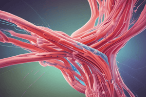
Muscle Tissue
Muscle Tissue
A type of tissue composed of specialized elongated cells arranged in parallel, which are responsible for contraction.
Function of Muscle Cells
Function of Muscle Cells
The main function of muscle cells is to contract, or shorten, in order to produce movement.
Muscle Contraction Mechanism
Muscle Contraction Mechanism
The interaction between thin and thick filaments within muscle cells is what allows for contraction.
Muscle Tissue Classification
Muscle Tissue Classification
Signup and view all the flashcards
Types of Striated Muscle
Types of Striated Muscle
Signup and view all the flashcards
Skeletal Muscle fibers
Skeletal Muscle fibers
Signup and view all the flashcards
Perimysium
Perimysium
Signup and view all the flashcards
Fatigue Resistance
Fatigue Resistance
Signup and view all the flashcards
Type IIb Muscle fibers
Type IIb Muscle fibers
Signup and view all the flashcards
Sarcomere
Sarcomere
Signup and view all the flashcards
A-band
A-band
Signup and view all the flashcards
I-band
I-band
Signup and view all the flashcards
Z-disc
Z-disc
Signup and view all the flashcards
What are intercalated discs?
What are intercalated discs?
Signup and view all the flashcards
What is the function of the transverse component of intercalated discs?
What is the function of the transverse component of intercalated discs?
Signup and view all the flashcards
What is the role of gap junctions in intercalated discs?
What is the role of gap junctions in intercalated discs?
Signup and view all the flashcards
Why are mitochondria important for heart muscle function?
Why are mitochondria important for heart muscle function?
Signup and view all the flashcards
What is the significance of glycogen granules in cardiac myocytes?
What is the significance of glycogen granules in cardiac myocytes?
Signup and view all the flashcards
Oxytocin and ADH during birth
Oxytocin and ADH during birth
Signup and view all the flashcards
Gap junctions in smooth muscle
Gap junctions in smooth muscle
Signup and view all the flashcards
Muscle-Tendon Junction
Muscle-Tendon Junction
Signup and view all the flashcards
Muscle fiber bundles
Muscle fiber bundles
Signup and view all the flashcards
Cardiac Muscle Structure
Cardiac Muscle Structure
Signup and view all the flashcards
Intercalated Discs function
Intercalated Discs function
Signup and view all the flashcards
Cardiac Muscle Orientation
Cardiac Muscle Orientation
Signup and view all the flashcards
What is ATP in muscle fibers used for?
What is ATP in muscle fibers used for?
Signup and view all the flashcards
What is creatine phosphate?
What is creatine phosphate?
Signup and view all the flashcards
What is anaerobic respiration?
What is anaerobic respiration?
Signup and view all the flashcards
What is aerobic respiration?
What is aerobic respiration?
Signup and view all the flashcards
What is muscle fatigue?
What is muscle fatigue?
Signup and view all the flashcards
What is the definition of oxygen debt?
What is the definition of oxygen debt?
Signup and view all the flashcards
What is a twitch contraction?
What is a twitch contraction?
Signup and view all the flashcards
What is the latent period in a muscle contraction?
What is the latent period in a muscle contraction?
Signup and view all the flashcards
What is the refractory period in a muscle contraction?
What is the refractory period in a muscle contraction?
Signup and view all the flashcards
What is an isotonic contraction?
What is an isotonic contraction?
Signup and view all the flashcards
What is an isometric contraction?
What is an isometric contraction?
Signup and view all the flashcards
What is smooth muscle?
What is smooth muscle?
Signup and view all the flashcards
What are gap junctions in smooth muscle?
What are gap junctions in smooth muscle?
Signup and view all the flashcards
What is peristalsis?
What is peristalsis?
Signup and view all the flashcards
What is the regulation of smooth muscle contraction?
What is the regulation of smooth muscle contraction?
Signup and view all the flashcards
What is the I band in muscle anatomy?
What is the I band in muscle anatomy?
Signup and view all the flashcards
What is Titin?
What is Titin?
Signup and view all the flashcards
What is Troponin in muscle cells?
What is Troponin in muscle cells?
Signup and view all the flashcards
What is Tropomyosin in muscle cells?
What is Tropomyosin in muscle cells?
Signup and view all the flashcards
How does muscle contraction work at the molecular level?
How does muscle contraction work at the molecular level?
Signup and view all the flashcards
What is Dystrophin?
What is Dystrophin?
Signup and view all the flashcards
What are the Z discs in muscle tissue?
What are the Z discs in muscle tissue?
Signup and view all the flashcards
What is the A band in muscle tissue?
What is the A band in muscle tissue?
Signup and view all the flashcards
What is Myosin in muscle tissue?
What is Myosin in muscle tissue?
Signup and view all the flashcards
What is Actin in muscle tissue?
What is Actin in muscle tissue?
Signup and view all the flashcards
What is the Sarcoplasmic Reticulum (SR) in muscle cells?
What is the Sarcoplasmic Reticulum (SR) in muscle cells?
Signup and view all the flashcards
What is Excitation-Contraction Coupling?
What is Excitation-Contraction Coupling?
Signup and view all the flashcards
Describe the steps of the Contraction Cycle in muscle.
Describe the steps of the Contraction Cycle in muscle.
Signup and view all the flashcards
What is the Sarcoplasmic Reticulum? (SR)
What is the Sarcoplasmic Reticulum? (SR)
Signup and view all the flashcards
Study Notes
Muscle Tissue Overview
- Muscle tissue is a specialized tissue comprised of elongated cells called myocytes.
- Myocytes are primarily responsible for contraction.
- Myofilaments (thin and thick) are the key structures mediating contraction.
- Thin filaments are composed of actin, a fibrous protein polymerized from globular actin subunits.
- Thick filaments are made of myosin II protein.
- Actin and myosin interact to cause muscle contraction.
- The cytoplasm of muscle cells is referred to as the sarcoplasm
- Other functions of actin and myosin include cytokinesis, exocytosis, and cell migration.
Muscle Tissue Classification
- Muscle tissue is categorized as striated or smooth.
- Striated muscle cells exhibit cross-striations.
- Smooth muscle cells lack cross-striations.
- Cross-striations are a consequence of the organized arrangement of actin and myosin myofilaments.
- Striated muscle is subdivided further, as skeletal, visceral and cardiac muscles.
- Skeletal muscle is attached to bone and responsible for movement.
- Visceral striated muscle is found in certain locations in the body and is often morphologically indistinct from skeletal muscle
- Cardiac muscle is found in the heart and is specialized for rhythmic contraction.
Skeletal Muscle Overview
- Skeletal muscle is composed of multinucleated cells called muscle fibers
- Muscle fibers are formed by the fusion of multiple individual myoblast cells.
- Muscle fibers vary in length.
Skeletal Muscle Structure
- Skeletal muscle fibers are organized and held together by connective tissues.
- Endomysium: connective tissue surrounding individual muscle fibers
- Perimysium: connective tissue surrounding bundles of muscle fibers (fascicles).
- Epimysium: connective tissue surrounding entire muscle bundles, forming muscles.
Skeletal Muscle Fiber Types
- Skeletal muscle fibers are generally categorized into three types, I, IIa, and IIb, with varying oxidative and glycolytic capabilities.
- Type I fibers are slow oxidative, appearing red and containing many mitochondria and a large quantity of myoglobin and cytochromes
- Type IIa fibers (fast oxidative)
- Type IIb fibers (fast glycolytic)
Myofibrils and Myofilaments
- Muscle fibers are composed of structural units called myofibrils, arranged longitudinally within the cells.
- Myofibrils are bundles of myofilaments (actin and myosin).
- Myofilaments are the actual contractile units in skeletal muscle.
Sarcomere Structure
- The sarcomere is the fundamental functional unit of a myofibril.
- Defined by the structure between successive Z lines
- A-bands and I-bands are visible under phase contrast microscopy.
- Z-discs traverse the I-bands.
- The M-line traverses and bisects the A-band.
Sarcomere Structure Details
- Myosin-containing thick filaments are located in the central part of the sarcomere, the A-band
- The thin actin filaments connect to the Z-lines and extend into the A-band.
- The I-band contains only thin filaments.
- Thin filaments are primarily composed of F-actin, tropomyosin, and troponin.
- Thick filaments consist only of myosin II.
Contractile Proteins
- Myosin is a motor protein with projections that allow it to "walk" along actin filaments.
- Myosin converts Adenosine triphosphate (ATP) into mechanical energy
- Thin filaments (actin), contain sites where myosin can attach
- Tropomyosin and troponin are accessory proteins associated with actin.
- In relaxed muscle, myosin binding sites on actin are blocked by tropomyosin.
- Calcium ion binding to troponin exposes the myosin-binding sites.
Structural Proteins (cont.)
- Titin stabilizes the position of myosin within the sarcomere.
- Titin accounts for much of the elasticity and extensibility of myofibrils.
- Dystrophin links thin filaments to the sarcolemma.
Contraction and Relaxation of Skeletal Muscle
- The sliding filament mechanism describes how muscle fibers contract.
- Myosin heads attach to and move along actin filaments.
- This progressively pulls the thin filaments towards the center of the sarcomere.
- Z-discs move closer together, shortening the sarcomere.
- The sarcomere shortening leads to the contraction of the entire muscle.
Contraction Cycle
- The contraction cycle begins with the SR releasing calcium ions into the muscle cell.
- Calcium ions bind to actin, opening myosin-binding sites.
- Myosin hydrolyzes ATP and reorients to generate force.
- Myosin heads attach to actin, and the power stroke rotates, sliding the filaments.
- Myosin detaches from actin when the next ATP binds.
- The cycle repeats as long as ATP is available and calcium ion level remains sufficient.
Excitation-Contraction Coupling
- An increase in calcium ions initiates contraction.
- Muscle cell membrane has calcium pumps to quickly return calcium ion levels.
- Decresing calcium ions initiates muscle relaxation.
- Action potentials trigger calcium release from the sarcoplasmic reticulum.
- Calcium movement away from myosin-binding sites causes muscle relaxation.
Muscle Metabolism
- Muscle contraction requires a substantial amount of ATP.
- ATP supplies the energy for contraction, and for pumping calcium ions back into the sarcoplasmic reticulum.
- Muscles have several mechanisms for ATP production: creatine phosphate, anaerobic respiration and aerobic respiration.
- ATP regeneration from creatine phosphate is immediate and fuels short-duration, high-intensity contractions.
- Anaerobic respiration occurs without oxygen, produces lactic acid, and supports moderate-intensity contractions.
- Aerobic respiration, requiring oxygen, occurs for prolonged activity and produces the most ATP per glucose molecule.
Muscle Fatigue
- Muscle fatigue is the inability to maintain muscle contraction.
- Factors like calcium ion release inadequacy, creatine phosphate depletion, glycogen depletion, oxygen insufficiency, buildup of lactic acid and ADP contribute to muscle fatigue
Oxygen Consumption After Exercise
- Oxygen consumption remains elevated after exercise to restore muscle cells to their resting state.
- Muscle cells use the additional oxygen to convert lactic acid to glycogen, synthesize ATP, and replenish oxygen stores in myoglobin.
Control of Muscle Tension - Twitch Contraction and Muscle Fiber Characteristics
- Twitch contraction is the response of a muscle fiber to a single action potential.
- The contraction period and relaxation period collectively define the duration of a twitch
- Latent period refers to the delay between stimulus and contraction
- Contraction period: duration of active cross-bridge activity (myosin pulling on actin).
- Relaxation period: duration of calcium reuptake into the sarcoplasmic reticulum.
- Isotonic contractions: muscle changes length while tension remains constant.
- Isometric contractions: muscle does not change length during contraction.
Smooth Muscle
- Smooth muscle is found in various tissues, and characterized by tapered ends and elongated fusiform cells.
- Smooth muscle cells often exhibit synchronous contractions due to gap junctions that connect the cells.
- Thin filaments of smooth muscle are associated with intermediate filaments (desmin and vimentin), which give the cells their structural support
- Labile thick myosin filaments that are easily lost during tissue preparation.
Smooth Muscle (cont'd)
- Smooth muscle is frequently regulated by the autonomic nervous system.
- Sometimes, smooth muscle will spontaneously contract or exhibit spontaneous activity
Muscle-Tendon Junction
- Oblique-cut skeletal muscle inserting into tendon is a common structural observation
Cardiac Muscle
- Cardiac muscle cells are striated similar to skeletal muscle
- Cardiac muscle fibers contain intercalated discs between cells, highly specialized attachments for coordinated contractions.
- cardiac muscle cells are not a syncitium (single large muscle fiber).
- Intercalated discs facilitate ionic flow between cells, enabling synchronized contractions.
- Cardiac muscle requires a large amount of ATP and has an abundance of mitochondria
- Cardiac muscle can spontaneously contract (pacemaker cells).
Cardiac Muscle Structure Details
- The sarcoplasmic reticulum (SR) in cardiac muscle is often simpler than in skeletal muscle.
- Cardiac myocytes have many large mitochondria with numerous cristae packed between the myofibrils.
- There are numerous glycogen granules between the myofibrils.
- The intercalated disc represents the attachment site between individual cardiac myocytes.
Intercalated Discs
- Components of the intercalated disc include inter-connecting transverse components and inter-connecting lateral components.
- Adhering junctions, forming part of the transverse component, structurally join the muscle cells and transmit the force generated during contractions.
- Gap junctions, which form part of the lateral component of the intercalated discs, allow ionic flow between the cells facilitating synchronized contraction in cardiac muscle.
- The SR network in cardiac muscle forms a single network along the sarcomere, extending from Z-line to Z-line.
- One T-tubule usually extends into the centre of cardiac muscle cells per sarcomere.
Studying That Suits You
Use AI to generate personalized quizzes and flashcards to suit your learning preferences.