Podcast
Questions and Answers
What is the ability of muscular tissue to respond to stimuli and trigger action potentials called?
What is the ability of muscular tissue to respond to stimuli and trigger action potentials called?
- Electrical excitability (correct)
- Extensibility
- Elasticity
- Contractility
Which term describes the ability of muscular tissue to contract forcefully when stimulated?
Which term describes the ability of muscular tissue to contract forcefully when stimulated?
- Elasticity
- Electrical excitability
- Extensibility
- Contractility (correct)
What characteristic allows muscular tissue to stretch without being damaged?
What characteristic allows muscular tissue to stretch without being damaged?
- Extensibility (correct)
- Contractility
- Elasticity
- Plasticity
Which property of muscular tissue enables it to return to its original length and shape after contraction?
Which property of muscular tissue enables it to return to its original length and shape after contraction?
If muscular tissue is unable to stretch and return to its original shape, which property is likely impaired?
If muscular tissue is unable to stretch and return to its original shape, which property is likely impaired?
What is the primary role of fascia in the body?
What is the primary role of fascia in the body?
How does fascia contribute to muscle function?
How does fascia contribute to muscle function?
Which of the following does fascia NOT do?
Which of the following does fascia NOT do?
What type of tissue is fascia categorized as?
What type of tissue is fascia categorized as?
Which statement about the function of fascia is correct?
Which statement about the function of fascia is correct?
What is the function of the epimysium in muscle structure?
What is the function of the epimysium in muscle structure?
Which layer of connective tissue surrounds groups of muscle fibers and forms bundles known as fascicles?
Which layer of connective tissue surrounds groups of muscle fibers and forms bundles known as fascicles?
What type of connective tissue is the endomysium classified as?
What type of connective tissue is the endomysium classified as?
Which layer of connective tissue is NOT correctly described in terms of its function?
Which layer of connective tissue is NOT correctly described in terms of its function?
Which connective tissue layer is the innermost, surrounding individual muscle fibers?
Which connective tissue layer is the innermost, surrounding individual muscle fibers?
Which connective tissue layer is responsible for encircling the entire muscle?
Which connective tissue layer is responsible for encircling the entire muscle?
What is the primary role of the perimysium in muscle structure?
What is the primary role of the perimysium in muscle structure?
What type of connective tissue forms the endomysium?
What type of connective tissue forms the endomysium?
Which layer of connective tissue has the least thickness compared to the others?
Which layer of connective tissue has the least thickness compared to the others?
Which connective tissue layer acts directly to separate individual muscle fibers from one another?
Which connective tissue layer acts directly to separate individual muscle fibers from one another?
What is the primary composition of a tendon?
What is the primary composition of a tendon?
What is the function of an aponeurosis in the body?
What is the function of an aponeurosis in the body?
Which of the following correctly identifies a specific example of a tendon?
Which of the following correctly identifies a specific example of a tendon?
Which statement is true regarding the attachment of tendons?
Which statement is true regarding the attachment of tendons?
What differentiates an aponeurosis from a tendon?
What differentiates an aponeurosis from a tendon?
What is the role of synovial fluid found in the cavity between the visceral and parietal layers of a tendon sheath?
What is the role of synovial fluid found in the cavity between the visceral and parietal layers of a tendon sheath?
Which layer of the tendon sheath is directly attached to the tendon itself?
Which layer of the tendon sheath is directly attached to the tendon itself?
Which statement accurately describes the parietal layer of a tendon sheath?
Which statement accurately describes the parietal layer of a tendon sheath?
What is the primary structure that encloses the tendons of the wrist and ankle?
What is the primary structure that encloses the tendons of the wrist and ankle?
Which of the following best describes the relationship between the visceral and parietal layers of a tendon sheath?
Which of the following best describes the relationship between the visceral and parietal layers of a tendon sheath?
Which component is NOT found within a muscle fibre?
Which component is NOT found within a muscle fibre?
What characteristic defines skeletal muscle fibres?
What characteristic defines skeletal muscle fibres?
The outer membrane surrounding each muscle fibre is known as what?
The outer membrane surrounding each muscle fibre is known as what?
Which statement correctly describes the nature of skeletal muscle fibres?
Which statement correctly describes the nature of skeletal muscle fibres?
What term is used to describe the cytoplasm found within muscle fibres?
What term is used to describe the cytoplasm found within muscle fibres?
What is the primary role of mitochondria in muscle cells?
What is the primary role of mitochondria in muscle cells?
What role does the sarcotubular system serve in muscle function?
What role does the sarcotubular system serve in muscle function?
What is the relationship between the endoplasmic reticulum and muscle contraction?
What is the relationship between the endoplasmic reticulum and muscle contraction?
Why do muscle cells require a large amount of energy?
Why do muscle cells require a large amount of energy?
How are mitochondria critical for muscle function?
How are mitochondria critical for muscle function?
What is the main function of T-tubules in muscle cells?
What is the main function of T-tubules in muscle cells?
What do terminal cisternae primarily do in muscle cells?
What do terminal cisternae primarily do in muscle cells?
How do T-tubules and the sarcoplasmic reticulum relate in muscle fibers?
How do T-tubules and the sarcoplasmic reticulum relate in muscle fibers?
In a triad structure of muscle cells, what does each triad consist of?
In a triad structure of muscle cells, what does each triad consist of?
Which statement about the location and structure of T-tubules is correct?
Which statement about the location and structure of T-tubules is correct?
What is the primary function of the Z discs in a sarcomere?
What is the primary function of the Z discs in a sarcomere?
Which structure in the sarcomere contains only thick filaments?
Which structure in the sarcomere contains only thick filaments?
In what part of the sarcomere would you find both actin and myosin filaments?
In what part of the sarcomere would you find both actin and myosin filaments?
Which of these statements accurately describes the M line?
Which of these statements accurately describes the M line?
What distinguishes the I bands from the A bands in a sarcomere?
What distinguishes the I bands from the A bands in a sarcomere?
Flashcards are hidden until you start studying
Study Notes
Electrical Excitability
- Muscular tissue can respond to stimuli and generate action potentials
Contractility
- The ability of muscular tissue to shorten forcefully when stimulated
Extensibility
- Muscular tissue can be stretched without damage
Elasticity
- Muscular tissue can return to its original shape after contraction
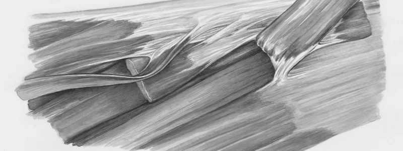
Fascia
- Dense sheet or broad band of irregular connective tissue
- Supports and surrounds muscles and organs
- Holds muscles with similar functions together
- Allows free movement of muscles
- Carries nerves, blood vessels and lymphatic vessels
- Fills spaces between muscles
Connective Tissue Layers of Muscle
- Epimysium: The outermost layer of connective tissue surrounding the entire muscle. It is composed of dense irregular connective tissue.
- Perimysium: This layer of dense irregular connective tissue surrounds groups of muscle fibers, separating them into bundles called fascicles.
- Endomysium: A thin sheath of areolar connective tissue that penetrates the interior of each fascicle, separating individual muscle fibers from one another.
Tendon
- Connects muscle to bone
- Composed of dense regular connective tissue
- Parallel bundles of collagen fibers
- Extends from all three connective tissue layers
- Example: Calcaneal (Achilles) tendon connects Gastrocnemius (calf) muscle to the heel bone
Aponeurosis
- Connective tissue extension
- Broad, flat layer of tendon
- Example: Epicranial aponeurosis connects the Occipitofrontalis muscle to the skull
Tendon Sheaths
-
Tendon sheaths are tubes of fibrous connective tissue that enclose certain tendons, notably those in the wrist and ankle.
-
These sheaths consist of two layers:
- Visceral layer: This inner layer directly attaches to the surface of the tendon.
- Parietal layer: The outer layer of the sheath is attached to the bone.
-
Between the visceral and parietal layers is a cavity filled with synovial fluid.
-
The synovial fluid acts as a lubricant, reducing friction as the tendons slide back and forth during movement.
Muscle Fibres
- Muscle fibres are the building blocks of skeletal muscles, forming long, thin, cylindrical cells.
- Each muscle fibre is surrounded by a membrane known as the sarcolemma.
- The cytoplasm within the muscle fibre is referred to as sarcoplasm.
- Muscle fibres are multinucleated, meaning they contain multiple nuclei.
- Muscle fibres have a striated appearance due to the arrangement of protein filaments within them.
- Myofibrils are the basic units of a muscle fibre, and are made up of protein filaments that give the striated appearance.
- The sarcoplasm contains a variety of cytoplasmic components essential for muscle function.
Cytoplasmic Components of Muscle Cells
- Muscle cells contain a large number of mitochondria, which are responsible for generating energy (ATP).
- This high energy demand is required for muscle contraction.
- Muscle cells also possess a specialized endoplasmic reticulum called the sarcotubular system.
- The sarcotubular system is extensive and plays a crucial role in muscle excitation and contraction.
T-Tubules
- Inward extensions of the sarcolemma (muscle cell membrane)
- Open to the exterior of the cell
- Contain extracellular fluid (ECF)
- Run transversely (across) to the myofibrils (muscle fibers)
- Transmit action potentials (electrical impulses) into the muscle fiber, triggering muscle contraction
Sarcoplasmic Reticulum
- Network of membranous sacs (like a network of tubes) that run parallel to the myofibrils
- Terminate in specialized structures called terminal cisternae
- Stores calcium ions (Ca2+) essential for muscle contraction
Triads
- Formed by two terminal cisternae and a T-tubule positioned adjacent to each other
- This close proximity facilitates the rapid release of calcium ions from the sarcoplasmic reticulum into the cytoplasm when a muscle fiber is stimulated.
Sarcomere Structure
- The sarcomere is the functional unit of muscle contraction.
- It is the portion of a myofibril between two adjacent Z discs.
- I bands are light bands that contain only actin filaments.
- A bands are dark bands that contain myosin filaments and the ends of actin filaments.
- The ends of actin filaments are anchored in Z discs.
- The H zone is a narrow region in the center of each A band that contains only thick filaments.
- The M line is a region in the center of the H zone that holds the thick filaments together.
Studying That Suits You
Use AI to generate personalized quizzes and flashcards to suit your learning preferences.


