Podcast
Questions and Answers
What is the role of tropomyosin in muscle contraction?
What is the role of tropomyosin in muscle contraction?
- It binds to actin to prevent contraction.
- It directly stimulates calcium release from the sarcoplasmic reticulum.
- It covers the binding sites on actin, blocking interaction. (correct)
- It binds to myosin and promotes contraction.
What occurs when calcium binds to troponin?
What occurs when calcium binds to troponin?
- Troponin stabilizes tropomyosin, maintaining its blocking position.
- Troponin undergoes a conformational change that exposes actin's binding sites. (correct)
- Calcium inhibits the binding of myosin to actin.
- Actin and myosin separate, leading to muscle relaxation.
What triggers the release of calcium from the sarcoplasmic reticulum?
What triggers the release of calcium from the sarcoplasmic reticulum?
- The action potential traveling down the T-tubules. (correct)
- Direct interaction of actin and myosin.
- The binding of acetylcholine to receptors on the muscle cell.
- Increased levels of tropomyosin in the cytoplasm.
Which of the following best describes the sliding filament mechanism?
Which of the following best describes the sliding filament mechanism?
Which process is necessary for muscle relaxation after contraction?
Which process is necessary for muscle relaxation after contraction?
What structural feature distinguishes smooth muscle from skeletal muscle?
What structural feature distinguishes smooth muscle from skeletal muscle?
What activates the phosphorylation of the myosin light chain in smooth muscle contraction?
What activates the phosphorylation of the myosin light chain in smooth muscle contraction?
What characterizes tonic smooth muscle?
What characterizes tonic smooth muscle?
What is a key characteristic of multiunit smooth muscle?
What is a key characteristic of multiunit smooth muscle?
What is the function of acetylcholinesterase at the neuromuscular junction?
What is the function of acetylcholinesterase at the neuromuscular junction?
Which of the following elements is not involved in the excitation-contraction coupling process?
Which of the following elements is not involved in the excitation-contraction coupling process?
How does the arrangement of myofilaments in smooth muscle differ from that of skeletal muscle?
How does the arrangement of myofilaments in smooth muscle differ from that of skeletal muscle?
What is a critical role of the sarcoplasmic reticulum in muscle fibers?
What is a critical role of the sarcoplasmic reticulum in muscle fibers?
What happens to calcium ion levels during muscle relaxation?
What happens to calcium ion levels during muscle relaxation?
What is the primary function of the thick filaments in skeletal muscle?
What is the primary function of the thick filaments in skeletal muscle?
Which protein is known for connecting myosin to the Z disc in skeletal muscle?
Which protein is known for connecting myosin to the Z disc in skeletal muscle?
What represents the contractile unit of a muscle fiber?
What represents the contractile unit of a muscle fiber?
What denotes the dark band in skeletal muscle?
What denotes the dark band in skeletal muscle?
Which component of the muscle fiber primarily facilitates contraction?
Which component of the muscle fiber primarily facilitates contraction?
What is the role of tropomyosin in thin filaments?
What is the role of tropomyosin in thin filaments?
Which part of the sarcomere contains only thick filaments?
Which part of the sarcomere contains only thick filaments?
What is the primary shape of actin molecules in a thin filament?
What is the primary shape of actin molecules in a thin filament?
Which muscle type is classified as unstratified and involuntary?
Which muscle type is classified as unstratified and involuntary?
What characteristic feature allows myofibrils to display striations?
What characteristic feature allows myofibrils to display striations?
Which part of the muscle contraction cycle involves the splitting of ATP?
Which part of the muscle contraction cycle involves the splitting of ATP?
How is skeletal muscle categorized in terms of nerve innervation?
How is skeletal muscle categorized in terms of nerve innervation?
Which of the following accurately describes the role of myosin in muscle contraction?
Which of the following accurately describes the role of myosin in muscle contraction?
Which part of a muscle fiber holds thick filaments together?
Which part of a muscle fiber holds thick filaments together?
What common function is shared by the thick and thin filaments during muscle contraction?
What common function is shared by the thick and thin filaments during muscle contraction?
Flashcards
Muscle fiber
Muscle fiber
A single skeletal muscle cell, characterized by its elongated, cylindrical shape and containing numerous myofibrils.
Myofibrils
Myofibrils
Cylindrical structures within muscle fibers, extending the entire length of the fiber. They are responsible for muscle contraction and contain the contractile elements of the muscle fiber.
Myosin
Myosin
The contractile proteins that make up the thick filaments in a myofibril. This protein molecule has two identical subunits shaped like a golf club with tails intertwined and globular heads that project outward.
Actin
Actin
Signup and view all the flashcards
Sarcomere
Sarcomere
Signup and view all the flashcards
A Bands
A Bands
Signup and view all the flashcards
I Bands
I Bands
Signup and view all the flashcards
H Zone
H Zone
Signup and view all the flashcards
M Line
M Line
Signup and view all the flashcards
Z Line
Z Line
Signup and view all the flashcards
Cross Bridges
Cross Bridges
Signup and view all the flashcards
Titin
Titin
Signup and view all the flashcards
Tropomyosin
Tropomyosin
Signup and view all the flashcards
Troponin
Troponin
Signup and view all the flashcards
Sliding Filament Mechanism
Sliding Filament Mechanism
Signup and view all the flashcards
Sarcoplasmic Reticulum
Sarcoplasmic Reticulum
Signup and view all the flashcards
Transverse Tubules (T-tubules)
Transverse Tubules (T-tubules)
Signup and view all the flashcards
Excitation-Contraction Coupling
Excitation-Contraction Coupling
Signup and view all the flashcards
Muscle Relaxation
Muscle Relaxation
Signup and view all the flashcards
Smooth Muscle
Smooth Muscle
Signup and view all the flashcards
Calmodulin
Calmodulin
Signup and view all the flashcards
Multiunit Smooth Muscle
Multiunit Smooth Muscle
Signup and view all the flashcards
Single-Unit Smooth Muscle
Single-Unit Smooth Muscle
Signup and view all the flashcards
Tonic Smooth Muscle
Tonic Smooth Muscle
Signup and view all the flashcards
Study Notes
Muscle Physiology Overview
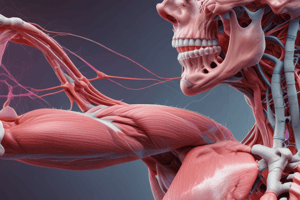
- Muscle tissue comprises roughly half of the human body's weight (men 40%, women 32%).
- Smooth and cardiac muscle make up approximately 10%.
- Muscles are categorized based on striations (alternating dark and light bands):
- Striated: skeletal and cardiac muscle.
- Unstriated: smooth muscle.
- Muscles are also categorized by innervation:
- Voluntary (somatic nervous system): skeletal muscles.
- Involuntary (autonomic nervous system): smooth and cardiac muscle.
Skeletal Muscle Structure
- A skeletal muscle cell is a muscle fiber.
- Muscle fibers are large, elongated, and cylinder-shaped.
- Multiple muscle fibers are bundled together by connective tissue.
- Each muscle fiber contains numerous myofibrils.
- Myofibrils are cylindrical and extend the length of the muscle fiber.
- Myofibrils consist of contractile elements (thick and thin filaments).
Skeletal Muscle Detail
- Myofibrils:
- Contractile elements within muscle fibers.
- Composed of repeating units called sarcomeres.
- Arranged in a regular pattern, creating striations (alternating dark and light bands).
- Dark Band (A Band):
- Composed of thick filaments (myosin) and overlapping thin filaments (actin).
- Light Band (I Band):
- Contains only thin filaments (actin).
- H Zone (of the A band):
- Lighter area within the center of the A band where thin filaments do not overlap thick filaments.
- Z Line (or Z disk):
- Found in the middle of each I band, a dense vertical line. The Z-line acts as the sarcomere border.
- M Line:
- Proteins that hold thick filaments together vertically.
- Sarcomere:
- The functional unit of muscle contraction.
- The region between two successive Z-lines.
Myosin
- Component of thick filament.
- Protein molecule with two identical subunits.
- Subunits resemble a golf club.
- Tail regions intertwine.
- Globular heads project outward at the ends, forming cross-bridges.
- Thick filaments have regularly spaced cross-bridges between thin and thick filaments.
- Cross bridges contain: An actin-binding site and A myosin ATPase (ATP-splitting) site.
Actin
- Primary structural component of thin filaments.
- Spherical shape.
- Thin filaments consist of actin molecules, tropomyosin, and troponin.
- Actin binding sites allow for cross-bridge interaction between actin and myosin.
Tropomyosin and Troponin
- Tropomyosin:
- Thread-like protein that lies end-to-end along the groove of actin's spiral.
- When not participating in cross-bridge forming, the tropomyosin covers actin's binding sites, preventing myosin interaction.
- Troponin:
- Made of three polypeptide units.
- One binds to tropomyosin; One binds to actin; and One binds with calcium ions (Ca2+).
- In the absence of Ca2+, troponin stabilizes the tropomyosin/actin blocking position.
- In the presence of Ca2+, troponin changes shape and pulls tropomyosin away.
- This exposure of the actin binding site allows for myosin interaction, enabling muscle contraction.
T-tubules
- Run perpendicularly from the surface of the muscle cell membrane into the central portions of the muscle fiber.
- Action potentials travel through T tubules.
- Action potentials stimulate release of calcium.
Sarcoplasmic Reticulum
- Modified endoplasmic reticulum.
- Interconnected compartments surrounding the myofibrils.
- Wraps around each A band and I band.
- Expand to form lateral sacs (terminal cisternae) at the ends of the segments.
Muscle Contraction
- Sliding Filament Mechanism: Thin filaments slide over thick filaments during contraction.
- This mechanism does not shorten the thick or thin filaments themselves. The sarcomere shortens.
- Cross-bridge interaction: Myosin cross-bridges attach to actin, pull thin filaments towards the center of the sarcomere, causing shortening This pulling action is fuelled by ATP.
Excitation–Contraction Coupling
- Stepwise process:
- Acetylcholine (ACh) release initiates the signal.
- Action potential propagates through T tubules.
- Calcium (Ca2+) released from the sarcoplasmic reticulum.
- Ca2+ binding changes the conformation of troponin, exposing actin binding sites.
- Myosin binds to actin initiating the power stroke and sliding filaments.
Muscle Relaxation
- Ca2+ is actively taken up into the sarcoplasmic reticulum.
- Tropomyosin returns to its blocking position over actin's binding sites preventing further myosin interaction.
- Muscle contraction ceases.
Muscle Types
- Skeletal muscle: Voluntary muscle attached to bones for movement, striated and multi-nucleated.
- Smooth muscle: Mostly involuntary, found in internal organs and blood vessels; non-striated and single-nucleated. Smooth muscles operate differently from skeletal muscles, regulating through phosphorylation of myosin and calcium binding mechanisms. Different forms of smooth muscle include multi-unit and single-unit subtypes.
- Cardiac muscle: Involuntary muscle of the heart; striated and single-nucleated with intercalated discs (gap junctions). Cardiac muscles exhibit pacemaker activity in the absence of neuronal stimulation.
Studying That Suits You
Use AI to generate personalized quizzes and flashcards to suit your learning preferences.


