Podcast
Questions and Answers
What type of muscle attachment involves tendons bridging the gap between muscle ends and bony attachment?
What type of muscle attachment involves tendons bridging the gap between muscle ends and bony attachment?
- Fleshy attachment
- Tendinous attachment
- Indirect attachment (correct)
- Direct attachment
Which muscle is an example of an indirect attachment to bone?
Which muscle is an example of an indirect attachment to bone?
- Pectoralis major
- Triceps brachii
- Biceps brachii (correct)
- Brachialis
Where is the origin of a muscle typically located?
Where is the origin of a muscle typically located?
- At the end where the muscle contracts
- At the stationary end of the muscle (correct)
- At the movable end of the muscle
- In the middle of the muscle belly
What describes a direct (fleshy) muscle attachment to bone?
What describes a direct (fleshy) muscle attachment to bone?
Which of the following is the thicker, middle region of muscle called?
Which of the following is the thicker, middle region of muscle called?
What is the primary function of tendons in muscle attachments?
What is the primary function of tendons in muscle attachments?
Which part of the muscle is attached to the more movable end?
Which part of the muscle is attached to the more movable end?
Which muscle is NOT an example of a direct attachment to bone?
Which muscle is NOT an example of a direct attachment to bone?
Intercalated discs are structures that keep cells of which type of muscle together?
Intercalated discs are structures that keep cells of which type of muscle together?
What is true about smooth muscle?
What is true about smooth muscle?
Which function is NOT associated with skeletal muscles?
Which function is NOT associated with skeletal muscles?
Which of the following is a function of muscles related to stability?
Which of the following is a function of muscles related to stability?
What percentage of body heat can skeletal muscles produce?
What percentage of body heat can skeletal muscles produce?
What role do sphincters play in the muscular system?
What role do sphincters play in the muscular system?
Which characteristic distinguishes smooth muscle cells?
Which characteristic distinguishes smooth muscle cells?
How does muscle tissue contribute to glycemic control?
How does muscle tissue contribute to glycemic control?
What role does a synergist muscle play during a joint action?
What role does a synergist muscle play during a joint action?
Which muscle group acts to oppose the prime mover?
Which muscle group acts to oppose the prime mover?
What is the primary function of a fixator muscle?
What is the primary function of a fixator muscle?
Which of the following describes the prime mover in muscle action?
Which of the following describes the prime mover in muscle action?
How do antagonistic pairs function during movement?
How do antagonistic pairs function during movement?
What is the function of a muscle described as a prime mover during a soccer kick?
What is the function of a muscle described as a prime mover during a soccer kick?
Which muscle can be categorized as a synergist to the biceps brachii?
Which muscle can be categorized as a synergist to the biceps brachii?
Which option best defines the primary action of the antagonist muscle?
Which option best defines the primary action of the antagonist muscle?
What is the primary characteristic of intrinsic muscles?
What is the primary characteristic of intrinsic muscles?
Which of the following best illustrates an extrinsic muscle?
Which of the following best illustrates an extrinsic muscle?
What does the innervation of a muscle refer to?
What does the innervation of a muscle refer to?
What is a plexus in the context of muscles?
What is a plexus in the context of muscles?
Which cranial nerve number corresponds to the olfactory nerve?
Which cranial nerve number corresponds to the olfactory nerve?
Which of the following names describes the action of a muscle?
Which of the following names describes the action of a muscle?
How does muscle naming aid in understanding muscle function?
How does muscle naming aid in understanding muscle function?
What is a recommended strategy for studying muscles effectively?
What is a recommended strategy for studying muscles effectively?
What best characterizes the flexor digitorum superficialis?
What best characterizes the flexor digitorum superficialis?
Which of the following best describes extrinsic muscles in the context of the fingers?
Which of the following best describes extrinsic muscles in the context of the fingers?
Which muscle is located at the front of the head and is involved in raising the eyebrows?
Which muscle is located at the front of the head and is involved in raising the eyebrows?
Which muscle is primarily responsible for chewing?
Which muscle is primarily responsible for chewing?
Which of these muscles is located on the posterior side of the shoulder?
Which of these muscles is located on the posterior side of the shoulder?
Which muscles are categorized as the adductors of the thigh?
Which muscles are categorized as the adductors of the thigh?
Which muscle is located in the lateral compartment of the leg?
Which muscle is located in the lateral compartment of the leg?
Which muscle is responsible for flexing the elbow?
Which muscle is responsible for flexing the elbow?
Which of the following muscles assists in shoulder stabilization and rotation?
Which of the following muscles assists in shoulder stabilization and rotation?
Which muscle is responsible for the flexion and adduction of the thigh?
Which muscle is responsible for the flexion and adduction of the thigh?
Which muscle is part of the rotator cuff and is responsible for lateral rotation of the arm?
Which muscle is part of the rotator cuff and is responsible for lateral rotation of the arm?
Which two muscles are primarily responsible for extending the knee?
Which two muscles are primarily responsible for extending the knee?
Which muscle functions primarily in the plantar flexion of the foot?
Which muscle functions primarily in the plantar flexion of the foot?
Which abdominal muscle is located deepest under the other abdominal muscles?
Which abdominal muscle is located deepest under the other abdominal muscles?
Which muscle is responsible for the flexion of the wrist?
Which muscle is responsible for the flexion of the wrist?
Which group of muscles is primarily involved in the stabilization of the pelvis during walking?
Which group of muscles is primarily involved in the stabilization of the pelvis during walking?
Which muscle performs the action of extension of the elbow?
Which muscle performs the action of extension of the elbow?
Flashcards
What are intercalated discs?
What are intercalated discs?
Intercalated discs are specialized junctions that connect cardiac muscle cells, allowing for coordinated contraction and communication.
What makes smooth muscle different?
What makes smooth muscle different?
Smooth muscle lacks striations, meaning it doesn't have the striped appearance of skeletal muscle, and it is involuntary, meaning it contracts without conscious control.
Describe the structure of smooth muscle cells.
Describe the structure of smooth muscle cells.
Smooth muscle cells are relatively short, spindle-shaped (fusiform) with a thick middle and tapered ends. They have a single centrally located nucleus.
What is visceral muscle?
What is visceral muscle?
Signup and view all the flashcards
Name 4 functions of muscles.
Name 4 functions of muscles.
Signup and view all the flashcards
How do muscles attach to bones?
How do muscles attach to bones?
Signup and view all the flashcards
What is the difference between a muscle's origin and insertion?
What is the difference between a muscle's origin and insertion?
Signup and view all the flashcards
Describe the role of a prime mover.
Describe the role of a prime mover.
Signup and view all the flashcards
What is the function of a synergist?
What is the function of a synergist?
Signup and view all the flashcards
How does an antagonist muscle work?
How does an antagonist muscle work?
Signup and view all the flashcards
What is the role of a fixator muscle?
What is the role of a fixator muscle?
Signup and view all the flashcards
Explain the difference between intrinsic and extrinsic muscles.
Explain the difference between intrinsic and extrinsic muscles.
Signup and view all the flashcards
What does muscle innervation mean?
What does muscle innervation mean?
Signup and view all the flashcards
What are spinal nerves?
What are spinal nerves?
Signup and view all the flashcards
What is a plexus?
What is a plexus?
Signup and view all the flashcards
What are cranial nerves?
What are cranial nerves?
Signup and view all the flashcards
How are muscles named?
How are muscles named?
Signup and view all the flashcards
What is the frontalis muscle?
What is the frontalis muscle?
Signup and view all the flashcards
What does the orbicularis oculi do?
What does the orbicularis oculi do?
Signup and view all the flashcards
Describe the function of the zygomaticus major.
Describe the function of the zygomaticus major.
Signup and view all the flashcards
What is the primary function of the orbicularis oris?
What is the primary function of the orbicularis oris?
Signup and view all the flashcards
What is the platysma muscle responsible for?
What is the platysma muscle responsible for?
Signup and view all the flashcards
What is the masseter muscle used for?
What is the masseter muscle used for?
Signup and view all the flashcards
What is the sternocleidomastoid muscle?
What is the sternocleidomastoid muscle?
Signup and view all the flashcards
What are the primary functions of the trapezius muscle?
What are the primary functions of the trapezius muscle?
Signup and view all the flashcards
Describe the location and functions of the deltoid muscle.
Describe the location and functions of the deltoid muscle.
Signup and view all the flashcards
What does the pectoralis major muscle do?
What does the pectoralis major muscle do?
Signup and view all the flashcards
What are the actions of the biceps brachii muscle?
What are the actions of the biceps brachii muscle?
Signup and view all the flashcards
Study Notes
Intercalated Discs
- Intercalated Discs are structures that keep cells of cardiac muscle together
Smooth Muscle
- Smooth Muscle lacks striations and is involuntary
- Relatively short, fusiform cells (thick in middle, tapered at ends)
- One centrally located nucleus
- Visceral Muscle - forms layers of digestive, respiratory, and urinary tract: propels contents through an organ, regulates diameter of blood vessels
Functions of Muscles
- Movement
- Move from place to place
- Movement of body parts and body contents in breathing, circulation, feeding and digestion, defecation, urination, and childbirth
- Role in communication: speech, writing, nonverbal communications
- Stability
- Maintain posture by preventing unwanted movements
- Antigravity muscles: resist pull of gravity and prevent us from falling or slumping over
- Stabilize joints
- Control of openings and passageways
- Sphincters: internal muscular rings that control the movement of food, bile, blood, and other materials within the body
- Heat production by skeletal muscles
- As much as 85% of our body heat
- Glycemic control
- Regulation of blood glucose concentrations within its normal range
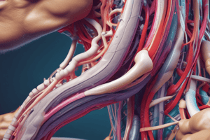
Muscle Attachments
- Indirect attachment to bone
- Tendons bridge the gap between muscle ends and bony attachment
- Collagen fibers of the endo-, peri-, and epimysium continue into the tendon
- From there into the periosteum and the matrix of bone
- Very strong structural continuity from muscle to bone
- Biceps brachii, Achilles tendon
- Direct (fleshy) attachment to bone
- Little separation between muscle and bone
- Muscle seems to immerge directly from bone
- Margins of brachialis, lateral head of triceps brachii
Muscle Origins and Insertions
- Origin
- Bony attachment at stationary end of muscle
- Belly
- Thicker, middle region of muscle between origin and insertion
- Insertion
- Bony attachment to mobile end of muscle
Functional Groups of Muscles
- Action - the effects produced by a muscle
- To produce or prevent movement
- Prime mover (agonist)
- Muscle that produces most of force during a joint action
- Synergist
- Muscle that aids the prime mover
- Stabilizes the nearby joint
- Modifies the direction of movement
- Antagonist
- Opposes the prime mover
- Relaxes to give prime mover control over action
- Preventing excessive movement and injury
- Antagonistic pairs - muscles that act on opposite sides of a joint
- Fixator
- Muscle that prevents movement of bone
Intrinsic and Extrinsic Muscles
- Intrinsic muscles - entirely contained within a region, such as the hand
- Both its origin and insertion there
- Extrinsic muscles – act on a designated region, but has its origin elsewhere
- Fingers: extrinsic muscles in the forearm
Muscle Innervation
- Innervation of a muscle - refers to identity of the nerve that stimulates it
- Enables diagnosis of nerve, spinal cord, and brainstem injuries from their effects on muscle function
- Spinal nerves arise from the spinal cord
- Emerge through intervertebral foramina
- Immediately branch into a posterior and anterior ramus
- Innervate muscles below the neck
- Plexus: weblike network of spinal nerves adjacent to the vertebral column
- Cranial nerves arise from the base of the brain
- Emerge through skull foramina
- Innervate the muscles of the head and neck
- Numbered CN I to CN XII
How Muscles Are Named
- Latin names
- Depressor labii inferioris – depresses the bottom lip
- flexor digiti minimi brevis – short muscle that flexes the smallest finger
- Describes distinctive aspects of the structure, location, or action of a muscle
A Learning Strategy
-
Examine models, cadavers, dissected animals, or a photographic atlas to get visual images of the muscle
-
When studying a particular muscle, palpate it on yourself if possible
-
Locate origins and insertions of muscles on an articulated skeleton
-
Study derivation of each muscle name
- Usually describes the muscle’s location, appearance, origin, insertion, or action
-
Say the names aloud to yourself or a study partner, and spell them correctly### Anterior View
-
Superficial Muscles:
- Frontalis: Located on forehead, raises eyebrows.
- Orbicularis oculi: Located around eye, closes eye.
- Zygomaticus major: Located on cheek, lifts corner of mouth for smiling.
- Orbicularis oris: Located around mouth, closes mouth.
- Platysma: Located on neck, pulls down corner of mouth.
- Masseter: Located on cheek, elevates mandible for chewing.
- Sternocleidomastoid: Located on side of neck, flexes and rotates head.
- Trapezius: Located on upper back and neck, elevates and retracts scapula, rotates scapula, extends head.
- Deltoid: Located on shoulder, abducts arm, flexes and extends arm.
- Pectoralis major: Located on chest, flexes and adducts arm.
- Biceps brachii: Located on front of upper arm, flexes elbow, supinates forearm.
- Brachioradialis: Located on forearm, flexes elbow.
- Flexor carpi radialis: Located on forearm, flexes wrist, abducts wrist.
- Palmaris longus: Located on forearm, flexes wrist.
- Flexor carpi ulnaris: Located on forearm, flexes wrist, adducts wrist.
- Sartorius: Located on thigh, flexes, abducts, and externally rotates hip; flexes knee.
- Rectus femoris: Located on front of thigh, extends knee, flexes hip.
- Vastus lateralis: Located on side of thigh, extends knee.
- Vastus medialis: Located on inside of thigh, extends knee.
- Tibialis anterior: Located on shin, dorsiflexes ankle, inverts foot.
- Extensor digitorum longus: Located on shin, extends toes, dorsiflexes ankle.
Posterior View
- Superficial Muscles:
- Occipitalis: Located on back of head, retracts scalp.
- Splenius capitis: Located on back of neck, extends and rotates head.
- Trapezius: Located on upper back and neck, elevates and retracts scapula, rotates scapula, extends head.
- Latissimus dorsi: Located on back, extends, adducts, and internally rotates arm.
- Gluteus maximus: Located on buttocks, extends hip, laterally rotates hip.
- Semitendinosus: Located on back of thigh, extends hip, flexes knee.
- Biceps femoris: Located on back of thigh, extends hip, flexes knee.
- Gastrocnemius: Located on calf, plantar flexes foot, flexes knee.
- Soleus: Located on calf, plantar flexes foot.
- Extensor hallucis longus: Located on shin, extends big toe, dorsiflexes ankle, inverts foot.
- Fibularis longus: Located on outside of calf, plantar flexes foot, everts foot.
Deep Muscles (Anterior View)
- Deep Muscles:
- Pectoralis minor: Located on chest, depresses and protracts scapula.
- Coracobrachialis: Located on upper arm, flexes and adducts arm.
- Biceps brachii: Located on front of upper arm, flexes elbow, supinates forearm.
- Brachialis: Located on upper arm, flexes elbow.
- Flexor digitorum superficialis: Located on forearm, flexes middle phalanges of fingers.
- Flexor carpi ulnaris: Located on forearm, flexes wrist, adducts wrist.
- Pronator teres: Located on forearm, pronates forearm, flexes elbow.
- Flexor pollicis longus: Located on forearm, flexes thumb, pronates forearm.
- Flexor digitorum profundus: Located on forearm, flexes distal phalanges of fingers.
- Pronator quadratus: Located on forearm, pronates forearm.
- Extensor digitorum (cut): Located on forearm, extends fingers, dorsiflexes ankle.
- Extensor carpi ulnaris (cut): Located on forearm, extends wrist, adducts wrist.
- Rectus abdominis: Located on abdomen, flexes and rotates lumbar spine.
- External abdominal oblique: Located on abdomen, flexes and rotates lumbar spine.
- Internal abdominal oblique: Located on abdomen, flexes and rotates lumbar spine.
- Transverse abdominis: Located on abdomen, compresses abdominal cavity.
- Adductors:
- Adductor longus: Located on medial thigh, adducts, flexes, and rotates hip.
- Adductor magnus: Located on medial thigh, adducts and extends hip.
- Gracilis: Located on medial thigh, adducts, flexes, and medially rotates hip.
- Pectineus: Located medial thigh, adducts, flexes, and laterally rotates hip.
- Tensor fasciae latae: Located on hip, flexes, abducts, and medially rotates hip.
Deep Muscles (Posterior View)
- Deep Muscles:
- Semispinalis capitis: Located on back of neck, extends and rotates head.
- Splenius capitis: Located on back of neck, extends and rotates head.
- Levator scapulae: Located on neck, elevates and depresses scapula, rotates scapula.
- Supraspinatus: Located on shoulder, abducts arm, externally rotates arm.
- Infraspinatus: Located on shoulder, externally rotates arm, adducts arm.
- Teres minor: Located on shoulder, externally rotates arm, adducts arm.
- Teres major: Located on shoulder, extends, adducts, and internally rotates arm.
- Triceps brachii: Located on back of upper arm, extends elbow.
- Rhomboideus minor: Located on back, retracts and elevates scapula.
- Rhomboideus major: Located on back, retracts and elevates scapula.
- Serratus posterior inferior: Located on back, depresses ribs.
- Erector spinae: Located on back, extends and rotates vertebral column.
- Lateral rotators:
- Piriformis (located on buttock, laterally rotates and abducts the hip)
- Obturator internus (located on pelvic wall, laterally rotates and abducts the hip)
- Obturator externus (located on pelvic wall, laterally rotates and abducts the hip)
- Gemelli (located on buttocks, laterally rotates and abducts the hip)
- Quadratus femoris (located on buttocks, laterally rotates and abducts the hip)
- Gluteus medius: Located on buttocks, abducts and medially rotates hip.
- Gluteus minimus: Located on buttocks, abducts and medially rotates hip.
- Vastus intermedius: Located on thigh, extends knee.
- Tibialis posterior: Located on shin, plantar flexes ankle, inverts foot.
- Flexor digitorum longus: Located on shin, flexes toes, plantar flexes ankle.
- Flexor hallucis longus: Located on shin, flexes big toe, plantar flexes ankle.
- Soleus (cut): Located on calf, plantar flexes foot.
- Calcaneal tendon: (attached to the heel, known as Achilles tendon)
Studying That Suits You
Use AI to generate personalized quizzes and flashcards to suit your learning preferences.