Podcast
Questions and Answers
A ______ is a learned series of movements that combine to produce a smooth, efficient action.
A ______ is a learned series of movements that combine to produce a smooth, efficient action.
motor skill
______ motor skills include actions like lifting one's head, rolling over, and walking.
______ motor skills include actions like lifting one's head, rolling over, and walking.
gross
Gross motor development usually precedes fine motor development because large muscles develop before ______ ones.
Gross motor development usually precedes fine motor development because large muscles develop before ______ ones.
smaller
______ motor skills involve precise movements such as using the pincer grasp or writing.
______ motor skills involve precise movements such as using the pincer grasp or writing.
______ is a specialized skill where there is no dominance between body symmetries, allowing tasks with fine motor skills to be performed with either extremity.
______ is a specialized skill where there is no dominance between body symmetries, allowing tasks with fine motor skills to be performed with either extremity.
Most voluntary movements are initiated by the cerebral cortex and involve function patterns stored in lower brain areas like the spinal cord, brain stem, basal ganglia and ______.
Most voluntary movements are initiated by the cerebral cortex and involve function patterns stored in lower brain areas like the spinal cord, brain stem, basal ganglia and ______.
______ motor neurons, located in the ventral horn of the spinal cord and brainstem, initiate skeletal muscle contraction.
______ motor neurons, located in the ventral horn of the spinal cord and brainstem, initiate skeletal muscle contraction.
The spatial and temporal patterns of lower motor neuron activation are determined by local circuits within the spinal cord and brainstem, which receive input from sensory neurons and mediate ______ reflexes.
The spatial and temporal patterns of lower motor neuron activation are determined by local circuits within the spinal cord and brainstem, which receive input from sensory neurons and mediate ______ reflexes.
______ motor neurons modulate the activity of lower motor neurons by influencing the local circuitry in the spinal cord and brainstem.
______ motor neurons modulate the activity of lower motor neurons by influencing the local circuitry in the spinal cord and brainstem.
Local circuit neurons and lower motor neurons are located within the gray matter of the spinal cord and the ______ of the brainstem.
Local circuit neurons and lower motor neurons are located within the gray matter of the spinal cord and the ______ of the brainstem.
______ circuit neurons provide much of the coordination between different muscle groups essential for organized movement.
______ circuit neurons provide much of the coordination between different muscle groups essential for organized movement.
Upper motor neuron pathways arising in the cortex are essential for the initiation of ______ movements.
Upper motor neuron pathways arising in the cortex are essential for the initiation of ______ movements.
Upper motor neurons originating in the brainstem regulate muscle tone and orient the eyes, head, and body concerning vestibular, somatic, auditory, and ______ sensory information.
Upper motor neurons originating in the brainstem regulate muscle tone and orient the eyes, head, and body concerning vestibular, somatic, auditory, and ______ sensory information.
The ______ functions as a servomechanism, detecting and attenuating the difference between an intended movement and the movement actually performed.
The ______ functions as a servomechanism, detecting and attenuating the difference between an intended movement and the movement actually performed.
Patients with cerebellar damage exhibit incoordination with persistent errors in controlling the direction and amplitude of ongoing ______.
Patients with cerebellar damage exhibit incoordination with persistent errors in controlling the direction and amplitude of ongoing ______.
The ______ ganglia prevent upper motor neurons from initiating unwanted movements and prepare the motor circuits for the initiation of movements.
The ______ ganglia prevent upper motor neurons from initiating unwanted movements and prepare the motor circuits for the initiation of movements.
Motor signals are transmitted directly from the cerebral cortex to the spinal cord through the ______ tract.
Motor signals are transmitted directly from the cerebral cortex to the spinal cord through the ______ tract.
The supplementary motor cortex is involved with the execution of sequences of movements, the attainment of motor skills, and the selection of movements based on incoming ______ information.
The supplementary motor cortex is involved with the execution of sequences of movements, the attainment of motor skills, and the selection of movements based on incoming ______ information.
The pre-motor cortex contributes about 30% of the neurons that enter the corticospinal tract but it’s more active during the ______ rather than the execution of movements.
The pre-motor cortex contributes about 30% of the neurons that enter the corticospinal tract but it’s more active during the ______ rather than the execution of movements.
The primary motor area (M1) is where voluntary/conscious ______ is initiated.
The primary motor area (M1) is where voluntary/conscious ______ is initiated.
A lesion to M1 causes a loss of voluntary movement of the contralateral body part corresponding to the damaged motor ______ area.
A lesion to M1 causes a loss of voluntary movement of the contralateral body part corresponding to the damaged motor ______ area.
The premotor area is located just anterior to M1 and has a role in motor ______, or praxis.
The premotor area is located just anterior to M1 and has a role in motor ______, or praxis.
Lesion to the premotor area causes ______ or motor planning difficulties such as the inability to understand the demands of a task.
Lesion to the premotor area causes ______ or motor planning difficulties such as the inability to understand the demands of a task.
The motor association area has a role in the ______ planning of movement.
The motor association area has a role in the ______ planning of movement.
The ______ motor area is part of the premotor area and has a role in the bilateral control of posture.
The ______ motor area is part of the premotor area and has a role in the bilateral control of posture.
Located in the middle frontal gyrus, the frontal eye field is responsible for visual ______.
Located in the middle frontal gyrus, the frontal eye field is responsible for visual ______.
The functions of the motor cortex are controlled mainly by nerve signals from the ______ system but also, to some degree, from other sensory systems such as hearing and vision.
The functions of the motor cortex are controlled mainly by nerve signals from the ______ system but also, to some degree, from other sensory systems such as hearing and vision.
Subcortical fibers arrive through the ______ from the opposite cerebral hemisphere connecting corresponding areas of the cortices in the two sides of the brain.
Subcortical fibers arrive through the ______ from the opposite cerebral hemisphere connecting corresponding areas of the cortices in the two sides of the brain.
Tracts from the ventrolateral and ventroanterior nuclei of the thalamus provide signals that are necessary for coordination among the motor control functions of the motor cortex, basal ganglia, and ______.
Tracts from the ventrolateral and ventroanterior nuclei of the thalamus provide signals that are necessary for coordination among the motor control functions of the motor cortex, basal ganglia, and ______.
The brain stem is an extension of the spinal cord upward into the cranial cavity, containing motor and sensory nuclei that perform motor and sensory functions for the face and ______ regions.
The brain stem is an extension of the spinal cord upward into the cranial cavity, containing motor and sensory nuclei that perform motor and sensory functions for the face and ______ regions.
Aside from the areas in the cerebral cortex that stimulates muscle contraction, two other brain structures are also essential for normal motor function: the cerebellum and the ______.
Aside from the areas in the cerebral cortex that stimulates muscle contraction, two other brain structures are also essential for normal motor function: the cerebellum and the ______.
The cerebellum plays a major role in the ______ of motor activities and in rapid, smooth progression from one muscle movement to the next.
The cerebellum plays a major role in the ______ of motor activities and in rapid, smooth progression from one muscle movement to the next.
The basal ganglia help to plan and control complex patterns of muscle movement, controlling relative intensities of the separate movements, directions of movements, and ______ of multiple successive and parallel movements.
The basal ganglia help to plan and control complex patterns of muscle movement, controlling relative intensities of the separate movements, directions of movements, and ______ of multiple successive and parallel movements.
The cerebellum receives continuously updated information about the desired sequence of muscle contractions from the brain motor control areas; it also receives continuous ______ information from the peripheral parts of the body.
The cerebellum receives continuously updated information about the desired sequence of muscle contractions from the brain motor control areas; it also receives continuous ______ information from the peripheral parts of the body.
In close association with the cerebral cortex and corticospinal motor control system, the basal ganglia receives most of their input signals from the cerebral cortex itself and also return almost all their ______ signals back to the cortex.
In close association with the cerebral cortex and corticospinal motor control system, the basal ganglia receives most of their input signals from the cerebral cortex itself and also return almost all their ______ signals back to the cortex.
An example of a pattern that requires Basal Ganglia intervention is the writing of letters of the alphabet. When there is serious damage to the Basal Ganglia, the cortical system of motor control can no longer provide these patterns, instead, writing becomes ______.
An example of a pattern that requires Basal Ganglia intervention is the writing of letters of the alphabet. When there is serious damage to the Basal Ganglia, the cortical system of motor control can no longer provide these patterns, instead, writing becomes ______.
The overall effect of the Basal Ganglia is to coordinate movements by using excitatory neurons (______) and inhibitory neurons (GABA).
The overall effect of the Basal Ganglia is to coordinate movements by using excitatory neurons (______) and inhibitory neurons (GABA).
Glutamate excitatory neurons project from the Thalamus to motor regions of the Cerebral Cortex and ______ movement.
Glutamate excitatory neurons project from the Thalamus to motor regions of the Cerebral Cortex and ______ movement.
At the spinal level, programmed in the spinal cord are local patterns of movement for all muscle areas of the body, for instance, programmed ______ reflexes that pull any part of the body away from a source of pain.
At the spinal level, programmed in the spinal cord are local patterns of movement for all muscle areas of the body, for instance, programmed ______ reflexes that pull any part of the body away from a source of pain.
The motor cortex level provides most of the activating motor signals to the ______.
The motor cortex level provides most of the activating motor signals to the ______.
At the cerebral cortex level, the cerebellum operates in association with the cortex to provide many accessory motor functions, especially to provide extra motor ______ for turning on muscle contraction rapidly at the start of a movement.
At the cerebral cortex level, the cerebellum operates in association with the cortex to provide many accessory motor functions, especially to provide extra motor ______ for turning on muscle contraction rapidly at the start of a movement.
The receptors of ______ are the hair cells of the semicircular canals, utricle, and saccule located in the inner ear.
The receptors of ______ are the hair cells of the semicircular canals, utricle, and saccule located in the inner ear.
Flashcards
Motor Skill
Motor Skill
A learned series of movements that combine to produce a smooth, efficient action.
Gross Motor Skills
Gross Motor Skills
Skills involving large muscle movements like lifting one's head, crawling, and walking.
Fine Motor Skills
Fine Motor Skills
Skills involving small muscle movements, especially hand-eye coordination, like writing or using small objects.
Ambidexterity
Ambidexterity
Signup and view all the flashcards
Lower Motor Neurons
Lower Motor Neurons
Signup and view all the flashcards
Upper Motor Neurons
Upper Motor Neurons
Signup and view all the flashcards
Cerebellum
Cerebellum
Signup and view all the flashcards
Basal Ganglia
Basal Ganglia
Signup and view all the flashcards
Supplementary Motor Cortex
Supplementary Motor Cortex
Signup and view all the flashcards
Premotor Cortex
Premotor Cortex
Signup and view all the flashcards
Primary Motor Area (M1)
Primary Motor Area (M1)
Signup and view all the flashcards
Apraxia
Apraxia
Signup and view all the flashcards
Ideational Praxis
Ideational Praxis
Signup and view all the flashcards
Ideomotor Planning I
Ideomotor Planning I
Signup and view all the flashcards
Ideomotor Planning II
Ideomotor Planning II
Signup and view all the flashcards
Frontal Eye Field
Frontal Eye Field
Signup and view all the flashcards
Incoming fiber pathways to the Motor Cortex
Incoming fiber pathways to the Motor Cortex
Signup and view all the flashcards
Brain Stem
Brain Stem
Signup and view all the flashcards
Cerebellum's Role
Cerebellum's Role
Signup and view all the flashcards
Basal Ganglia's Role
Basal Ganglia's Role
Signup and view all the flashcards
Direct Pathway
Direct Pathway
Signup and view all the flashcards
Indirect Pathway
Indirect Pathway
Signup and view all the flashcards
Hindbrain Level Functions
Hindbrain Level Functions
Signup and view all the flashcards
Spinal Level
Spinal Level
Signup and view all the flashcards
Cerebellum
Cerebellum
Signup and view all the flashcards
Receptors of Equilibrium
Receptors of Equilibrium
Signup and view all the flashcards
Study Notes
- A motor skill is a learned sequence of movements resulting in a smooth, efficient action.
Gross Motor Skills
- Gross motor skills involve large muscle movements, such as lifting the head, rolling over, sitting up, balancing, crawling, and walking.
- Development follows a pattern, with large muscles developing before smaller ones and progressing from top to bottom.
- Gross motor development is the foundation for developing skills in other areas like fine motor skills.
- By the age of two, most children can stand, walk, run, and walk up stairs.
Fine Motor Skills
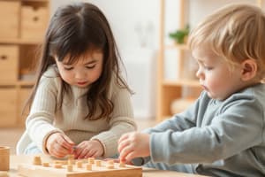
- Fine motor skills involve manipulating small objects, transferring items from hand to hand, and hand-eye coordination tasks.
- They often require precise movements for delicate tasks.
- Examples include using the pincer grasp, cutting, coloring, writing, and threading beads.
- Fine motor development refers to skills using smaller muscle groups.
Ambidexterity
- Ambidexterity is a skill where there is no dominance between body symmetries, allowing fine motor skills to be performed with either extremity.
- The most common example is writing with either hand.
Voluntary Movements
- Most voluntary movements, initiated by the cerebral cortex, activate functional patterns stored in lower brain areas, including the spinal cord, brain stem, basal ganglia, and cerebellum.
- These lower centers then send control signals to the muscles.
- The cortex has a direct pathway to anterior motor neurons for fine, dexterous movements of fingers and hands.
Skeletal Muscle Contraction
- Skeletal muscle contraction is initiated by lower motor neurons in the spinal cord and brainstem.
- These neurons are located in the ventral horn of the spinal cord gray matter and in the motor nuclei of the cranial nerves.
- They send axons to skeletal muscles via ventral roots and spinal peripheral nerves or cranial nerves.
- Spatial and temporal patterns of activation are determined by local circuits within the spinal cord and brainstem.
- These circuits receive sensory input and mediate sensorimotor reflexes, also maintaining interconnections for coordinated behaviors.
- Descending pathways from higher centers modulate lower motor neuron activity by influencing local circuitry.
- Upper motor neurons are located in brainstem centers like the vestibular nuclei, superior colliculus, reticular formation, and cerebral cortex.
- Upper motor neurons initiate and guide involuntary and voluntary movements by synapsing on local circuit neurons, which connect with lower motor neurons.
- Lower motor neurons are the final common pathway for transmitting information from various sources to skeletal muscles.
- The motor system consists of four interactive subsystems:
- Lower motor neurons that innervate skeletal muscles and local circuit neurons.
- Upper motor neurons in the brainstem or cerebral cortex synapse with local circuit neurons or lower motor neurons.
- The cerebellum detects and attenuates motor errors.
- The basal ganglia prevent unwanted movements and prepare motor circuits for initiating movements.
Motor Signals
- Motor signals are transmitted from the cerebral cortex to the spinal cord directly through the corticospinal tract and indirectly through accessory pathways involving the basal ganglia, cerebellum, and brain stem nuclei.
- Direct pathways are more involved with discrete and detailed movements.
- The non-primary motor cortex is divided into the supplementary motor cortex, and the pre-motor cortex:
- Supplementary motor cortex is involved in the execution of movement sequences, motor skill attainment, and movement selection based on sensory input.
- Pre-motor cortex contributes to the corticospinal tract and is active during motor planning.
The Cerebrum's Role in Motor Control
- Primary motor area (M1) in the precentral gyrus initiates voluntary movement; corticospinal tracts originate here.
- Lesions to M1 cause loss of voluntary movement in the contralateral body part corresponding to the damaged motor homunculus area and a loss of the ability to implement a specific motor plan. Premotor Area:
- Located anterior to M1; involved in motor planning or praxis.
- Lesions to the premotor area cause apraxia or motor planning difficulties.
- Apraxia involves either the inability to understand task demands or the inability to access the appropriate motor plan.
Motor Association Area
- Also called the prefrontal area, located in the anterior frontal lobe.
- Involved in the cognitive planning of movement.
- Lesions cause the loss of stored motor plans, resulting in the inability to cognitively understand how to carry out a previously known motor task.
- The premotor area may compensate for damage to the motor association area.
Praxis
- Praxis involves three processes:
- Ideational praxis: the ability to cognitively understand the motor demands of a task, largely a function of the motor association area.
- Ideomotor planning I: the ability to access the appropriate motor plan, commonly stored in the premotor area.
- Ideomotor planning II: the ability to implement the appropriate motor plan, commonly involving M1.
- The supplementary motor area is part of the premotor area.
- Located inside the medial longitudinal fissure, it controls bilateral posture.
- Lesions may result in loss of bilateral control of posture; other areas like the cerebellum or vestibular system may compensate.
- The frontal eye field is located in the middle frontal gyrus, anterior to the premotor area.
- It is responsible for visual saccades.
- Lesions result in deviation of the eyes to the same side of the lesion.
Incoming Motor Cortex Fiber Pathways
- The motor cortex is controlled by nerve signals from the somatosensory system and, to some degree, from other sensory systems like hearing and vision.
- Once sensory information is received, the motor cortex operates in association with the basal ganglia and cerebellum to excite appropriate motor actions.
- Key incoming fiber pathways include:
- Subcortical fibers from adjacent regions of the cerebral cortex, especially from somatosensory areas of the parietal cortex, adjacent areas of the frontal cortex anterior to the motor cortex, and the visual and auditory cortices.
- Subcortical fibers arriving through the corpus callosum from the opposite cerebral hemisphere, connecting corresponding areas of the cortices in the two sides of the brain.
- Somatosensory fibers arriving directly from the ventrobasal complex of the thalamus, sending cutaneous tactile signals and joint and muscle signals from the peripheral body.
- Tracts from the ventrolateral and ventroanterior nuclei of the thalamus, which receive signals from the cerebellum and basal ganglia, coordinating motor control functions.
- Fibers from the intralaminar nuclei of the thalamus, controlling the general level of excitability of the motor cortex.
Role of the Brain Stem
- The brain stem consists of the medulla, pons, and mesencephalon.
- It contains motor and sensory nuclei for the face and head regions, similar to the spinal cord for the neck down.
- It performs special control functions, including control of respiration, the cardiovascular system, partial control of gastrointestinal function, control of stereotyped movements, control of equilibrium, and control of eye movements.
- It serves as a way station for command signals from higher neural centers.
Cerebellum and Basal Ganglia Contributions
- The cerebellum and basal ganglia are essential for normal motor function.
- They function in association with other motor control systems, not independently.
- The cerebellum plays a key role in the timing of motor activities and smooth progression from one movement, controlling muscle contraction intensity and interplay between agonist and antagonist muscle groups.
- The basal ganglia help plan and control complex movement patterns, controlling movement intensities, directions, and sequencing for complex motor goals.
Cerebellum and Its Motor Functions
- Electrical excitation of the cerebellum doesn't cause conscious sensation or motor movement.
- Removal of the cerebellum causes abnormal body movements.
- The cerebellum is vital during rapid muscular activities like running, typing, and talking.
- Loss of the cerebellum can cause incoordination, without paralysis.
- The cerebellum helps sequence motor activities, monitoring and adjusting body movements to conform to motor signals from the cerebral motor cortex.
- The cerebellum receives updated information about muscle contraction sequences and sensory information from the body's periphery.
- It compares actual movements with intended movements and transmits corrective signals to adjust muscle activation.
- The cerebellum aids the cerebral cortex in planning the next sequential movement in advance.
- The cerebellar circuit learns from mistakes, adjusting the excitability of neurons to improve subsequent muscle contractions.
Basal Ganglia and Their Motor Functions
- Basal ganglia work with the cerebral cortex and corticospinal motor control system.
- Basal ganglia receive input signals from, and return output signals to, the cerebral cortex.
- Principal role: works with the corticospinal system to control complex motor activity patterns.
- Damage to the basal ganglia impairs the cortical system's ability to provide patterns; writing becomes crude.
- Patterns that require basal ganglia intervention include cutting paper, hammering nails, shooting a basketball, throwing a baseball, shoveling dirt, vocalization, controlled eye movements, and other skilled subconscious movements.
- Basal ganglia coordinate using excitatory (glutamate) and inhibitory (GABA) neurons.
- The basal ganglia work in two distinct pathways:
- Direct pathway: initiates movement which turns on the motor cortex by disinhibiting thalamic control of motor planning.
- Indirect pathway: terminates movement by turning off the motor cortex by activating inhibition. which results in the inhibition of activity in the indirect pathway, which leads to the facilitation of movement.
- Glutamate excitatory neurons from the thalamus stimulate movement by projecting to motor regions of the cerebral cortex.
- Neurons from the GPi and SNpr steadily release GABA, inhibiting thalamic neurons and suppressing unwanted movement.
- When a movement is wanted, information is sent from the cortex to the striatum (CSP), where glutamate neurons excite striatum neurons, releasing GABA in the GPi and SNpr, stopping the inhibition of thalamic neurons involved in the desired movement, thus opening the gate for the movement to occur.
- The SNpc modulates the direct pathway by sending neurons to the striatum through the nigrostriatal pathway, where they release dopamine, facilitating activity in the direct pathway.
- The indirect pathway is involved with inhibiting movement: cortex projections activate GABA neurons in the striatum causing the activation of GABA neurons which exert an inhibitory effect on glutamate neurons in the subthalamic nucleus which activate GABA neurons and inhibit thalamic neurons from traveling to the motor regions of the cerebral cortex to stimulate movement
- Activity from these two pathways inhibits these thalamic neurons and thus inhibits movement; the activity in the indirect pathway antagonizes the activity of the direct pathway and acts to keep unwanted movements from occurring
- SNpc modulate the activity of the indirect pathway through dopamine release.
- Dopamine depletion causes difficulties in initiating movement.
The Spinal Level
- The spinal cord has local movement patterns for all muscle areas, such as withdrawal reflexes from pain.
- Complex rhythmical motions like walking and reciprocal motions are programmed in the cord.
- High levels of motor control can command these patterns into action or inhibit them.
The Hindbrain Level
- The hindbrain provides two major functions for motor control:
- Maintenance of axial tone for standing.
- Continuous modification of muscle tone based on vestibular apparatus information for maintaining equilibrium.
The Motor Cortex Level
- The motor cortex provides activating motor signals to the spinal cord.
- It issues sequential and parallel commands to activate cord patterns, change intensities, or modify timing.
- Corticospinal system can bypass cord patterns with higher-level patterns from the brain stem or cerebral cortex.
- Cortical patterns are complex and can be learned; cord patterns are mainly determined by heredity.
Associated Functions of the Cerebellum
- The cerebellum enhances the stretch reflex by transmitting it through the cerebellum and back to the cord.
- The cerebellum makes postural movements (especially from the equilibrium system) smooth and continuous at the brain stem level.
- At the cerebral cortex level, the cerebellum provides extra motor force at the start of a movement, turns on antagonist muscles to stop the movement, and programs muscle contractions for smooth transitions between rapid movements.
- Cerebellum mainly functions when muscle movements must be rapid; slow movements can occur without it.
Associated Functions of the Basal Ganglia
- The basal ganglia help the cortex execute subconscious patterns of movement and plan multiple parallel and sequential movement patterns to accomplish a task.
- Motor patterns that require the basal ganglia include writing, throwing a ball, and typing.
- The basal ganglia also allow action in response to stimuli.
Equilibrium
- The receptors are the hair cells in the semicircular canals, utricle, and saccule located in the inner ear.
- The semicircular canals respond to angular acceleration and deceleration (rotation) and linear acceleration and deceleration.
- The utricle and saccule respond to gravity and changes in head position.
Studying That Suits You
Use AI to generate personalized quizzes and flashcards to suit your learning preferences.