Podcast
Questions and Answers
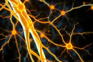
Alpha motor neurons innervate intrafusal muscle fibers.
Alpha motor neurons innervate intrafusal muscle fibers.
False (B)
Lower motor neurons originate in the anterior horn of the spinal cord or motor cranial nuclei.
Lower motor neurons originate in the anterior horn of the spinal cord or motor cranial nuclei.
True (A)
Renshaw cells are inhibitory and receive collateral branches from motor axons.
Renshaw cells are inhibitory and receive collateral branches from motor axons.
True (A)
Lateral inhibition enhances the spread of signals to adjacent neurons.
Lateral inhibition enhances the spread of signals to adjacent neurons.
Gamma efferent neurons give off type B gamma nerve fibers
Gamma efferent neurons give off type B gamma nerve fibers
Tactile sensors are predominantly located in the fingertips and tongue.
Tactile sensors are predominantly located in the fingertips and tongue.
Stereognosis involves integrating signals from adjacent receptors into a chemical pattern.
Stereognosis involves integrating signals from adjacent receptors into a chemical pattern.
Synthetic senses are formed at the cortical level through the combination of basic sensations.
Synthetic senses are formed at the cortical level through the combination of basic sensations.
Proprioceptive sensations are exclusively carried by type C nerve fibers to the brain.
Proprioceptive sensations are exclusively carried by type C nerve fibers to the brain.
Dynamic proprioception, also known as kinesthesia, involves conscious recognition of body movement, including evaluating rates of movement .
Dynamic proprioception, also known as kinesthesia, involves conscious recognition of body movement, including evaluating rates of movement .
Voluntary movements, such as playing the piano, are improved with practice and are purposeful and goal-oriented.
Voluntary movements, such as playing the piano, are improved with practice and are purposeful and goal-oriented.
Mixed pattern movements are purely voluntary acts that never involve any repetitive reflexive action.
Mixed pattern movements are purely voluntary acts that never involve any repetitive reflexive action.
The efferent fibers of a reflex arc enter the spinal cord via the dorsal roots.
The efferent fibers of a reflex arc enter the spinal cord via the dorsal roots.
Reflexes are voluntary, rapid, and stereotyped movements.
Reflexes are voluntary, rapid, and stereotyped movements.
Sensory signals entering the spinal cord travel solely to the brain stem and cerebral cortex, bypassing local segmental motor responses.
Sensory signals entering the spinal cord travel solely to the brain stem and cerebral cortex, bypassing local segmental motor responses.
Occlusion of the middle cerebral artery will cause contralateral spastic paresis and impaired sensation of the face and lower extremities.
Occlusion of the middle cerebral artery will cause contralateral spastic paresis and impaired sensation of the face and lower extremities.
Damage to the supplementary motor cortex results in the patient being unable to perform the sequence of a particular movement.
Damage to the supplementary motor cortex results in the patient being unable to perform the sequence of a particular movement.
The premotor cortex assembles the details of the mental planning of a motor act received from the primary motor area.
The premotor cortex assembles the details of the mental planning of a motor act received from the primary motor area.
The supplementary motor cortex is positioned superior to the primary motor cortex on the lateral side of the frontal lobe.
The supplementary motor cortex is positioned superior to the primary motor cortex on the lateral side of the frontal lobe.
Damage to the premotor cortex can lead to motor apraxia, a defect in motor performance without paralysis, because the selection of a particular movement is impaired.
Damage to the premotor cortex can lead to motor apraxia, a defect in motor performance without paralysis, because the selection of a particular movement is impaired.
Flashcards
Classes of Movement
Classes of Movement
Three categories of CNS motor functions: reflexes, voluntary, and mixed movements.
Reflexes
Reflexes
Involuntary, rapid responses to stimuli involving a reflex arc; examples include knee jerk and eye blink.
Reflex Arc
Reflex Arc
The functional unit of reflex action consisting of receptors, neurons, and effectors that mediate reflexes.
Voluntary Movement
Voluntary Movement
Signup and view all the flashcards
Spinal Cord Reflexes
Spinal Cord Reflexes
Signup and view all the flashcards
Tactile sensors
Tactile sensors
Signup and view all the flashcards
Proprioceptive sensations
Proprioceptive sensations
Signup and view all the flashcards
Static proprioceptive sensation
Static proprioceptive sensation
Signup and view all the flashcards
Dynamic proprioceptive sensation
Dynamic proprioceptive sensation
Signup and view all the flashcards
Pain sensation
Pain sensation
Signup and view all the flashcards
Lower Motor Neurons
Lower Motor Neurons
Signup and view all the flashcards
Alpha Motor Neurons
Alpha Motor Neurons
Signup and view all the flashcards
Gamma Motor Neurons
Gamma Motor Neurons
Signup and view all the flashcards
Interneurons
Interneurons
Signup and view all the flashcards
Renshaw Cells
Renshaw Cells
Signup and view all the flashcards
Middle Cerebral Artery (MCA)
Middle Cerebral Artery (MCA)
Signup and view all the flashcards
Anterior Cerebral Artery (ACA)
Anterior Cerebral Artery (ACA)
Signup and view all the flashcards
Supplementary Motor Cortex
Supplementary Motor Cortex
Signup and view all the flashcards
Premotor Cortex
Premotor Cortex
Signup and view all the flashcards
Motor Apraxia
Motor Apraxia
Signup and view all the flashcards
Study Notes
Central Nervous System Physiology
- The central nervous system (CNS) coordinates activities of other systems to maintain homeostasis
- The CNS stores experiences (memories) and establishes response patterns
- The CNS has three functional levels:
- Spinal cord level: Conduit for signals from body periphery to the brain and vice versa, reflex control centers
- Lower brain level (subcortical level): Controls subconscious activities ; e.g., arterial pressure, respiration, equilibrium, emotions
- Higher brain level (cortical level): Determines specific brain functions; e.g. operations, memory, thought processes
- Neuronal pools: Collections of interconnected neurons with specific organization characteristics
Sensory Functions of the CNS
- Somatosensory system: Detects sensory stimuli from skin, muscles, tendons, and joints
- Types of sensory receptors:
- Mechanoreceptors: Detect mechanical deformation (touch, pressure, vibration, itch, stereognosis, equilibrium, and proprioception)
- Thermoreceptors: Detect temperature changes
- Nociceptors (pain receptors): Detect tissue damage
- Photoreceptors: Detect light
- Chemoreceptors: Detect chemicals (taste, smell, O2, CO2 concentration, osmolality)
- Sensory units: Single sensory axons with branches forming a receptive field
- Adaptation / Desensitization of Receptors
- Tonic receptors: Adapt slowly (pressure, etc)
- Phasic receptors: Adapt quickly (touch)
- Tactile sensations: Detection of touch, pressure, itch, tickle, vibration, stereognosis (touch to determine object)
- Proprioception: Sensation of body position and movement
- Pain: Unpleasant sensory and emotional experience
Somatosensory Functions of the CNS
- Pain sensation:
- Acute pain (fast): Occurs rapidly after injury (e.g., skin burn).
- Chronic pain (slow): Develops gradually after injury or condition, often described as throbbing or burning.
- Mechanism:
- Pain signals travel from sensory receptors to the spinal cord, then to the brain.
- The brainstem and higher brain centers modulate pain perception through descending pathways.
- Types of pain receptors:
- Mechanosensitive: Activated by mechanical pressure or damage.
- Thermosensitive: Activated by extreme temperature changes.
- Chemosensitive: Activated by chemical substances released at sites of injury (e.g., substance P, lactic acid).
- Central inhibition of pain:
- Presynaptic inhibition: Inhibits the transmission of pain signals before they reach the synapse.
- Postsynaptic Inhibition: Activates inhibitory interneurons that inhibit neurons responsible for pain signalling.
- Referred pain: Pain felt in a location distant from the site of damage due to common sensory pathways within the spinal cord.
Higher Interpretation of Sensory Signals
- Primary sensory areas: Specific areas in the cerebral cortex where simple sensory inputs are received (e.g., primary auditory, visual, and somatic sensory areas).
- Sensory association areas: Areas that integrate sensory information to form a more complex perception from the primary sensory areas (e.g., sensory association areas for touch interpretation).
- Wernicke's area: An important language area located in the temporal lobe, where it interprets the spoken word, combining information from sensory association areas.
Somatomotor Functions of the CNS
- Ascending tracts: Convey sensory information from the body to the brain
- Descending tracts: Convey motor commands from the brain to the body
- Lateral pathways: Control voluntary movements of distal muscles (corticospinal tract, rubrospinal tract)
- Ventromedial pathways: Control posture, muscle tone, and gross movements of the neck, trunk, and proximal limbs (anterior corticospinal tract, tectospinal tract, medial reticulospinal tracts)
- Pyramidal tract: Originates in the motor cortex and directly controls voluntary movement of skeletal muscles.
- Extrapyramidal tract: Regulates involuntary movements and posture, including the basal ganglia and cerebellum.
Cerebellum
- The cerebellum is a structure located below the occipital lobe that functions in:
- Executing smooth and coordinated movements
- Regulating posture, balance, and equilibrium.
- Coordinating sequences of muscle actions.
- Motor learning.
- The cerebellum receives input from the spinal cord, brainstem, and motor cortex.
Spinal Cord Reflexes
- Spinal cord reflexes are involuntary stereotyped patterns of response to stimuli.
- Reflex arc: A pathway mediating the reflex, including sensory receptors, afferent neurons, motor neurons, and effectors.
- Stretch reflex: A monosynaptic reflex that maintains muscle tone and resists sudden changes in muscle length
- Golgi tendon reflex (inverse stretch reflex)
The Brainstem
- Midbrain (mesencephalon): Contains structures involved in auditory, visual, and postural reflexes, eye movements, as well as contains descending motor tracts
- Pons (metencephalon): Contains structures involved in regulating breathing, sleep-wake cycle, and motor nuclei for cranial nerves
- Medulla oblongata (myelencephalon): Contains control centers for vital functions; e.g heart rate, blood pressure, and breathing
The Hypothalamus
- The hypothalamus is a major integrating center of the autonomic nervous system, including hormonal, emotional and visceral functions such as:
- Autonomic regulation
- Endocrine regulation
- Thermoregulation
- Regulation of food intake
- Regulation of water balance (thirst mechanism)
- Biological clock
- Regulation of emotions
- Circadian rhythm
The Limbic System
- The limbic system comprises structures from the cerebrum and diencephalon that participate in emotional behavior, memory, and motivation.
- Major components: amygdala, hippocampus, cingulate gyrus.
- Hypothalamus plays a major role
The Cerebrospinal Fluid (CSF)
- CSF is a clear, colorless fluid that circulates in and around the brain and spinal cord.
- Functions:
- Protection: Cushions the brain and spinal cord from impacts.
- Buoyancy: Reduces the weight of the brain.
- Removal of metabolic waste: It helps flush out metabolic byproducts from the CNS.
- Compartments of CSF:
- Ventricles within the brain and their connecting channels.
- Subarachnoid space surrounding the brain and spinal cord
Blood-Brain Barrier (BBB)
- The blood-brain barrier selectively restricts passage of substances from the blood into the interstitial fluid surrounding the neurons.
- Structures that make up the BBB: tight junctions between endothelial cells of the brain capillaries, and end feet of astrocytes.
Studying That Suits You
Use AI to generate personalized quizzes and flashcards to suit your learning preferences.