Podcast
Questions and Answers
What are the major structural elements of the cytoskeleton?
What are the major structural elements of the cytoskeleton?
- Actin, myosin, tropomyosin
- Microfilaments, microtubules, intermediate filaments (correct)
- Ribosomes, endoplasmic reticulum, Golgi apparatus
- DNA, RNA, proteins
What are microtubules composed of?
What are microtubules composed of?
- Tubulin subunits (correct)
- Myosin subunits
- Keratin subunits
- Actin subunits
Which bacterial cytoskeletal element is involved in DNA segregation and cell shape?
Which bacterial cytoskeletal element is involved in DNA segregation and cell shape?
- Crescentin
- Actin-like MreB (correct)
- Myosin-like ParM
- Tubulin-like FtsZ
What are microfilaments essential components of?
What are microfilaments essential components of?
What has research shown about the cytoskeleton?
What has research shown about the cytoskeleton?
What are intermediate filaments composed of?
What are intermediate filaments composed of?
What are the protein building blocks of microtubules?
What are the protein building blocks of microtubules?
What contributes to the dynamic instability of microtubules?
What contributes to the dynamic instability of microtubules?
Where do microtubules originate from?
Where do microtubules originate from?
What is the function of microtubule-associated proteins (MAPs)?
What is the function of microtubule-associated proteins (MAPs)?
What type of polymerization do microtubules undergo?
What type of polymerization do microtubules undergo?
What is the role of microtubules in cancer treatment?
What is the role of microtubules in cancer treatment?
Which type of proteins bind at regular intervals along a microtubule wall?
Which type of proteins bind at regular intervals along a microtubule wall?
Which proteins promote depolymerization of microtubules?
Which proteins promote depolymerization of microtubules?
What do some microtubule-binding proteins use ATP for?
What do some microtubule-binding proteins use ATP for?
Which proteins act at the ends of microtubules and promote the peeling of subunits from the ends?
Which proteins act at the ends of microtubules and promote the peeling of subunits from the ends?
What is the function of katanin in relation to microtubules?
What is the function of katanin in relation to microtubules?
Which proteins prevent the polymerization of tubulin heterodimers?
Which proteins prevent the polymerization of tubulin heterodimers?
What do microtubule-binding proteins use ATP for, in some cases?
What do microtubule-binding proteins use ATP for, in some cases?
What type of proteins regulate microtubule structure?
What type of proteins regulate microtubule structure?
What is the main function of microfilaments?
What is the main function of microfilaments?
What is the structural core of microvilli composed of?
What is the structural core of microvilli composed of?
What does actin polymerize to form?
What does actin polymerize to form?
What do G-actin molecules polymerize to form?
What do G-actin molecules polymerize to form?
How are F-actin filaments structured?
How are F-actin filaments structured?
What is the orientation of all the actin monomers in the microfilament filament?
What is the orientation of all the actin monomers in the microfilament filament?
What are the fundamental subunits of intermediate filament proteins?
What are the fundamental subunits of intermediate filament proteins?
How long is the central rodlike domain of intermediate filament proteins?
How long is the central rodlike domain of intermediate filament proteins?
What role do intermediate filaments play in tissues?
What role do intermediate filaments play in tissues?
What is the structure of the basic unit of intermediate filaments?
What is the structure of the basic unit of intermediate filaments?
Which cytoskeletal element resists bending when a cell is compressed?
Which cytoskeletal element resists bending when a cell is compressed?
Which proteins connect intermediate filaments, microfilaments, and microtubules?
Which proteins connect intermediate filaments, microfilaments, and microtubules?
What is the primary function of microfilaments?
What is the primary function of microfilaments?
What is the composition of a tetrameric protofilament of intermediate filaments?
What is the composition of a tetrameric protofilament of intermediate filaments?
What is the name of the protein that binds to actin microfilaments in a distinctive arrowhead pattern?
What is the name of the protein that binds to actin microfilaments in a distinctive arrowhead pattern?
What is the plus end of microfilaments called?
What is the plus end of microfilaments called?
Which protein binds a large amount of free G-actin?
Which protein binds a large amount of free G-actin?
What is the function of ADF/cofilin?
What is the function of ADF/cofilin?
Which protein is important in forming loose networks of crosslinked actin filaments?
Which protein is important in forming loose networks of crosslinked actin filaments?
What complex is activated by proteins like WASP and WAVE/Scar to form a dendritic network of actin?
What complex is activated by proteins like WASP and WAVE/Scar to form a dendritic network of actin?
What does contractility refer to in the context of cell motility?
What does contractility refer to in the context of cell motility?
What is the primary function of microfilament-based motility?
What is the primary function of microfilament-based motility?
What do molecular motors couple ATP hydrolysis to?
What do molecular motors couple ATP hydrolysis to?
What are microtubules and microfilaments likened to in the context of motile systems?
What are microtubules and microfilaments likened to in the context of motile systems?
What is the function of motor proteins at the molecular level?
What is the function of motor proteins at the molecular level?
What is an example of microtubule-based motility?
What is an example of microtubule-based motility?
What is the structure that gives myofibrils a pattern of alternating dark and light bands?
What is the structure that gives myofibrils a pattern of alternating dark and light bands?
Which protein constitutes a calcium-sensitive switch that activates contraction in striated muscle?
Which protein constitutes a calcium-sensitive switch that activates contraction in striated muscle?
What is the primary component of thick filaments in skeletal muscle?
What is the primary component of thick filaments in skeletal muscle?
What does the sliding filament model propose as the cause of muscle contraction?
What does the sliding filament model propose as the cause of muscle contraction?
What is the arrangement of myosin in a thick filament?
What is the arrangement of myosin in a thick filament?
What is the function of tropomyosin in skeletal muscle?
What is the function of tropomyosin in skeletal muscle?
What gives rise to the observed shortening of sarcomeres during contraction?
What gives rise to the observed shortening of sarcomeres during contraction?
What constitutes the thin filaments in sarcomeres?
What constitutes the thin filaments in sarcomeres?
What are kinesins and dyneins?
What are kinesins and dyneins?
What is the role of dyneins in intracellular movement?
What is the role of dyneins in intracellular movement?
What is the function of myosins in cellular processes?
What is the function of myosins in cellular processes?
What causes cilia and flagella to bend, generating forces for movement?
What causes cilia and flagella to bend, generating forces for movement?
What is the primary responsibility of type II myosins?
What is the primary responsibility of type II myosins?
What are skeletal muscles composed of?
What are skeletal muscles composed of?
What did Chargaff observe about the amount of nucleotides in DNA?
What did Chargaff observe about the amount of nucleotides in DNA?
What did Watson and Crick propose about the structure of DNA?
What did Watson and Crick propose about the structure of DNA?
What was known about the bases in DNA at physiological pH?
What was known about the bases in DNA at physiological pH?
What did Erwin Chargaff study in DNA?
What did Erwin Chargaff study in DNA?
What was Chargaff's most striking observation about the base composition of DNA?
What was Chargaff's most striking observation about the base composition of DNA?
What did Watson and Crick build to determine the structure of DNA?
What did Watson and Crick build to determine the structure of DNA?
What did Chargaff show about the DNA from different cells of a given species?
What did Chargaff show about the DNA from different cells of a given species?
What is the significance of Chargaff's rules?
What is the significance of Chargaff's rules?
What did Chargaff's rules reveal about the nucleotide bases?
What did Chargaff's rules reveal about the nucleotide bases?
What did Watson and Crick determine about the structure of DNA?
What did Watson and Crick determine about the structure of DNA?
Who developed the double helix model of DNA?
Who developed the double helix model of DNA?
How many base pairs are there per helical turn in the double helix model of DNA?
How many base pairs are there per helical turn in the double helix model of DNA?
What is the distance per nucleotide pair in the double helix model of DNA?
What is the distance per nucleotide pair in the double helix model of DNA?
What type of pairing adheres to Chargaff’s rules in DNA?
What type of pairing adheres to Chargaff’s rules in DNA?
How is DNA length measured?
How is DNA length measured?
What is the biological significance of Z-DNA?
What is the biological significance of Z-DNA?
What mediates the interconversion between relaxed and supercoiled forms of DNA?
What mediates the interconversion between relaxed and supercoiled forms of DNA?
What induces strand separation (denaturation) in DNA?
What induces strand separation (denaturation) in DNA?
What is the challenge of DNA packaging in eukaryotes?
What is the challenge of DNA packaging in eukaryotes?
What are plasmids in bacteria?
What are plasmids in bacteria?
Flashcards are hidden until you start studying
Study Notes
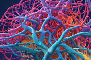
Microtubules: Structure, Assembly, and Function
- Microtubules (MTs) are the largest structural elements of the cytoskeleton, involved in various cell functions.
- Two types of microtubules exist: cytoplasmic microtubules and their functions in maintaining axons, formation of spindles, and cell shape.
- Tubulin heterodimers are the protein building blocks of microtubules, consisting of protofilaments with α and β-tubulin.
- Protofilaments have inherent polarity with plus and minus ends, and microtubules form through reversible polymerization of tubulin dimers.
- Microtubule assembly involves nucleation, elongation, and critical concentration of tubulin dimers.
- Addition of tubulin dimers occurs more quickly at the plus ends of microtubules, leading to treadmilling.
- GTP hydrolysis contributes to the dynamic instability of microtubules, involving GTP-tubulin and dynamic instability with catastrophe and rescue phases.
- Microtubules originate from microtubule-organizing centers (MTOCs) and centrosomes containing γ-tubulin ring complexes (γ-TuRCs).
- MTOCs organize and polarize microtubules within cells, nucleating and anchoring MTs with fixed polarity.
- Microtubules facilitate various cellular functions, including maintaining cell shape and facilitating intracellular transport.
- Microtubule-based drugs such as taxanes and vinca alkaloids target microtubule dynamics for cancer treatment.
- Microtubule-associated proteins (MAPs) regulate microtubule dynamics and stability, influencing their functions in the cell.
Cytoskeleton Proteins: Actin and Intermediate Filaments
- Myosin subfragment 1 (S1) binds to actin microfilaments (MFs) in a distinctive arrowhead pattern
- The plus end of microfilaments is called the barbed end, and the minus end is called the pointed end
- Actin monomers in the cytosol bind ATP and are converted to ADP after complexing ATP
- Microfilaments show polarity with more rapid addition or loss of G-actin at the plus end
- Cells can dynamically assemble actin into various structures using actin-binding proteins
- Thymosin β4 binds a large amount of free G-actin, while profilin competes for G-actin binding
- ADF/cofilin binds ADP-G-actin and F-actin to increase turnover of ADP-actin at the minus end
- Capping proteins like CapZ and tropomodulins regulate the growth of microfilaments
- Filamin is important in forming loose networks of crosslinked actin filaments
- Actin may be bundled by proteins like α-actinin and fascin into highly organized arrays
- Microfilaments are connected to the plasma membrane by crosslinks made of myosin I and calmodulin
- Actin can form a dendritic network through the Arp2/3 complex, activated by proteins like WASP and WAVE/Scar
Microtubule and Microfilament-Based Intracellular Movement
- Microtubules act as tracks for organelle and vesicle transport inside cells
- Kinesins and dyneins are motor proteins that move along microtubules
- Kinesins move toward the plus ends of microtubules and are involved in fast axonal transport
- Dyneins move cargo toward the minus ends of microtubules and are linked to cargo by dynactin protein complexes
- Microtubule motors are crucial for shaping the endomembrane system and transporting vesicles
- Cilia and flagella are involved in cellular movement and consist of axoneme connected to a basal body
- Doublet sliding within the axoneme causes cilia and flagella to bend, generating forces for movement
- Myosins, a large superfamily of ATP-dependent motors, interact with and exert force on actin microfilaments
- Myosins function in muscle contraction, cell movement, phagocytosis, and vesicle transport
- Type II myosins are well understood and responsible for pulling arrays of actin filaments together, resulting in cell contraction
- Muscle contraction is a familiar example of mechanical work mediated by intracellular filaments
- Skeletal muscles are composed of parallel muscle fibers responsible for voluntary movement
DNA Structure and Packaging
- The double helix model of DNA was developed by Watson and Crick, with critical evidence from X-ray diffraction data by Rosalind Franklin
- The model revealed 10 base pairs per helical turn and a 0.34 nm distance per nucleotide pair
- Purine-pyrimidine pairing adheres to Chargaff’s rules, with hydrogen bonding between complementary bases (adenine-thymine, guanine-cytosine)
- The double helix model suggested a mechanism for DNA replication, where the strands could act as templates for new complementary strands
- DNA length is measured in base pairs (bp), with larger stretches in kilobases (kb)
- DNA can form right-handed (BDNA) and left-handed (Z-DNA) helices, with Z-DNA's biological significance not fully understood
- DNA can be interconverted between relaxed and supercoiled forms, with positive and negative supercoils
- Supercoiling occurs in both linear and circular DNA, mediated by topoisomerases which induce and relax supercoils
- Strand separation (denaturation) can be induced by raising temperature or pH, while reformation (renaturation) occurs by lowering the temperature to permit hydrogen bonds to reform
- DNA packaging is a challenge for all forms of life, especially in eukaryotes where DNA interacts with histones to form chromatin
- Bacterial chromosomes are bound to proteins, negatively supercoiled, and folded into loops held in place by RNAs and proteins
- Bacteria may contain plasmids, small circular DNA molecules with genes for replication and cellular functions, including virulence factors and metabolic enzymes.
Studying That Suits You
Use AI to generate personalized quizzes and flashcards to suit your learning preferences.